Case Reports
. 2023 Dec 4;220(12):e20231045.
doi: 10.1084/jem.20231045. Epub 2023 Oct 31.

First-in-human therapy with Treg produced from thymic tissue (thyTreg) in a heart transplant infant

Affiliations
Case Reports

First-in-human therapy with Treg produced from thymic tissue (thyTreg) in a heart transplant infant

Esther Bernaldo-de-Quirós et al. J Exp Med. .

Abstract

Due to their suppressive capacity, regulatory T cells (Tregs) have attracted growing interest as an adoptive cellular therapy for the prevention of allograft rejection, but limited Treg recovery and lower quality of adult-derived Tregs could represent an obstacle to success. To address this challenge, we developed a new approach that provides large quantities of Tregs with high purity and excellent features, sourced from thymic tissue routinely removed during pediatric cardiac surgeries (thyTregs). We report on a 2-year follow-up of the first patient treated worldwide with thyTregs, included in a phase I/II clinical trial evaluating the administration of autologous thyTreg in infants undergoing heart transplantation. In addition to observing no adverse effects that could be attributed to thyTreg administration, we report that the Treg frequency in the periphery was preserved during the 2-year follow-up period. These initial results are consistent with the trial objective, which is to confirm safety of the autologous thyTreg administration and its capacity to restore the Treg pool.


Conflict of interest statement

Disclosures: E. Bernaldo-de-Quirós reported a patent to WO2019/166658A1 pending. M. Pion reported a patent to WO2019/166658A1 issued. R. Correa-Rocha reported personal fees from THYTECH outside the submitted work; in addition, R. Correa-Rocha had a patent to WO2019/166658A1 pending. No other disclosures were reported.

Figures

None
Graphical abstract
Figure S1.
Figure S1.
Gating strategy of thyTreg cells. Flow cytometry gating strategy and representative dot plots for immune cell populations and subpopulations. After gating on singlets, lymphocytes (thyTreg cells) were identified by size (forward scatter [FSC]) and complexity (side scatter [SSC]). Within this population, viable thyTreg were identified. Within viable thyTreg cells, we determined the purity in terms of CD25 and FOXP3 expression (CD25+FOXP3+). Fluorescence minus one (FMO) of FOXP3 is shown to determine background signal.
Figure 1.
Figure 1.
Characteristics of the infused thyTreg product. (A and B) Frequency of phenotypic/functionality (A) and homing (B) markers within viable cells of the thyTreg product infused to patient thy1-01 (purple) in comparison with the expression in peripheral Tregs analyzed on PBMCs (gray) from a blood sample collected immediately pretransplant (pre-Tx) from the same patient. (C) Quantification of cytokine release capacity of thyTreg in culture supernatant of infused thyTreg product. Anti-inflammatory cytokine IL-10 in blue; proinflammatory cytokines in red. (D–F) Suppressive capacity of infused thyTreg product, defined as % inhibition of CD4+ T cell (D) and CD8+ T cell (E) proliferation at the indicated ratios. Stability of FOXP3 expression in the thyTreg product under control (CT, blue) or Th1 (orange) and Th17 (green) proinflammatory conditions (F).
Figure S2.
Figure S2.
Additional characterization and phenotypic and functional stability of the infused thyTreg product. (A) Demethylation level of six genome regions located in four genes (calculated as the mean of demethylation of the CpGs contained in the region) within thyTreg cell product and thyTconv of patient thy1-01 (female). FOXP3-ADS783 corresponds to the TSDR region of FOXP3. An aliquot of the thyTreg cell product (thy1-01) was restimulated under control (CT, blue) or under Th1 (orange) and Th17 (green) proinflammatory conditions and evaluated after 3 days of culture. (B) The frequency of FOXP3 in viable cells is representative of the phenotypic stability. (C and D) Summary of quantitation of secreted IFN-γ (C) and IL-17A (D) by thyTreg or restimulated PBMC (n = 3) under proinflammatory conditions. (E and F) Summary of the suppressive capacity as inhibition of CD4+ (E) and CD8+ (F) T cell proliferation of thyTreg at ratio 1:1 in control or Th1 and Th17 switching conditions.
Figure 2.
Figure 2.
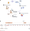
Schematic representation of the clinical trial (NCT04924491). (A and B) Graphic summary of the thyTreg infusion performed in patient thy1-01 (A) and peripheral blood sampling chronogram employed for both treated patient and control cohort (B).
Figure 3.
Figure 3.
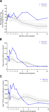
Peripheral Treg values in thyTreg-treated patients in comparison with age-matched control cohort. (A) Percentage of change from the pretransplant (pre-Tx) sample (0) in Treg frequency within CD4+ T cells of the patient treated and controls . The dotted line represents the 0% change from pretransplant values. (B and C) Treg (B) and total CD4+ T (C) absolute counts (cells per μl of blood) in thy1-01 and control cohort. Mean ± 95% confidence interval in the control cohort (n = 4) is represented as a solid gray line. Whole peripheral blood samples from patient thy1-01 (blue) and controls (gray) at all time points were considered.
Figure S3.
Figure S3.
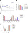
Treg frequencies and characterization of the Treg pool before and after infusion. (A) Treg frequencies within CD4+ T cells of the patient treated (thy1-01) and controls. Mean ± 95% confidence interval in the control cohort (n = 4) is represented as a solid gray line. Whole peripheral blood samples from patient thy1-01 (blue) and controls (gray) at all time points were considered. Peripheral blood Treg cell compartment was analyzed in patient thy1-01 by flow cytometry at preinfusion (Pre-inf; 8 days after transplant), 1 day after infusion (Post-inf 1 day), 1 mo, 6 mo, 1 year, and 2 years after infusion in patient thy1-01. (B) Frequency of CD25+FOXP3+ Treg on CD4+ T cells analyzed on PBMCs from blood samples. (C and D) Frequency of phenotypic/functionality (C) and homing (D) markers within viable CD25+FOXP3+ Treg cells analyzed on PBMCs from blood samples at those time points during the follow-up.

References

    1. Arroyo Hornero, R., Betts G.J., Sawitzki B., Vogt K., Harden P.N., and Wood K.J.. 2017. CD45RA distinguishes CD4+CD25+CD127-/low TSDR demethylated regulatory T cell subpopulations with differential stability and susceptibility to tacrolimus-mediated inhibition of suppression. Transplantation. 101:302–309. 10.1097/TP.0000000000001278 - DOI - PMC - PubMed
    1. Bernaldo-de-Quirós, E., Cózar B., López-Esteban R., Clemente M., Gil-Jaurena J.M., Pardo C., Pita A., Pérez-Caballero R., Camino M., Gil N., et al. . 2022a. A novel GMP protocol to produce high-quality Treg cells from the pediatric thymic tissue to Be employed as cellular therapy. Front. Immunol. 13:893576. 10.3389/fimmu.2022.893576 - DOI - PMC - PubMed
    1. Bernaldo-de-Quirós, E., Pion M., Martínez-Bonet M., and Correa-Rocha R.. 2022b. A new generation of cell therapies employing regulatory T cells (Treg) to induce immune tolerance in pediatric transplantation. Front. Pediatr. 10:862807. 10.3389/fped.2022.862807 - DOI - PMC - PubMed
    1. Bernaldo-de-Quirós, E., López-Abente J., Camino M., Gil N., Panadero E., López-Esteban R., Martínez-Bonet M., Pion M., and Correa-Rocha R.. 2021. The presence of a marked imbalance between regulatory T cells and effector T cells reveals that tolerance mechanisms could Be compromised in heart transplant children. Transpl. Direct. 7:e693. 10.1097/TXD.0000000000001152 - DOI - PMC - PubMed
    1. Brunstein, C.G., Miller J.S., McKenna D.H., Hippen K.L., DeFor T.E., Sumstad D., Curtsinger J., Verneris M.R., MacMillan M.L., Levine B.L., et al. . 2016. Umbilical cord blood-derived T regulatory cells to prevent GVHD: Kinetics, toxicity profile, and clinical effect. Blood. 127:1044–1051. 10.1182/blood-2015-06-653667 - DOI - PMC - PubMed

Publication types