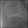
Leadless epicardial pacing at the left ventricular apex: an animal study
- PMID: 37906433
- PMCID: PMC10616611
- DOI: 10.1093/europace/euad303
Leadless epicardial pacing at the left ventricular apex: an animal study
Abstract
Aims: State-of-the-art pacemaker implantation technique in infants and small children consists of pace/sense electrodes attached to the epicardium and a pulse generator in the abdominal wall with a significant rate of dysfunction during growth, mostly attributable to lead failure. In order to overcome lead-related problems, feasibility of epicardial implantation of a leadless pacemaker at the left ventricular apex in a growing animal model was studied.
Methods and results: Ten lambs (median body weight 26.8 kg) underwent epicardial implantation of a Micra transcatheter pacing system (TPS) pacemaker (Medtronic Inc., Minneapolis, USA). Using a subxyphoid access, the Micra was introduced through a short, thick-walled tube to increase tissue contact and to prevent tilting from the epicardial surface. The Micra's proprietary delivery system was firmly pressed against the heart, while the Micra was pushed forward out of the sheath allowing the tines to stick into the left ventricular apical epimyocardium. Pacemakers were programmed to VVI 30/min mode. Pacemaker function and integrity was followed for 4 months after implantation. After implantation, median intrinsic R-wave amplitude was 5 mV [interquartile range (IQR) 2.8-7.5], and median pacing impedance was 2235 Ω (IQR 1725-2500), while the median pacing threshold was 2.13 V (IQR 1.25-2.9) at 0.24 ms. During follow-up, 6/10 animals had a significant increase in pacing threshold with loss of capture at maximum output at 0.24 ms in 2/10 animals. After 4 months, median R-wave amplitude had dropped to 2.25 mV (IQR 1.2-3.6), median pacing impedance had decreased to 595 Ω (IQR 575-645), and median pacing threshold had increased to 3.3 V (IQR 1.8-4.5) at 0.24 ms. Explantation of one device revealed deep penetration of the Micra device into the myocardium.
Conclusion: Short-term results after epicardial implantation of the Micra TPS at the left ventricular apex in lambs were satisfying. During mid-term follow-up, however, pacing thresholds increased, resulting in loss of capture in 2/10 animals. Penetration of one device into the myocardium was of concern. The concept of epicardial leadless pacing seems very attractive, and the current shape of the Micra TPS makes the device unsuitable for epicardial placement in growing organisms.
Keywords: Children; Epicardial pacing; Leadless pacemaker; Micra.
© The Author(s) 2023. Published by Oxford University Press on behalf of the European Society of Cardiology.
Conflict of interest statement
Conflict of interest: None declared.
Figures

References
-
- Haghjoo M, Nikoo MH, Fazelifar AF, Alizadeh A, Emkanjoo Z, Sadr-Ameli MA. Predictors of venous obstruction following pacemaker or implantable cardioverter-defibrillator implantation, a contrast venographic study on 100 patients admitted for generator change, lead revision, or device upgrade. Europace 2007;9:328–32. - PubMed
-
- Brugada J, Blom N, Sarquella-Brugada G, Blomstrom-Lundqvist C, Deanfield J, Janousek Jet al. . Pharmacological and non-pharmacological therapy for arrhythmias in the pediatric population: EHRA and AEPC-arrhythmia working group joint consensus statement. Europace 2013;15:1337–82. - PubMed
-
- Glikson M, Nielsen JC, Kronborg MB, Michowitz Y, Auricchio A, Barbash IMet al. . 2021 ESC guidelines on cardiac pacing and cardiac resynchronization therapy. Europace 2022;24:71–164. - PubMed
-
- Fortescue EB, Berul CI, Cecchin F, Walsh EP, Triedman JK, Alexander ME. Patient, procedural, and hardware factors associated with pacemaker lead failures in pediatrics and congenital heart disease. Heart Rhythm 2004;1:150–9. - PubMed
-
- Backhoff D, Betz T, Eildermann K, Paul T, Zenker D, Bonner Met al. . Epicardial implantation of a leadless pacemaker in a lamb model. Pacing Clin Electrophysiol 2020;43:1481–5. - PubMed
Publication types
MeSH terms
Grants and funding
LinkOut - more resources
Full Text Sources
Medical
Miscellaneous

