. 2023 Nov 28;42(11):113327.
doi: 10.1016/j.celrep.2023.113327. Epub 2023 Oct 30.

The epithelial Na+ channel UNC-8 promotes an endocytic mechanism that recycles presynaptic components to new boutons in remodeling neurons

Affiliations

The epithelial Na+ channel UNC-8 promotes an endocytic mechanism that recycles presynaptic components to new boutons in remodeling neurons

Andrea Cuentas-Condori et al. Cell Rep. .

Abstract

Circuit refinement involves the formation of new presynaptic boutons as others are dismantled. Nascent presynaptic sites can incorporate material from recently eliminated synapses, but the recycling mechanisms remain elusive. In early-stage C. elegans larvae, the presynaptic boutons of GABAergic DD neurons are removed and new outputs established at alternative sites. Here, we show that developmentally regulated expression of the epithelial Na+ channel (ENaC) UNC-8 in remodeling DD neurons promotes a Ca2+ and actin-dependent mechanism, involving activity-dependent bulk endocytosis (ADBE), that recycles presynaptic material for reassembly at nascent DD synapses. ADBE normally functions in highly active neurons to accelerate local recycling of synaptic vesicles. In contrast, we find that an ADBE-like mechanism results in the distal recycling of synaptic material from old to new synapses. Thus, our findings suggest that a native mechanism (ADBE) can be repurposed to dismantle presynaptic terminals for reassembly at new, distant locations.

Keywords: C. elegans; CP: Cell biology; CP: Neuroscience; DEG/ENaC; bulk endocytosis; circuit refinement; synaptic remodeling.


Conflict of interest statement

Declaration of interests The authors declare no competing interests.

Figures

Figure 1.
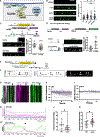
Figure 1.. UNC-8 localizes to remodeling DD presynaptic boutons
(A) Re-location of presynaptic boutons (green) from ventral to dorsal DD neurites during early larval development (late L1 and L2 stages). Arrowheads denote the DD commissure. (B) Split-GFP strategy to tag endogenous UNC-8 in DD neurons. The unc-8 locus was genetically engineered to produce a C-terminal UNC-8::GFP11×7 fusion protein. An extrachromosomal array drives the complementary GFP1-10 peptide in DD neurons under the flp-13 promoter (Pflp-13). (C) Ventral UNC-8::GFP puncta were evaluated (left) at developmental time points (arrows): early L1 (16 hours post lay [hpl]) and late L1 (24 hpl) and L2 stages (32 hpl) (right) in remodeling DD axons (see STAR Methods). (D) Ventral UNC-8::GFP puncta at the L2 stage in the wild type (left) vs. unc-104/Kif1A mutant (right). Scale bar, 2 μm. (E) UNC-8::GFP density (UNC-8 puncta/10 μm) at each time point shows progressive upregulation in the wild type (left; early L1 [1.28 ± 1.1, n = 26] and late L1 [2.93 ± 1.8, n = 34] and L2 [5.31 ± 1.7, n = 39] stages) but not in an unc-104/Kif1A mutant (right; late L1 [2.66 ± 1.8, n = 25] and L2 [3.31 ± 1.5, n = 20] stages). Data are mean ± SD. Kruskal-Wallis test. **p < 0.01, ****p < 0.0001; NS, not significant. All comparisons are relative to the 24 hpl time point in the wild type. (F) Accumulation of ventral UNC-8::GFP in DD cell soma of the unc-104/Kif1A mutant. The key denotes relative UNC-8::GFP signal intensity. Scale bar, 2 μm (30 hpl, L2). (G) Dual-color imaging during remodeling (late L1) detects UNC-8::GFP puncta (green) and neighboring mCherry::RAB-3 (magenta) at ventral presynaptic DD boutons. Scale bar, 2 μm. Inset scale bar, 1 μm. (H) Cholinergic DA motor neuron presynaptic boutons (magenta) provide excitatory input to DD GABAergic motor neurons (green) in the dorsal nerve cord. Optogenetically activated Chrimson in DA neurons (magenta) evokes Ca++ transients in postsynaptic DD neurons (I–N). GCaMP signals were collected from DD presynaptic boutons (ROI). (I) Chrimson activation in DA neurons (magenta bars) evokes GCaMP transients in wild-type (WT) DD presynaptic boutons. Gray vertical bars depict ~1-s lag before imaging. (J) Initial GCaMP signal (0.02 ± 0.3, n = 32) is elevated (0.25 ± 0.5, n = 32) after Chrimson activation. Non-parametric paired Wilcoxon test, **p = 0.002. (K) Examples of upregulated GCaMP signals after Chrimson activation in the WT. (L) Chrimson activation in DA neurons fails to evoke a GCaMP response in DD neurons of the unc-8 mutant. (M and N) GCaMP signal (0.06 ± 0.4, n = 32) does not increase after DA activation (0.07 ± 0.5, n = 32). Non-parametric paired Wilcoxon test, p = 0.0803. (K and N) A dashed line denotes DD cell soma. Scale bar, 4 μm. All animals were grown with ATR.
Figure 2.
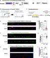
Figure 2.. Presynaptic TAX-6/CaN elevates Ca++ transients in remodeling DD boutons
(A) Split-GFP strategy for labeling endogenous TAX-6/CaN in DD neurons. Three copies of GFP11 were inserted at the tax-6 locus to produce TAX-6:: GFP11×3. Pflp-13 drives GFP1-10 expression from an extrachromosomal array in DD neurons. (B) Left: Before DD remodeling, in L1 larvae, TAX-6::GFP is enriched (arrowheads) in the ventral DD neurite but relocated to the dorsal cord (right) after remodeling, at the L4 stage. Scale bar, 2 μm. (C) Quantification of TAX-6::GFP detects initial enrichment in the ventral cord (V = 0.79 ± 0.1 vs. D = 0.2 ± 0.1, n = 29) in L1 larvae and later enrichment on the dorsal side at the L4 stage (V = 0.27 ± 0.1, n = 21 vs. D = 0.73 ± 0.1, n = 21). Data are mean ± SD. One-way ANOVA with Tukey’s multiple comparisons test, ****p < 0.0001. (D and E) Top: relocation of TAX-6 (green) from ventral (L1 stage) to dorsal (L4 stage) DD neurites. (Bottom: co-localization (arrowheads) of endogenous TAX-6::GFP (green) and active-zone protein mRuby::CLA-1s (magenta) (D) on the ventral side before remodeling (L1 stage) and (E) on the dorsal side (L4 stage) after remodeling. Line scans show co-localization of TAX-6 (green) and CLA-1s (magenta) from insets. Scale bar, 2 μm. (F) Chrimson was expressed in presynaptic DA neurons (magenta) to evoke Ca++ transients in DD neurons (G–I). (G–I) Chrimson activation in DA neurons (G, magenta bar) elevates GCaMP fluorescence in remodeling DD presynaptic boutons (ROI) (H) (before activation = −0.02 ± 0.2, n = 44; after activation = 0.14 ± 0.4, n = 44). Non-parametric paired Wilcoxon test, ****p < 0.0001. Gray vertical bars depict ~1-s lag before imaging. GCaMP fluorescence is elevated after Chrimson activation in the WT (0.32 ± 0.3, n = 32) in comparison with tax-6 mutants (0.18 ± 0.2, n = 44) (I). Data are mean ± SD. Mann-Whitney test, *p = 0.0109. All experiments were performed with ATR.
Figure 3.
Figure 3.. Cell-autonomous dynamin activity is required for presynaptic disassembly in remodeling DD neurons
(A) During ADBE, synaptic activity elevates intracellular Ca++ to activate CaN/TAX-6. Dephosphorylation of dynamin by CaN promotes the formation of the dynamin-syndapin complex for membrane localization to drive branched-actin polymerization for bulk endosome formation and local SV recycling. (B) Left: UNC-8 overexpression (OE) in VD neurons induces presynaptic disassembly. Shown are control VD neurons (gray, VD11) with SNB-1::GFP puncta (arrowheads) vs. anterior region of UNC-8(OE) VD neurons (magenta, VD10) with fewer SNB-1::GFP puncta (dashed line). Right: paired analysis of neighboring VD neurons: control VDs (196 ± 139 a.u., n = 12) and UNC-8(OE) VD neurons (56.7 ± 67 a.u., n = 12). Wilcoxon matched-pairs signed-rank test, **p = 0.0024. (C) TAX-6 is required for UNC-8-dependent removal of presynaptic domains. Left: tax-6 RNAi-treated UNC-8(OE) VD neuron (magenta, VD5) vs. adjacent control VD neuron (gray, VD6), both with SNB-1::GFP puncta (arrowheads). Right: paired analysis of neighboring VD neurons treated with tax-6 RNAi: control VDs (126 ± 76 a.u., n = 17) and UNC-8(OE) VD neurons (86 ± 99 a.u., n = 17). Wilcoxon matched-pairs signed-rank test, p = 0.16. Scale bars, 10 μm. L4 stage. (D) Transgenic strategy for cell-specific RNAi (csRNAi) knockdown of dyn-1/dynamin. (E) Top left: control DD neuron (gray) shows perinuclear SNB-1::GFP (arrows). Bottom left: dyn-1(csRNAi)-expressing DD neurons (magenta) labeled with cytosolic mCherry, nuclear GFP (green arrowhead), and perinuclear SNB-1::GFP (arrows) retain ventral SNB-1::GFP puncta (white arrowheads). Right: dyn-1(csRNA)-treated DD neurons retain a greater fraction of ventral SNB-1::GFP fluorescence (0.31 ± 0.2, n = 18) than controls (0.15 ± 0.1, n = 18). Data are mean ± SD. Unpaired t test, ***p = 0.0006. Scale bar, 10 μm. (F) Schematic of DYN-1 protein domains (see text). A magenta bar denotes the putative C-terminal dynamin phospho-box sequence with phosphorylatable residues (S53, Y55, S59) in the WT and converted to alanine (A) in phospho-resistant dyn-1. (G) Top: transgenic strategy for OE of dynamin (DYN-1) and phospho-resistant DYN-1 in DD neurons (STAR Methods). Center: a control DD neuron (gray) shows robust SNB-1::GFP puncta (white arrowheads). See inset for cell soma and line tracing. Bottom: DD neuron that overexpresses phospho-resistant DYN-1 (dyn-1AAA (OE)) is labeled with nucleus-localized TagRFP (magenta) (inset) and shows fewer SNB-1::GFP puncta than control DD neurons (white arrowheads). Scale bar, 10 μm; late L1 stage. (H) Top: SNB-1::GFP in DD neurons was imaged during remodeling (arrow) to assess whether ventral (bottom left) OE of phospho-resistant DYN-1 (dyn-1AAA(OE)) accelerates SNB-1::GFP removal (0.61 ± 0.3, n = 21) vs. control DD neurons (0.92 ± 0.5, n = 62). Bottom right: OE of WT dynamin (dyn-1(OE)) (1.47 ± 0.7, n = 21) does not result in fewer SNB-1::GFP puncta vs. control DD neurons (1.21 ± 0.6, n = 34). Data are mean ± SD. Unpaired t test. **p = 0.0055, NS (not significant), p = 0.137. See inset for cell soma and line tracing.
Figure 4.
Figure 4.. Branched-actin polymerization functions downstream of UNC-8 to remodel DD boutons
(A) F-BAR proteins associated with the plasma membrane, like syndapin, recruit the wave regulatory complex (WRC) via SH3 domains to activate Arp2/3 for branched-actin polymerization. The WRC components Sra1/GEX-2, Nap1/GEX-3, Abi2/ABI-1, WAVE1/WVE-1 and HSPC300/Y57G11C.1147 are conserved in C. elegans. The VCA domain (V, verprolin homology domain; C, central domain; A, acidic domain) of WAVE1 interacts with Arp2/3 to promote its activation. (B) SDPN-1 and UNC-8 function in a common genetic pathway. Left: residual endogenous GFP::RAB-3 puncta (arrowheads) in ventral DD neurites in the WT, unc-8 and sdpn-1 single mutants, and unc-8; sdpn-1 double mutants in early L4 larvae. Right: ventral GFP::RAB-3 density is not significantly different among unc-8 (1.02 ± 0.5, n = 51), sdpn-1 (1.22 ± 0.9, n = 46) and unc-8; sdpn-1 double mutants (1.13 ± 0.9, n = 62) but is elevated in comparison with the WT (0.69 ± 0.6, n = 73). Data are mean ± SD. Kruskal-Wallis test. ** p < 0.01. * p = 0.019. (C) Top: OE of the WRC-VCA domain (VCA(OE)) and cytosolic mCherry (magenta) in remodeling DD neurons labeled with SNB-1::GFP (green). Bottom left: ventral SNB-1::GFP puncta (arrowheads) in VCA(OE) and control DD neurons. Scale bar, 5 μm. Bottom right: proportion of ventral SNB-1::GFP fluorescence in control DDs (0.56 ± 0.1, n = 26) vs. VCA(OE) (0.40 ± 0.1, n = 42) during remodeling. Data are mean ± SD. Unpaired t test, ****p < 0.0001. (D) Top: arx-5 sense and antisense transcripts co-expressed with either cytosolic mCherry or nucleus-localized GFP. Bottom left: arx-5(csRNAi) knockdown DDs with nuclear GFP (green arrowhead) and cytosolic mCherry (magenta) vs. unlabeled control DDs (gray). Bottom center: ventral SNB-1::GFP (white arrowheads) in control and arx-5(csRNAi) DD cells. Bottom right: proportion of ventral SNB-1::GFP fluorescence in control DDs (0.19 ± 0.1, n = 24) vs. arx-5(csRNAi) (0.43 ± 0.3, n = 24) DDs of L4 larvae. Data are mean ± SD. Unpaired t test, ****p < 0.0001. Scale bar, 10 μm. (E) Ventral DD neurites imaged during remodeling (gray arrow) to monitor actin dynamics with LifeAct::mCherry (magenta) and SVs with GFP::RAB-3 (green). (F) GFP::RAB-3 (green) associated with LifeAct::mCherry (magenta). Shown are GFP::RAB-3 clusters (arrowheads). Scale bar, 5 μm. (G–K) Elevated actin dynamics during DD remodeling in controls but not in unc-8 mutants. (G) During remodeling, LifeAct::mCherry associates with stable and transient GFP::RAB-3 puncta in control animals. Arrows and arrowheads point to locations of dynamic GFP::RAB-3 puncta. (G′) Line scans from kymographs show dynamic LifeAct::mCherry fluorescence during remodeling; n = 6 boutons from the kymograph in (G). (G′′) Line scans of LifeAct::mCherry and GFP::RAB-3 from kymographs (arrowheads in G) showing reduced GFP::RAB-3 signal with transient elevation of LifeAct::mCherry (asterisk). (H and I) LifeAct::mCherry fluorescence from GFP::RAB-3 regions (normalized to t0 = 0) in (H) WT (n = 12 videos) and (I) unc-8 mutants (n = 11 videos). (J) Fewer transient GFP::RAB-3 events in unc-8 mutants (1.2 ± 0.8, n = 11 videos) vs. WT (3.2 ± 2.4, n = 12 videos). Data are mean ± SD. t test, *p < 0.05. (K) unc-8 mutants retain more GFP::RAB-3 puncta (2.37 ± 0.8, n = 19 snapshots) than the WT (1.74 ± 0.7, n = 20 snapshots) during the remodeling window (L2 stage). Data are mean ± SD. t test, *p < 0.05.
Figure 5.
Figure 5.. UNC-8 promotes the formation of endosome-like structures in remodeling DD neurons
(A) L1 larvae were subjected to high-pressure freezing (HPF) during DD remodeling (27 hpl), and serial sections were imaged by transmission electron microscopy (TEM). (B and C) Reconstructions of (B) WT and (C) unc-8 mutant DD2 neurons with enlargements shown below. Neuronal membrane, transparent white; SVs, blue; dense core vesicles, green; dense projections, gray; endocyte-like structures, magenta. (D) Representative TEM sections containing presynaptic densities (stars) and endosome-like structures (arrowheads) from reconstructed WT and unc-8 mutant DD2 neurons. (E) Percentage of TEM sections containing endosome-like structures in reconstructed DD2 neurons. Numbers denote fractions of sections with endocyte-like structures for the WT (133/396) and unc-8 (55/349) mutant. Fisher’s exact test, ****p < 0.00001.
Figure 6.
Figure 6.. UNC-8 promotes RAB-3 recycling from ventral to dorsal DD boutons
(A) Endogenous labeling of RAB-3 with photoconvertible protein Dendra2 in DD neurons. Pflp-13 drives flippase in DD neurons to fuse Dendra2 to the endogenous RAB-3 protein. (B) UV irradiation of ventral DD neurites (box) produces photoconverted Dendra2::RAB-3 (magenta). (C) Photoconversion before DD remodeling and imaging after remodeling at the L3 stage. (D) Photoconverted Dendra2::RAB-3 in the dorsal nerve cord of WT and unc-8 animals in L3 larvae after DD remodeling. An asterisk denotes autofluorescence. Scale bar, 10 μm. (E) unc-8 (32.3% ± 25.6%, n = 14) mutants recycle less photoconverted Dendra2::RAB-3 to the dorsal nerve cord than the WT (82.0% ± 40.8%, n = 12). Data are mean ± SD. Ordinary one-way ANOVA with Dunnett’s multiple comparison test. ***p = 0.0009. (F) GFP::RAB-3 puncta in dorsal DD neurites in the WT and unc-8 mutants at the L4 stage. (G) Quantification of dorsal GFP::RAB-3 density in the WT (3.14± 0.36, n = 21) and unc-8 mutants (2.91± 0.34, n = 24). Data are mean± SD. Unpaired t test, *p = 0.046. (H) Photoconverted Dendra2::RAB-3 in the ventral nerve cords of the WT and unc-8 mutants at the L3 stage. Scale bar, 10 μm. (I) WT (1.14% ± 2.1%, n = 12) and unc-8 mutants (1.32% ± 2.9%, n = 14) lack photoconverted signal on the ventral cord at the L3 stage. Data are mean ± SD. Mann-Whitney test, p = 0.46.
Figure 7.
Figure 7.. RAB-11 promotes recycling of RAB-3 from old to new presynaptic boutons in remodeling DD neurons
(A) csRNAi of rab-11 in DD neurons by co-expression of rab-11 sense transcripts with nucleus-localized TagRFP (magenta) and rab-11 antisense transcript with nucleus-localized GFP (green). Non-fluorescent DD neurons (gray) served as controls. (B) Left: photoconverted Dendra2::RAB-3 (magenta) in control and rab-11(csRNAi) DD neurons in dorsal and ventral DD neurites. Arrowheads denote ventral retention of photoconverted Dendra2::RAB-3 in rab-11(csRNAi) neurons, and asterisks labels nucleus-localized TagRFP. Right: line scans of photoconverted Dendra2::RAB-3 dorsal (top) and ventral (bottom) DD neurites of control and rab-11(csRNAi) knockdown cells. Scale bar, 10 μm. (C) rab-11(csRNAi) DD cells recycle less photoconverted signal to the dorsal cord (37.7% ± 24.8%, n = 10) than controls (74.0% ± 36.2%, n = 13). Horizontal lines show median and 25% to 75% distribution. Unpaired t test, **p = 0.0064. (D) rab-11(csRNAi) DD cells retain more photoconverted signal on the ventral side (9.96% ± 10.5%, n = 10) than controls (1.09% ± 1.8%, n = 13). Horizontal lines show median and 25% to 75% distribution. Mann-Whitney test, *p = 0.0269. (E) Left: control VD neurons (gray) with SNB-1::GFP puncta (arrowheads) and UNC-8(OE) VD cells (magenta) with fewer SNB-1::GFP puncta (dashed line). Right: paired analysis of ventral SNB-1::GFP fluorescence in neighboring cells, control (607 ± 688 a.u., n = 13) vs. UNC-8(OE) VD neurons (94.8 ± 129 a.u., n = 13). Wilcoxon matched-pairs signed-rank test, ***p = 0.0002. Scale bar, 10 μm. (F) Left: SNB-1::GFP puncta (arrowheads) in control (gray, VD5) and UNC-8(OE) VD neurons (magenta, VD4) with rab-11 RNAi. Scale bar, 10 μm. Right: paired analysis of ventral SNB-1::GFP in neighboring cells, control VDs (439 ± 341 a.u., n = 23) and UNC-8(OE) VD neurons (360 ± 500 a.u., n = 23). Wilcoxon matched-pairs signed-rank test, p = 0.211.

References

    1. Hong YK, Park S, Litvina EY, Morales J, Sanes JR, and Chen C (2014). Refinement of the Retinogeniculate Synapse by Bouton Clustering. Neuron 84, 332–339. 10.1016/j.neuron.2014.08.059. - DOI - PMC - PubMed
    1. Berry KP, and Nedivi E (2016). Experience-Dependent Structural Plasticity in the Visual System. Annu. Rev. Vis. Sci. 2, 17–35. 10.1146/annurev-vision-111815-114638. - DOI - PMC - PubMed
    1. Tennant KA, Taylor SL, White ER, and Brown CE (2017). Optogenetic rewiring of thalamocortical circuits to restore function in the stroke injured brain. Nat. Commun. 8, 15879. - PMC - PubMed
    1. Carrillo J, Cheng SY, Ko KW, Jones TA, and Nishiyama H (2013). The long-term structural plasticity of cerebellar parallel fiber axons and its modulation by motor learning. J. Neurosci. 33, 8301–8307. 10.1523/JNEUROSCI.3792-12.2013. - DOI - PMC - PubMed
    1. Cuentas-Condori A, and Miller DM (2020). Synaptic remodeling, lessons from C. elegans. J. Neurogenet. 34, 307–322. 10.1080/01677063.2020.1802725. - DOI - PMC - PubMed

Publication types

Substances