. 2023 Oct 31;14(1):6952.
doi: 10.1038/s41467-023-42427-8.

Cryo-EM structure of human O-GlcNAcylation enzyme pair OGT-OGA complex

Affiliations

Cryo-EM structure of human O-GlcNAcylation enzyme pair OGT-OGA complex

Ping Lu et al. Nat Commun. .

Abstract

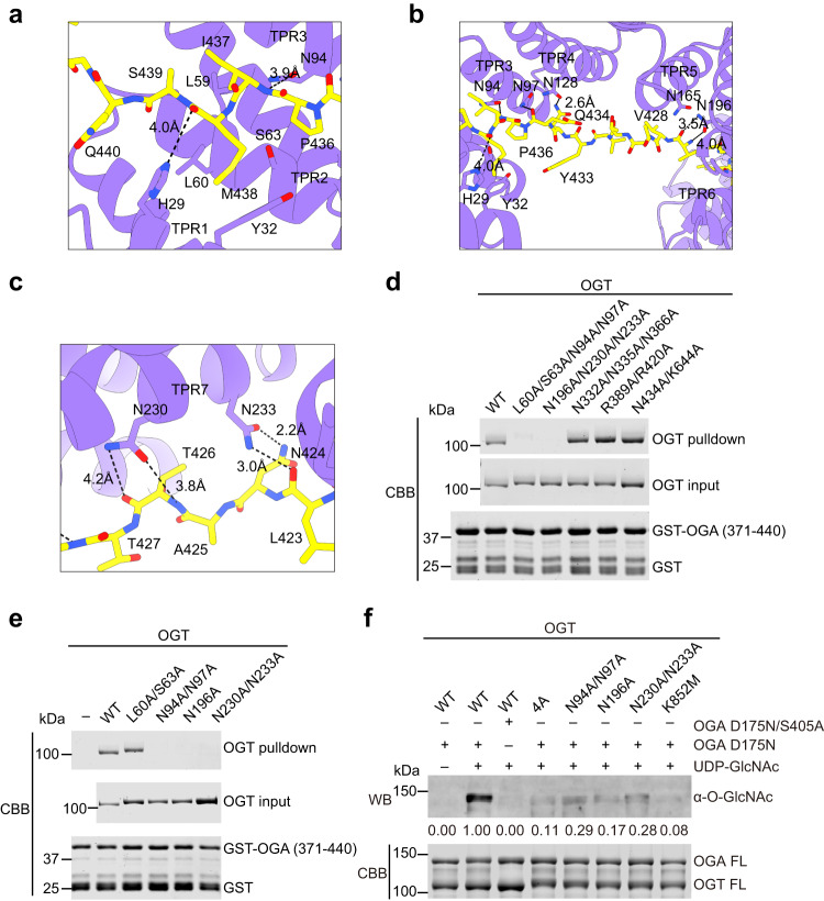
O-GlcNAcylation is a conserved post-translational modification that attaches N-acetyl glucosamine (GlcNAc) to myriad cellular proteins. In response to nutritional and hormonal signals, O-GlcNAcylation regulates diverse cellular processes by modulating the stability, structure, and function of target proteins. Dysregulation of O-GlcNAcylation has been implicated in the pathogenesis of cancer, diabetes, and neurodegeneration. A single pair of enzymes, the O-GlcNAc transferase (OGT) and O-GlcNAcase (OGA), catalyzes the addition and removal of O-GlcNAc on over 3,000 proteins in the human proteome. However, how OGT selects its native substrates and maintains the homeostatic control of O-GlcNAcylation of so many substrates against OGA is not fully understood. Here, we present the cryo-electron microscopy (cryo-EM) structures of human OGT and the OGT-OGA complex. Our studies reveal that OGT forms a functionally important scissor-shaped dimer. Within the OGT-OGA complex structure, a long flexible OGA segment occupies the extended substrate-binding groove of OGT and positions a serine for O-GlcNAcylation, thus preventing OGT from modifying other substrates. Conversely, OGT disrupts the functional dimerization of OGA and occludes its active site, resulting in the blocking of access by other substrates. This mutual inhibition between OGT and OGA may limit the futile O-GlcNAcylation cycles and help to maintain O-GlcNAc homeostasis.


Conflict of interest statement

The authors declare no competing interests.

Figures

Fig. 1
Fig. 1. Cryo-EM structures of human OGT and OGT–OGA complex.
a The dynamic protein O-GlcNAcylation cycle mediated by OGT and OGA. b Domains and motifs of human OGT and OGA. TPR, tetratricopeptide repeat. c Human HEK293 cells with the endogenous OGT locus tagged with the HaloTag were treated with the HaloPROTAC3 compound for different duration times. Total cell lysates were blotted with the indicated antibodies. The experiment was repeated thrice with similar results. d Cryo-EM map of human OGT dimer (left panel) and the structure of the OGT dimer fitted into the EM map (right panel). GTD, glycosyltransferase family B domain. e Close-up views of the OGT dimerization interface. f O-GlcNAcylation of recombinant TAB1 by OGT WT or its monomeric 4A mutant in the presence of UDP-GlcNAc. The relative O-GlcNAc levels were quantified and indicated below. The experiment was repeated thrice with similar results. g Cryo-EM map of human OGT-OGA complex (left panel) and the structure of the OGT-OGA complex fitted into the EM map. GHD, glycoside hydrolase domain; IDR, intrinsically disordered region. h Superimposition of the structures of the OGT-OGA complex and the OGT dimer (colored in gray and with only one protomer shown). i Cryo-EM density of the UDP and NAG in OGT-OGA complex. Source data are provided as a Source Data file.
Fig. 2
Fig. 2. OGA binding by human OGT.
a The schematic of human OGT TPR domain with the 13.5 tetratricopeptide repeats colored in purple. The distal, medial, and proximal units are indicated as shown. The residues at position 6 (top row, conserved Asparagine residues) and 9 (bottom row) in each unit are highlighted in red and blue, respectively. The canonical Asn Ladders are usually located at the inner concave of the TPR domain. b Extensive interactions between human OGT and OGA. XLID-related residues L254, A259, R284, A319, and E339 are shown as sticks and labeled in the structure of the OGT-OGA complex. c Binding between full-length OGT and the indicated GST-OGA fragments. The input and proteins bound to beads were analyzed by SDS-PAGE followed by Coomassie blue staining. An asterisk indicates a contaminating protein. The experiment was repeated thrice with similar results. d Close-up view of OGT catalytic pocket (colored in purple) interacting with residues 402–407 of OGA (colored in yellow). Source data are provided as a Source Data file.
Fig. 3
Fig. 3. Molecular details in OGA recognition by OGT TPR domain.
a Close-up view of OGT TPR 1–3 units (colored in purple) interacting with OGA (colored in yellow). b Close-up view of OGT TPR 3-6 units interacting with OGA. c Close-up view of OGT TPR7 interacting with OGA. d Binding between GST-OGA (residues 371–440) and the indicated OGT mutant proteins. The input proteins and proteins bound to GST beads were analyzed by SDS-PAGE and Coomassie staining. The experiment was repeated thrice with similar results. e Binding between GST-OGA (residues 371–440) and OGT WT and the indicated TPR mutants. The experiment was repeated thrice with similar results. f O-GlcNAcylation assay of the catalytically inactive OGA mutant (D175N or D175N/S405A) by OGT wild type (WT) and the indicated mutants in the presence or absence of UDP-GlcNAc. The relative O-GlcNAc levels were quantified and indicated below. The experiment was repeated thrice with similar results. Source data are provided as a Source Data file.
Fig. 4
Fig. 4. Inhibition of OGT by OGA.
a, b Close-up views of an OGA segment (colored in yellow) interacting with the intervening domain (ID), the UDP-binding pocket, and the active site of OGT (colored in blue and gray). c O-GlcNAcylation assay of TAB1 by the indicated OGT and OGA proteins in the presence of UDP-GlcNAc. The experiment was repeated thrice with similar results. d O-GlcNAcylation assay of TAB1 added with different doses of OGA as a competitor. The relative O-GlcNAc levels were quantified and indicated below the gels in (c, d). The experiment was repeated thrice with similar results. Source data are provided as a Source Data file.
Fig. 5
Fig. 5. Inhibition of OGA by OGT.
a Cartoon drawing of the crystal structure of the human OGA dimer (colored in cyan and pink) bound to a TAB1 peptide (PDB code: 5VVU). b Superimposition of the structures of the OGA molecules in the OGA dimer (as in a) and the OGT-OGA complex, showing the steric clashes between OGT and the second OGA molecule in the OGA dimer. c Close-up view of the superimposed structures in (b). d, e Close-up views of the interactions between OGT (colored in purple) and the GHD of OGA (colored in yellow). f Removal of TAB1 O-GlcNAcylation by OGA and its inhibition by OGT. O-GlcNAcylated TAB1 (G-TAB1) was incubated with OGA or the catalytically inactive OGA D175N mutant in the absence or presence of the indicated OGT proteins. The reaction mixtures were analyzed by SDS-PAGE, stained with Coomassie brilliant blue, and blotted with the anti-O-GlcNAc antibody. The relative O-GlcNAc levels were quantified and indicated below. The experiment was repeated thrice with similar results. g The quantitative OGA enzymatic assay using the artificial substrate PNP-GlcNAc with indicated p values. Data were presented as mean ± SD, n = 6 biologically independent samples per condition. P value was calculated from an unpaired two-tailed t test. Source data are provided as a Source Data file.
Fig. 6
Fig. 6. Working model of substrate recognition and modification by human OGT.
For clarity, only one OGT monomer (colored in blue) is shown. OGT binds to the intrinsically disordered regions (IDRs) (colored in yellow) of OGA-like substrates through its TPR domain. The superhelical turns of the TPR domain have to partially unwind to allow the IDR to access its lumen. The superhelical turns then reform and wrap around the IDR in a topological embrace, which lengthens the lifetime of the OGT-substrate complex for optimal O-GlcNAcylation. TPR tetratricopeptide repeat, GTD glycosyltransferase domain, IDR intrinsically disordered region.

Similar articles

Cited by

References

    1. Bond MR, Hanover JA. A little sugar goes a long way: the cell biology of O-GlcNAc. J. Cell Biol. 2015;208:869–880. doi: 10.1083/jcb.201501101. - DOI - PMC - PubMed
    1. Yang X, Qian K. Protein O-GlcNAcylation: emerging mechanisms and functions. Nat. Rev. Mol. Cell Biol. 2017;18:452–465. doi: 10.1038/nrm.2017.22. - DOI - PMC - PubMed
    1. Chatham JC, Zhang J, Wende AR. Role of O-linked N-acetylglucosamine protein modification in cellular (Patho)physiology. Physiol. Rev. 2021;101:427–493. doi: 10.1152/physrev.00043.2019. - DOI - PMC - PubMed
    1. Zhu Y, Hart GW. Nutrient regulation of the flow of genetic information by O-GlcNAcylation. Biochem. Soc. Trans. 2021;49:867–880. doi: 10.1042/BST20200769. - DOI - PubMed
    1. Ma J, Hou C, Wu C. Demystifying the O-GlcNAc code: a systems view. Chem. Rev. 2022;122:15822–15864. doi: 10.1021/acs.chemrev.1c01006. - DOI - PubMed

Publication types

MeSH terms