. 2023 Oct 31;13(1):18760.
doi: 10.1038/s41598-023-45402-x.

Breast density analysis of digital breast tomosynthesis

Affiliations

Breast density analysis of digital breast tomosynthesis

John Heine et al. Sci Rep. .

Abstract

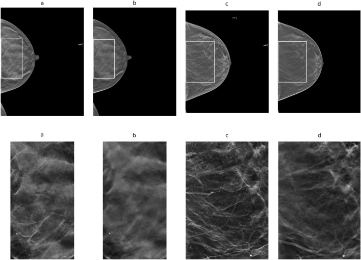
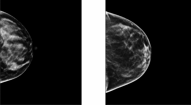
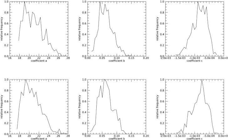
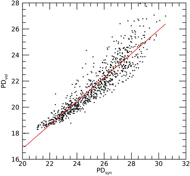
Mammography shifted to digital breast tomosynthesis (DBT) in the US. An automated percentage of breast density (PD) technique designed for two-dimensional (2D) applications was evaluated with DBT using several breast cancer risk prediction measures: normalized-volumetric; dense volume; applied to the volume slices and averaged (slice-mean); and applied to synthetic 2D images. Volumetric measures were derived theoretically. PD was modeled as a function of compressed breast thickness (CBT). The mean and standard deviation of the pixel values were investigated. A matched case-control (CC) study (n = 426 pairs) was evaluated. Odd ratios (ORs) were estimated with 95% confidence intervals. ORs were significant for PD: identical for volumetric and slice-mean measures [OR = 1.43 (1.18, 1.72)] and [OR = 1.44 (1.18, 1.75)] for synthetic images. A 2nd degree polynomial (concave-down) was used to model PD as a function of CBT: location of the maximum PD value was similar across CCs, occurring at 0.41 × CBT, and PD was significant [OR = 1.47 (1.21, 1.78)]. The means from the volume and synthetic images were also significant [ORs ~ 1.31 (1.09, 1.57)]. An alternative standardized 2D synthetic image was constructed, where each pixel value represents the percentage of breast density above its location. Several measures were significant and an alternative method for constructing a standardized 2D synthetic image was produced.


Conflict of interest statement

The authors declare no competing interests.

Figures

Figure 1
Figure 1
Image Illustrations: in the top row (a) C-View image for illustration-1; (b) respective central slice image; (c) C-View image for illustration-2; and (d) respective central slice image. Outlines in the top-row images are the largest rectangles that fit within the breast areas. These regions are shown in the second row with the same ordering for illustration purposes. Illustration-1 has 89 μm pixel spacing and illustration-2 has 107 μm pixel spacing.
Figure 2
Figure 2
Breast Density Detection: this shows the density detection for the illustrations: (a) C-view, illustration-1; (b) respective central slice image; (c) C-view, illustration-2; and (d) respective central slice.
Figure 3
Figure 3
Projected Standardized Synthetic Breast Density Images: these show the standardized, s(x,y), images for illustration-1 (left) and illustration-2 (right) resulting from Eq. (8). Pixel values represent the percentage of dense tissue in the breast volume above their locations.
Figure 4
Figure 4
Regression analysis with PDvol as a function of PDm This shows the scatter plot between the two measures (points) and regression line (solid red), The analysis gave: slope ≈ 1.002, intercept ≈ − 0.0201, and linear correlation ≈ 1.0. The [mean, standard deviations] for the distributions were [21.60, 1.98] for PDvol and [21.58, 1.98] for PDm.
Figure 5
Figure 5
PD slice Profiles: this shows PD (y-axis) by slice number using the normalized distance from the breast support surface (P on the x-axis) for illustration-1 (left) and illustration-2 (right). PD values per slice number (points) were fitted with a second-degree polynomial (solid). The slice distance increases as the distance increases from the breast support surface (left side of each plot).
Figure 6
Figure 6
DBT Volume Slice 2nd Degree Polynomial Coefficient Histograms: these show the normalized histograms for the fit-coefficients, (a,b,c), separated by case (top-row) and control status (bottom-row).
Figure 7
Figure 7
Regression analysis with PDvol as a function of PDsyn: this shows the scatter plot between the two measures (points) and regression line (red-solid): slope ≈ 0.92 and standard error ≈ 0.01, intercept ≈ − 1.4, and R ≈ 0.93. The [mean, standard deviations] for the distributions were [21.60, 1.98] for PDvol and [25.25, 2.01] for PDsyn.

Update of

Similar articles

Cited by

References

    1. Bodewes FTH, van Asselt AA, Dorrius MD, Greuter MJW, de Bock GH. Mammographic breast density and the risk of breast cancer: A systematic review and meta-analysis. Breast. 2022;66:62–68. doi: 10.1016/j.breast.2022.09.007. - DOI - PMC - PubMed
    1. Boyd NF, et al. Breast tissue composition and susceptibility to breast cancer. J. Natl. Cancer Inst. 2010;102:1224–1237. doi: 10.1093/jnci/djq239. - DOI - PMC - PubMed
    1. Bertrand KA, et al. Mammographic density and risk of breast cancer by age and tumor characteristics. Breast Cancer Res. 2013;15:R104. doi: 10.1186/bcr3570. - DOI - PMC - PubMed
    1. Butler R. Invited commentary: Breast cancer risk assessment and screening strategies-what’s new? Radiographics. 2020;40:937–940. doi: 10.1148/rg.2020190218. - DOI - PubMed
    1. Roman M, et al. Personalized breast cancer screening strategies: A systematic review and quality assessment. PLoS One. 2019;14:e0226352. doi: 10.1371/journal.pone.0226352. - DOI - PMC - PubMed

Publication types