. 2023 Oct 31;13(1):18685.
doi: 10.1038/s41598-023-46068-1.

Early-life origin of prostate cancer through deregulation of miR-206 networks in maternally malnourished offspring rats

Affiliations

Early-life origin of prostate cancer through deregulation of miR-206 networks in maternally malnourished offspring rats

Luiz M F Portela et al. Sci Rep. .

Abstract

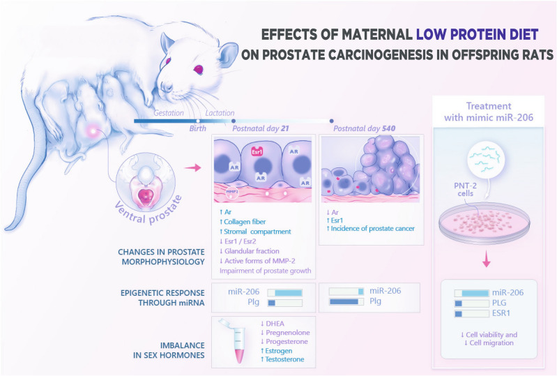
The Developmental Origins of Health and Disease (DOHaD) concept has provided the framework to assess how early life experiences can shape health and disease throughout the life course. While maternal malnutrition has been proposed as a risk factor for the developmental programming of prostate cancer (PCa), the molecular mechanisms remain poorly understood. Using RNA-seq data, we demonstrated deregulation of miR-206-Plasminogen (PLG) network in the ventral prostate (VP) of young maternally malnourished offspring. RT-qPCR confirmed the deregulation of the miR-206-PLG network in the VP of young and old offspring rats. Considering the key role of estrogenic signaling pathways in prostate carcinogenesis, in vitro miRNA mimic studies also revealed a negative correlation between miR-206 and estrogen receptor α (ESR1) expression in PNT2 cells. Together, we demonstrate that early life estrogenization associated with the deregulation of miR-206 networks can contribute to the developmental origins of PCa in maternally malnourished offspring. Understanding the molecular mechanisms by which early life malnutrition affects offspring health can encourage the adoption of a governmental policy for the prevention of non-communicable chronic diseases related to the DOHaD concept.


Conflict of interest statement

The authors declare no competing interests.

Figures

Figure 1
Figure 1
Representative macroscopic images of male offspring (ab and ef) and prostate (cd and gh) from Control (CTR) (c and g) and Gestational and Lactational Low Protein groups (GLLP) (d and h) at PND 21 (ad) and PND 540 (e–h). Note the smaller size of the animals in the GLLP in both ages. Scale bars: (a) and (b): 1 cm; (c) and (d): 0.1 cm; (e) and (f): 5 cm; (g) and (h): 0.5 cm.
Figure 2
Figure 2
Histological sections of the ventral prostate (VP) lobes from the CTR (a and c) and the GLLP group (b and d) stained with hematoxylin–eosin (HE) (a and b) or picrosirius red (c and d). Collagen quantification was represented in the bar graph (e). Quantification of the VP gelatinolytic activity of pro, intermediate, and active forms of MMP-2 (f) in the gelatin zymography gel of electrophoresis (g). Data are expressed as mean ± SD. *p < 0.05. Scale bar 50 µm.
Figure 3
Figure 3
Representative image of bioinformatics analyses. MicroRNome (GSE180674) and transcriptome (GSE180673) datasets from young rat VP were reanalyzed using the DESEq2 package. Differentially expressed targets were considered when p-value < 0.05 and Log2 Fold Change ≥|+ 0.66 ≤|− 0.66| (a). Up-set plot showing commonly deregulated miRNAs in the VP from the GLLP group and in patients with PCa (b). Heatmap showing the miRNAs expression profile shared between the VP from the GLLP group and PCa patients (c). Sequence alignment of commonly deregulated miRNAs in the VP from the GLLP group and in PCa samples. The sequences were obtained through the miRBase database (https://www.mirbase.org/) (d). Alluvial diagram showing the relationship between the main molecular pathways and enriched ontological terms for the predicted targets of miR-206, these analyzes were performed on the Enrichr platform (https://maayanlab.cloud/Enrichr/), considering p-value < 0.05 (e). Survival curves of PRAD patients (using TCGA data) show the impact of PLG, ZMAT3, and LFPL2 in the progression free PCa patients in altered (red) and unaltered (blue) risk (f). Immunohistochemical localization of PLG, ZMAT3, and LFPL2 in normal and PRAD tissues obtained from The Human Protein Atlas database (https://proteinatlas.org/) (g).
Figure 4
Figure 4
Functional validation of miR-206 in PNT2 benign prostatic cells. Morphologic aspects of the PNT2 cells in the Mock (a), Scrambled (b), and miR206-Mimic (c) groups after 24 h of treatment. Expression profile of miR-206 in PNT2 cells in both experimental groups after 24 h of treatment (d). PLG (e), ESR1 (f), ESR2 (g), and AR (h) expression levels in PNT2 cells after 24 h of treatment in both experimental groups. Representative images of wound healing assay after 24, 48, and 72 h (i). Cellular wound closure after the transfection of PNT2 cells with Mock and miR206-Mimic groups after 0 h, 24 h, 48 h, and 72 h post-wound (j). Cell viability assay (MTT) after 24, 48, and 72 h (k). Mock group: cells treated with lipofectamine. Scrambled: cells treated with negative control mimic. miR206-Mimic group: cells treated with miR-206 mimic. Data are expressed as mean ± SD. *p < 0.05. Scale bar: A, B and C 50 µm; detail in A, B and C: 10 µm; j: 200 µm.
Figure 5
Figure 5
In vivo validation of miR-206/Plg network and steroid hormone receptors (Androgen Receptor: Ar; Estrogen Receptor 1: Esr1; and Estrogen Receptor 2: Esr2) in the VP from the CTR and GLLP groups at PND 21 (ag) and 540 (hn). Histological sections of the ventral prostate at PND 21 in CTR (a) and GLLP (b) group showed the impairment of prostate development, and PND 540 in CTR (h) and GLLP (i) group showing prostate cancer (PCa) in the VP of GLLP group. Representative images of miR-206 (c and j) and Plg (d and k) gene expression. Hormone receptors gene expression Ar (e and l), Esr1 (f and m), and Esr2 (g and n) in VP of young and older offspring. Data are expressed as mean ± SD. *p < 0.05. Scale bar: 100 µm.
Figure 6
Figure 6
We analyzed and integrated RNAseq data of the ventral prostate to provide new insights into the association of maternal malnutrition with the deregulation of prostate developmental biology with long-lasting effects on prostate health. These analyses revealed that maternal exposure to a low protein diet permanently altered epigenetic markers involved in both prostate growth and carcinogenesis. After target prediction analysis, the deregulation of miRNA 206-ESR1-PLG network was confirmed in both ventral prostate lobes and in human prostate cell culture. Overall, these data, associated with an imbalance in sex steroid hormones observed in male offspring submitted to maternal malnutrition can be implicated in the developmental origins of prostate cancer in maternally malnourished old offspring.

References

    1. WHO, W. H. O. World Health Organization, Health Topics: Nutrition. (2021). Available at: https://www.who.int/health-topics/nutrition. (Accessed: 31st August 2021).
    1. Gluckman PD, Hanson MA, Mitchell MD. Developmental origins of health and disease: Reducing the burden of chronic disease in the next generation. Genome Med. 2010;2:14. doi: 10.1186/gm135. - DOI - PMC - PubMed
    1. Barker DJP, Osmond C. Infant mortality, childhood nutrition, and ischaemic heart disease in England and Wales. Lancet (London, England) 1986;1:1077–1081. doi: 10.1016/S0140-6736(86)91340-1. - DOI - PubMed
    1. Barker DJP, Osmond C, Thornburg KL, Kajantie E, Eriksson JG. A possible link between the pubertal growth of girls and prostate cancer in their sons. Am. J. Hum. Biol. 2012;24:406–410. doi: 10.1002/ajhb.22222. - DOI - PMC - PubMed
    1. Gardner WA. Hypothesis: The prenatal origins of prostate cancer. Hum. Pathol. 1995;26:1291–1292. doi: 10.1016/0046-8177(95)90291-0. - DOI - PubMed

Publication types