Review
. 2023 Oct 31:10.1111/bph.16274.
doi: 10.1111/bph.16274. Online ahead of print.

Nitric oxide and thiols: Chemical biology, signalling paradigms and vascular therapeutic potential

Affiliations
Review

Nitric oxide and thiols: Chemical biology, signalling paradigms and vascular therapeutic potential

Matthew R Dent et al. Br J Pharmacol. .

Abstract

Nitric oxide ( NO) interactions with biological thiols play crucial, but incompletely determined, roles in vascular signalling and other biological processes. Here, we highlight two recently proposed signalling paradigms: (1) the formation of a vasodilating labile nitrosyl ferrous haem (NO-ferrohaem) facilitated by thiols via thiyl radical generation and (2) polysulfides/persulfides and their interaction with NO. We also describe the specific (bio)chemical routes in which NO and thiols react to form S-nitrosothiols, a broad class of small molecules, and protein post-translational modifications that can influence protein function through catalytic site or allosteric structural changes. S-Nitrosothiol formation depends upon cellular conditions, but critically, an appropriate oxidant for either the thiol (yielding a thiyl radical) or NO (yielding a nitrosonium [NO+ ]-donating species) is required. We examine the roles of these collective NO/thiol species in vascular signalling and their cardiovascular therapeutic potential.

Keywords: NO-ferrohaem; S-nitrosothiols; labile haem; nitric oxide (•NO); nitrosation; nitrosylation; persulfides; polysulfides; thiyl radicals; vascular and cardiovascular disease.


Conflict of interest statement

CONFLICT OF INTEREST STATEMENT

MRD and AWD are co-inventors of a provisional patent application filed at the University of Pittsburgh (Application No. 63/420030) related to the creation and use of NO–ferrohaem in biomedical applications.

Figures

FIGURE 1
FIGURE 1
Formation of labile NO–ferrohaem via reductive nitrosylation. Traditional reductive nitrosylation (top, red) has been characterized at well-defined haem sites in haemoproteins and is a relatively slow process under physiological pH, temperature and NO concentrations. Nitrosyl ferric haem is in resonance with nitrosonium ferrous haem, an NO+ donor. The rate of NO–ferrohaem formation also increases at higher pH, consistent with nitrosation of an organic thiolate (RS) or hydroxide to generate S-nitrosothiol (RSNO) or nitrite (NO2), respectively. Critically, this mechanism requires two equivalents of NO to generate the signalling molecule NO–ferrohaem. In thiol-assisted reductive nitrosylation mechanism (bottom, green), only one NO equivalent is needed to generate labile NO–ferrohaem. The 1:1 stoichiometry of NO: haem thereby enables formation of NO–ferrohaem at low NO concentrations. Moreover, this reaction is significantly faster under physiological conditions than traditional reductive nitrosylation and results in formation of a thiyl radical (RS). This radical can react with a second NO to form RSNO (nitrosylation) in the presence of excess NO but can also react with other species including other thiyl radicals, glutathione, protein side chains, lipids and superoxide. See the main body of the text for more details.
FIGURE 2
FIGURE 2
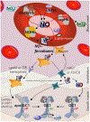
Proposed vascular signalling pathways of NO–ferrohaem. NO–ferrohaem is proposed to ‘shield’ NO from depletion by the NO-dioxygenation reaction in the presence oxyhaemoglobin (oxyHb) in red blood cells (RBCs, top). NO–ferrohaem may be generated in RBCs from the reaction of ‘free’ labile haem, which is found in many cells including RBCs, NO and glutathione. NO–ferrohaem also may be directly liberated from NO–haemoglobin (NO–Hb), which forms when NO, derived from deoxyhaemoglobin-facilitated nitrite reduction or erythrocytic eNOS, reacts with deoxyHb. While NO binding to Hb was previously considered a sequestration event that inhibits NO signalling, NO–Hb may in fact serve as a starting point for downstream signalling in vasodilation. Release of NO–ferrohaem would result in association with abundant serum albumin, which may be responsible for shuttling NO–ferrohaem into endothelial cells via gp60 or transferrin (TfR) transcytosis (left). Alternatively, albumin may present the NO–ferrohaem to known haem transporters such as haem-responsive gene 1 (HRG1) or feline leukaemia virus subgroup C receptor (FLVCR) (right). Access to the smooth muscle cell may follow similar routes, though if albumin is imported into the endothelium, it may release the NO–ferrohaem, which may then be shuttled by other known haem shuttling proteins such as GAPDH and enter the smooth muscle via haem transporters. NO–ferrohaem can then directly bind to the population of apo-GC1 and directly activate the protein, triggering cGMP production and vasodilation. Alternatively, an unknown oxidation event may trigger NO release from NO–ferrohaem, and NO could then activate haem-bound GC1 in the smooth muscle.
FIGURE 3
FIGURE 3
Selected biologically relevant reactions of NO and thiols that yield S-nitrosothiols (RSNOs). Not all reactions pictured here contribute equally to the formation of RSNO. Individual contributions of these reactions depend upon tissue-, cell-, organelle- and even membrane-specific localization and further depend upon cellular conditions (pH, oxygen tension, etc.). The rates of these reactions are also substrate and concentration dependent, with a strong dependence on NO concentration. Top: NO autoxidation, although second order in NO and thus unfavoured at low NO concentrations, is enhanced in membranes, owing to the increased solubility of NO and O2. This ‘lens’ effect in membranes results in accumulation of oxidizing reactive nitrogen species. Reactions highlighted in green are nitrosylations—that is, reactions where NO binds directly to ferric/ferrous iron to make a nitrosylated iron or reacts with a thiyl radical to generate RSNO. Thiyl radicals are generated from thiol oxidation by nitrogen dioxide (NO2), during formation of anionic iron(I) dinitrosyl iron complexes (DNICs) from labile ferrous iron and via thiol-facilitated NO–ferrohaem formation. Reactions highlighted in blue are nitrosation (or transnitrosation) reactions—that is, reactions where a nitrosonium (NO+) equivalent is donated to a nucleophilic substrate, such as thiolates, to generate RSNO. Nitrosating agents include dinitrogen trioxide (N2O3), nitrosyl ferric haemoproteins undergoing reductive nitrosylation, nitrous acid (HNO2), DNICs and RSNOs themselves, which can exchange NO+ with other thiols (transnitrosation). Persulfides are unique nucleophiles that carry out transnitrosation reactions that result in NO release: Transnitrosation from RSNO to R′SSH yields an unstable R′SSNO that rapidly undergoes homolytic cleavage to generate NO and a perthiyl radical, which readily dimerizes to yield a polysulfide. Finally, proteins such as thioredoxin (Trx) and glutaredoxin (Grx) catalyse the S-thiolation of RSNOs via thiolate (R′S) nucleophilic attack of the more electrophilic RSNO sulfur atom. This reaction results in formation of a mixed RSSR′ disulfide and the signalling agent nitroxyl (HNO).

References

    1. Akaike T, Ida T, Wei F-Y, Nishida M, Kumagai Y, Alam MM, Ihara H, Sawa T, Matsunaga T, Kasamatsu S, Nishimura A, Morita M, Tomizawa K, Nishimura A, Watanabe S, Inaba K, Shima H, Tanuma N, Jung M, … Motohashi H (2017). Cysteinyl-tRNA synthetase governs cysteine polysulfidation and mitochondrial bioenergetics. Nature Communications, 8, 1177. 10.1038/s41467-017-01311-y - DOI - PMC - PubMed
    1. Alapa M, Cui C, Shu P, Li H, Kholodovych V, & Beuve A (2021). Selective cysteines oxidation in soluble guanylyl cyclase catalytic domain is involved in NO activation. Free Radical Biology & Medicine, 162, 450–460. 10.1016/j.freeradbiomed.2020.11.001 - DOI - PMC - PubMed
    1. Alexander SPH, Fabbro D, Kelly E, Mathie A, Peters JA, Veale EL, Armstrong JF, Faccenda E, Harding SD, Pawson AJ, Southan C, Davies JA, Boison D, Burns KE, Dessauer C, Gertsch J, Helsby NA, Izzo AA, Koesling D, … Wong SS (2021). The Concise Guide to PHARMACOLOGY 2021/22: Enzymes. British Journal of Pharmacology, 178, S313–S411. - PubMed
    1. Alexander SPH, Kelly E, Mathie A, Peters JA, Veale EL, Armstrong JF, Faccenda E, Harding SD, Pawson AJ, Southan C, Buneman OP, Cidlowski JA, Christopoulos A, Davenport AP, Fabbro D, Spedding M, Striessnig J, Davies JA, Ahlers-Dannen KE, … Zolghadri Y (2021). The Concise Guide to PHARMACOLOGY 2021/22: Introduction and other protein targets. British Journal of Pharmacology, 178, S1–S26. - PMC - PubMed
    1. Armstrong PW, Pieske B, Anstrom KJ, Ezekowitz J, Hernandez AF, Butler J, Lam CSP, Ponikowski P, Voors AA, Jia G, McNulty SE, Patel MJ, Roessig L, Koglin J, & O’Connor CM (2020). Vericiguat in patients with heart failure and reduced ejection fraction. The New England Journal of Medicine, 382, 1883–1893. 10.1056/NEJMoa1915928 - DOI - PubMed

LinkOut - more resources