. 2024 Feb;20(2):1156-1165.
doi: 10.1002/alz.13541. Epub 2023 Oct 31.

Detection of TDP-43 seeding activity in the olfactory mucosa from patients with frontotemporal dementia

Affiliations

Detection of TDP-43 seeding activity in the olfactory mucosa from patients with frontotemporal dementia

Elena Fontana et al. Alzheimers Dement. 2024 Feb.

Abstract

Introduction: We assessed TAR DNA-binding protein 43 (TDP-43) seeding activity and aggregates detection in olfactory mucosa of patients with frontotemporal lobar degeneration with TDP-43-immunoreactive pathology (FTLD-TDP) by TDP-43 seeding amplification assay (TDP43-SAA) and immunocytochemical analysis.

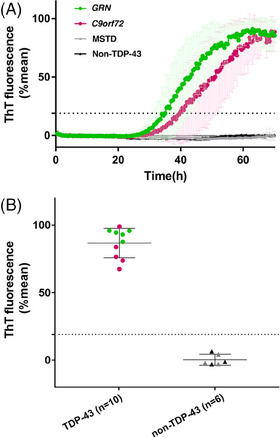
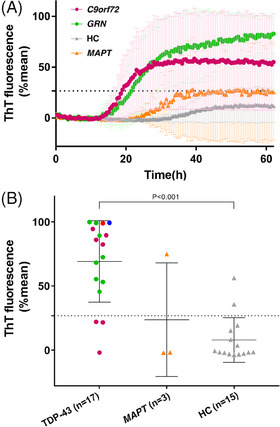
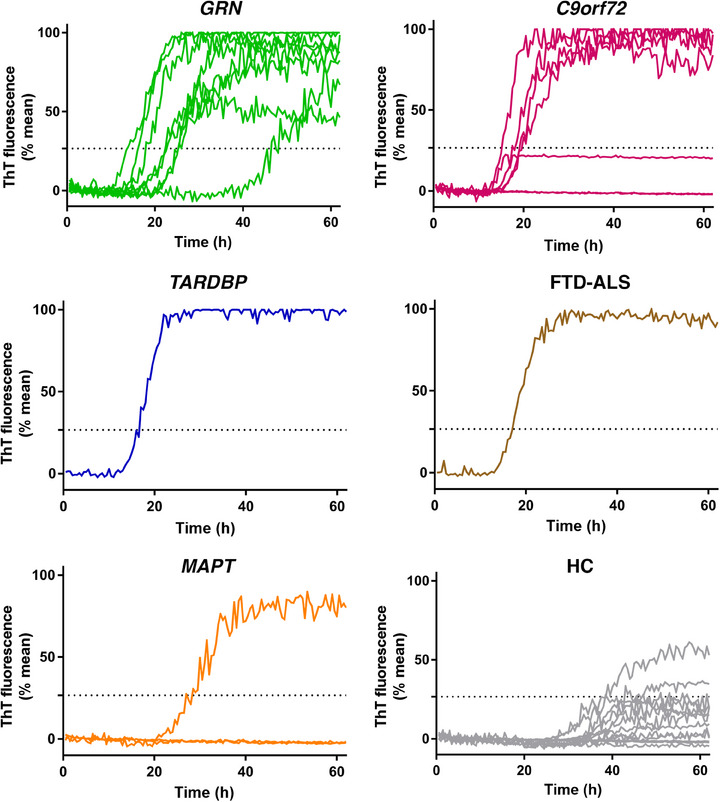
Methods: The TDP43-SAA was optimized using frontal cortex samples from 16 post mortem cases with FTLD-TDP, FTLD with tau inclusions, and controls. Subsequently, olfactory mucosa samples were collected from 17 patients with FTLD-TDP, 15 healthy controls, and three patients carrying MAPT variants.

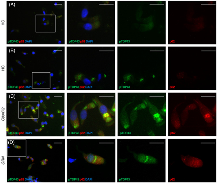
Results: TDP43-SAA discriminated with 100% accuracy post mortem cases presenting or lacking TDP-43 neuropathology. TDP-43 seeding activity was detectable in the olfactory mucosa, and 82.4% of patients with FTLD-TDP tested positive, whereas 86.7% of controls tested negative (P < 0.001). Two out of three patients with MAPT mutations tested negative. In TDP43-SAA positive samples, cytoplasmatic deposits of phosphorylated TDP-43 in the olfactory neural cells were detected.

Discussion: TDP-43 aggregates can be detectable in olfactory mucosa, suggesting that TDP43-SAA might be useful for identifying and monitoring FTLD-TDP in living patients.

Keywords: TAR DNA-binding protein 43; frontotemporal dementia; frontotemporal lobar degeneration; olfactory mucosa; seeding; seeding amplification assays.


Conflict of interest statement

AB was partially supported by Fondazione Cariplo (grant n° 2021‐1516), and by the Fondation pour la Recherche sur Alzheimer. BB was partially supported by ADDF, unrelated to the present work; received personal fees from Alector, UCB, AviadiBio, Lilly, and Wave Life Sciences; and is listed as an inventor on a pending patent on the use of non‐invasive brain stimulation to increase cognitive functions in patients with neurodegenerative disorders. BG was partially supported by a R01‐AG075802 grant. MB, EB, CS, LS, AC, SC, EB, FG, MI, AS, CF, MF, KLN, LC, HJG, MPC, MP, AP, SM, GL, and GZ report no disclosures. This work was supported by project funding from Target ALS Foundation. Author disclosures are available in the supporting information.

Figures

FIGURE 1
FIGURE 1
TDP‐43 seeding activity in frontal cortex of FTLD‐TDP and non–FTLD‐TDP. A, Curves of TDP43‐SAA from the frontal cortex of FTLD‐TDP (green line, GRN; purple line, C9orf72), MSTD (gray line), and non‐TDP‐43 (black line) cases. Curves derived from the average percentage of ThT fluorescence from four replicate reactions (normalized as described in the Methods section). Results are expressed as means of these averages (thick lines) and standard deviations (thin lines) as a function of SAA reaction time. B, Final average relative ThT fluorescence from four replicates readings obtained from frontal cortex of FTLD‐TDP (green dots, GRN; purple dots, C9orf72, i.e. TDP‐43) and non‐FTLD‐TDP (gray triangles, MSTD; black triangles, HC, i.e. non‐TDP‐43) at 60 hours. Bars represent the average ± standard deviation for each case. The dashed line marks the fluorescence threshold for positive results. C9orf72, chromosome 9 open reading frame 72 expansion; FTLD‐TDP, frontotemporal lobar degeneration with TDP‐43‐immunoreactive pathology; GRN, granulin variants; MSTD, multiple system tauopathy with dementia; SAA, seeding amplification assay; TDP‐43, TAR DNA‐binding protein–43‐immunoreactive pathology cases; ThT, thioflavin T
FIGURE 2
FIGURE 2
TDP‐43 seeding activity in olfactory mucosa of FTLD‐TDP and non–FTLD‐TDP cases. A, Curves of TDP43‐SAA from the olfactory mucosa of patients with FTLD‐TDP (green line = GRN; purple line = C9orf72), FTLD‐tau (orange line), and HC (gray line). Curves derived from the average percentage of ThT fluorescence from four replicate reactions (normalized as described in the methods section). Results are expressed as means of these averages (thick lines) and standard deviations (thin lines) as a function of SAA reaction time. B, Final average relative ThT fluorescence from four replicates readings obtained from the olfactory mucosa of FTLD‐TDP (green dots, GRN; purple dots, C9orf72; blue dot, TARDP; red dot, FTD‐ALS, i.e. TDP‐43), HC (gray triangles), and FTLD‐tau with MAPT variants (yellow triangles) at 60 hours. Bars represent the average ± standard deviation for each case. The dashed line marks the fluorescence threshold for positive results. C9orf72, chromosome 9 open reading frame 72 expansion; FTD‐ALS, frontotemporal dementia‐amyotrophic lateral sclerosis; FTLD‐tau; frontotemporal lobar degeneration with tau inclusions; FTLD‐TDP, frontotemporal lobar degeneration with TDP‐43‐immunoreactive pathology; GRN, granulin variants; HC, healthy controls; MAPT, microtubule associate protein tau variants; SAA, seeding amplification assay; TARDP, TAR DNA‐binding protein variants; TDP‐43, TAR DNA‐binding protein 43; ThT, thioflavin T
FIGURE 3
FIGURE 3
TDP‐43 seeding activity in olfactory mucosa in different subgroups. Representative curves of TDP43‐SAA from the olfactory mucosa, derived from the average percentage of ThT fluorescence from four replicate reactions (normalized as described in the Methods section) in each subject, grouped according to genetic trait or diagnosis. C9orf72, chromosome 9 open reading frame 72 expansion; FTD‐ALS, frontotemporal dementia‐amyotrophic lateral sclerosis; GRN, granulin variants; HC, healthy controls; MAPT, microtubule associate protein tau variants; SAA, seeding amplification assay; TARDP, TAR DNA‐binding protein variants; TDP‐43, TAR DNA‐binding protein 43; ThT, thioflavin T
FIGURE 4
FIGURE 4
Distribution pattern of TDP‐43 aggregates. Cells obtained by olfactory brushing stained with phosphorylated TDP‐43 (pTDP43, green), p62 (red), and DAPI (blue). A and B, Two representative healthy controls (HC). C, One representative patient with C9orf72 repeat expansion (C9orf72). D, One representative patient with granulin variant (GRN). See Results section for details. Original 40X images (scale bar 25 μM) on the left, with insets on the three right columns. C9orf72, chromosome 9 open reading frame 72 expansion; TDP‐43, TAR DNA‐binding protein 43

References

    1. Cairns NJ, Bigio EH, Mackenzie IRA, et al. Neuropathologic diagnostic and nosologic criteria for frontotemporal lobar degeneration: consensus of the Consortium for Frontotemporal Lobar Degeneration. Acta Neuropathol. 2007;114(1):5‐22. http://link.springer.com/10.1007/s00401‐007‐0237‐2 - DOI - PMC - PubMed
    1. Mackenzie IR, Neumann M. Reappraisal of TDP‐43 pathology in FTLD‐U subtypes. Acta Neuropathol. 2017;134(1):79‐96. doi:10.1007/s00401-017-1716-8 - DOI - PubMed
    1. Gorno‐Tempini ML, Hillis AE, Weintraub S, et al. Classification of primary progressive aphasia and its variants. Neurology. 2011;76(11):1006‐1014. http://eutils.ncbi.nlm.nih.gov/entrez/eutils/elink.fcgi?dbfrom=pubmed&id... - PMC - PubMed
    1. Rascovsky K, Hodges JR, Knopman D, et al. Sensitivity of revised diagnostic criteria for the behavioural variant of frontotemporal dementia. Brain. 2011;134(Pt 9):2456‐2477. doi:10.1093/brain/awr179 - DOI - PMC - PubMed
    1. Logroscino G, Piccininni M, Graff C, et al. Incidence of syndromes associated with frontotemporal lobar degeneration in 9 European countries. JAMA Neurol. 2023;80(3):279. doi:10.1001/jamaneurol.2022.5128. Published online January 30, 2023. - DOI - PMC - PubMed

Publication types

MeSH terms