

Cryo-EM structure of a Shigella podophage reveals a hybrid tail and novel decoration proteins
- PMID: 37909043
- PMCID: PMC10842012
- DOI: 10.1016/j.str.2023.10.007
Cryo-EM structure of a Shigella podophage reveals a hybrid tail and novel decoration proteins
Abstract
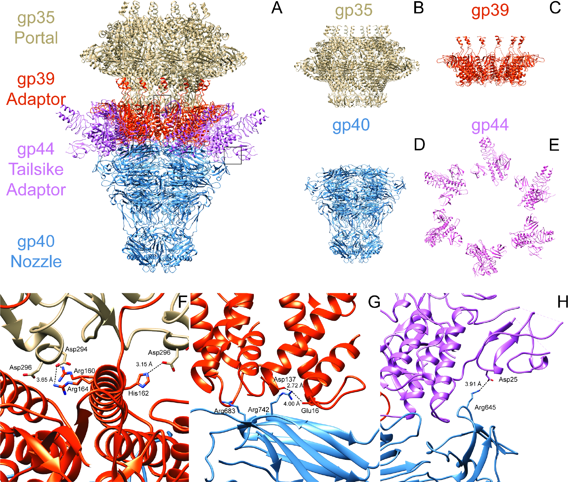
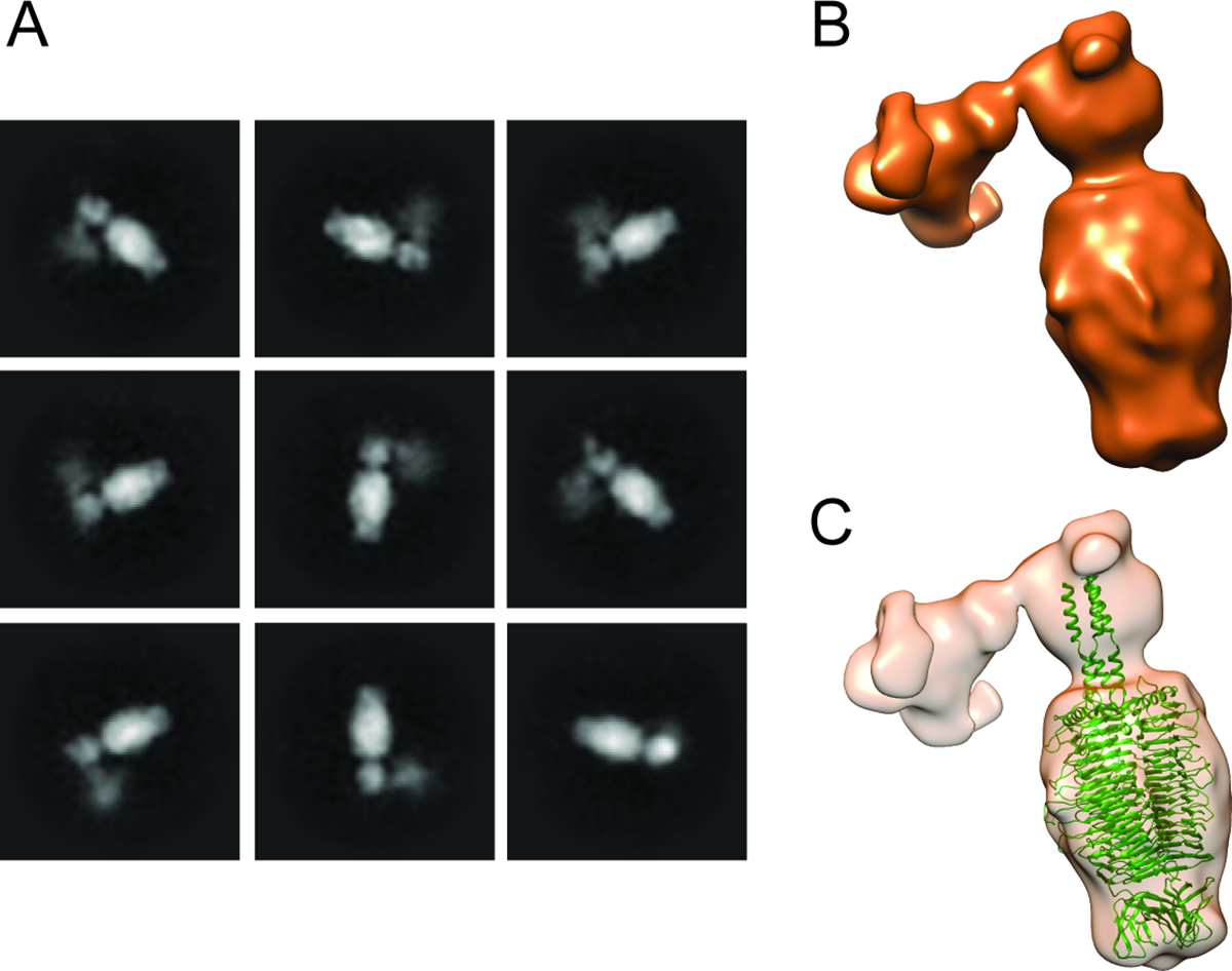
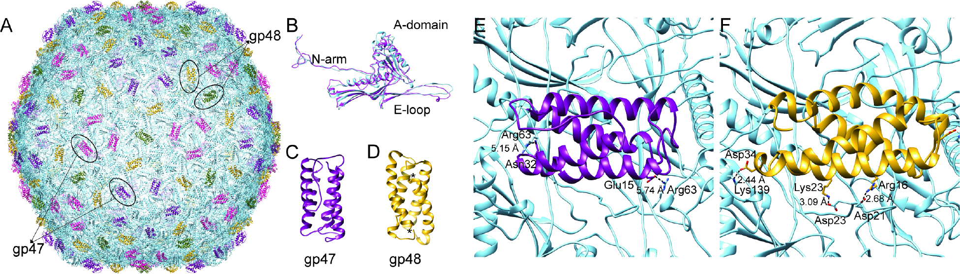
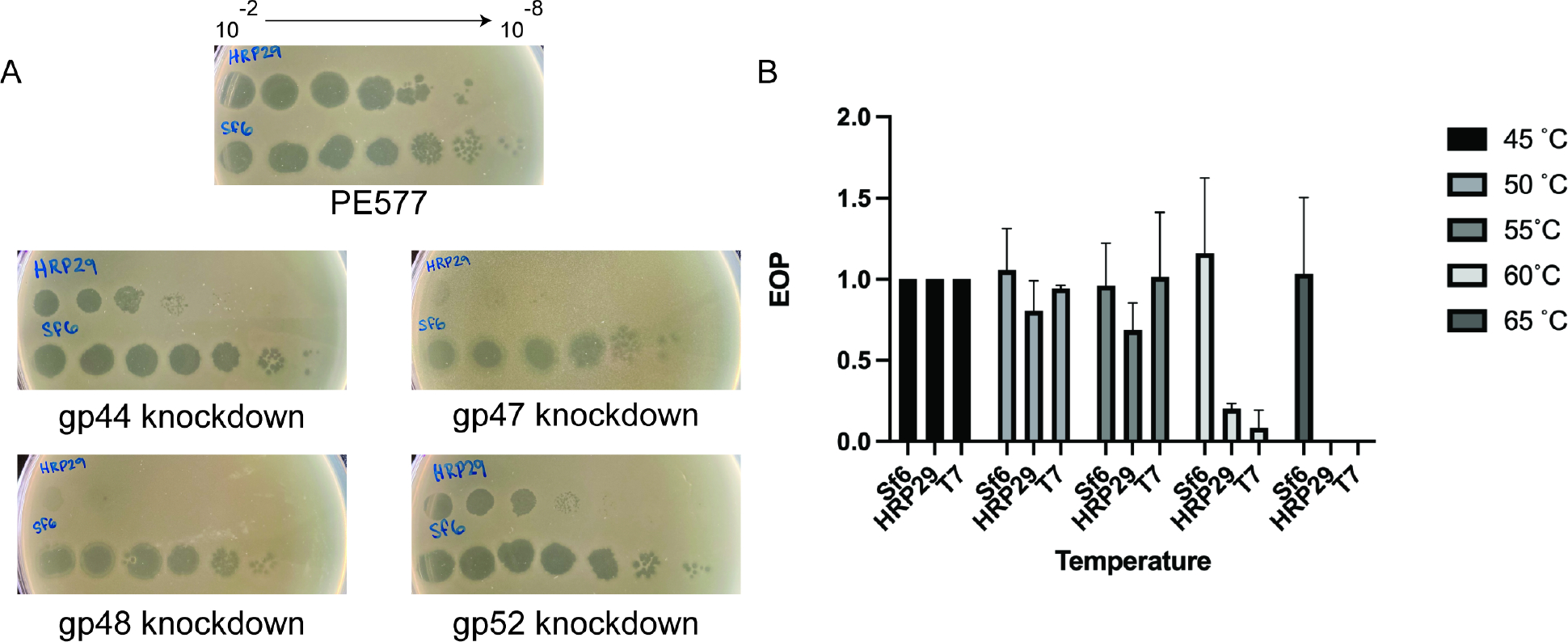
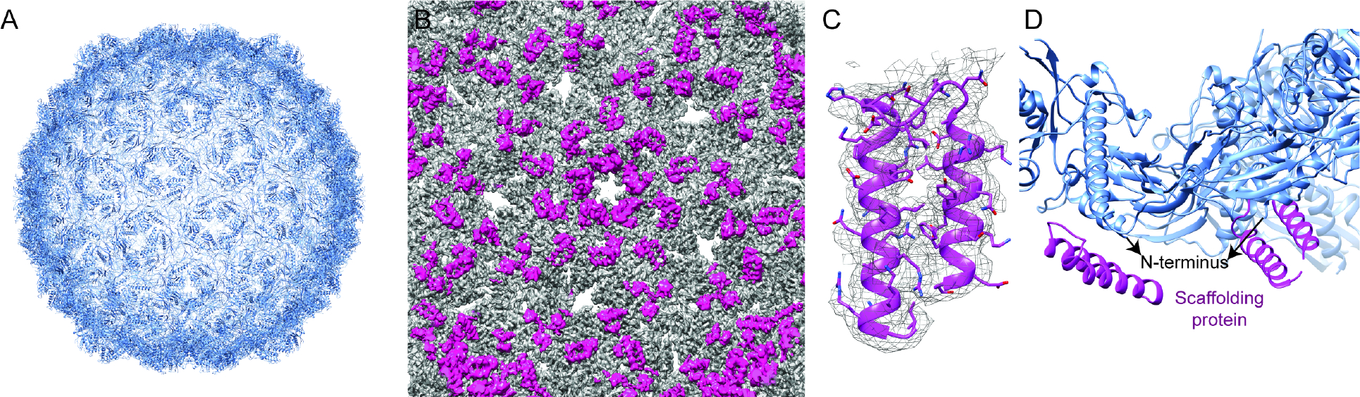
There is a paucity of high-resolution structures of phages infecting Shigella, a human pathogen and a serious threat to global health. HRP29 is a Shigella podophage belonging to the Autographivirinae family, and has very low sequence identity to other known phages. Here, we resolved the structure of the entire HRP29 virion by cryo-EM. Phage HRP29 has a highly unusual tail that is a fusion of a T7-like tail tube and P22-like tailspikes mediated by interactions from a novel tailspike adaptor protein. Understanding phage tail structures is critical as they mediate hosts interactions. Furthermore, we show that the HRP29 capsid is stabilized by two novel, and essential decoration proteins, gp47 and gp48. Only one high resolution structure is currently available for Shigella podophages. The presence of a hybrid tail and an adapter protein suggests that it may be a product of horizontal gene transfer, and may be prevalent in other phages.
Keywords: bacteriophage; capsid; cryo-EM; virion; virus.
Copyright © 2023 Elsevier Ltd. All rights reserved.
Conflict of interest statement
Declaration of interests The authors declare no competing interests.
Figures

Similar articles
-
Multiple masks of a Shigella podophage.Structure. 2024 Jan 4;32(1):1-2. doi: 10.1016/j.str.2023.12.002. Structure. 2024. PMID: 38181725
-
The structure of Shigella virus Sf14 reveals the presence of two decoration proteins and two long tail fibers.Commun Biol. 2025 Feb 12;8(1):222. doi: 10.1038/s42003-025-07668-x. Commun Biol. 2025. PMID: 39939755 Free PMC article.
-
Structural Study of the Cobetia marina Bacteriophage 1 (Carin-1) by Cryo-EM.J Virol. 2023 Apr 27;97(4):e0024823. doi: 10.1128/jvi.00248-23. Epub 2023 Mar 21. J Virol. 2023. PMID: 36943070 Free PMC article.
-
Major tail proteins of bacteriophages of the order Caudovirales.J Biol Chem. 2022 Jan;298(1):101472. doi: 10.1016/j.jbc.2021.101472. Epub 2021 Dec 8. J Biol Chem. 2022. PMID: 34890646 Free PMC article. Review.
-
Cryo-EM reveals infection steps of single-stranded RNA bacteriophages.Prog Biophys Mol Biol. 2021 Mar;160:79-86. doi: 10.1016/j.pbiomolbio.2020.07.011. Epub 2020 Aug 22. Prog Biophys Mol Biol. 2021. PMID: 32841651 Review.
Cited by
-
Structure and infection dynamics of mycobacteriophage Bxb1.Cell. 2025 May 29;188(11):2925-2942.e17. doi: 10.1016/j.cell.2025.03.027. Epub 2025 Apr 15. Cell. 2025. PMID: 40239650
-
Ejectosome of Pectobacterium bacteriophage ΦM1.PNAS Nexus. 2024 Sep 19;3(9):pgae416. doi: 10.1093/pnasnexus/pgae416. eCollection 2024 Sep. PNAS Nexus. 2024. PMID: 39351541 Free PMC article.
-
Elucidating double stranded DNA viral scaffolding protein structures through advances in cryogenic electron microscopy data processing.Curr Opin Struct Biol. 2025 Jun 16;94:103081. doi: 10.1016/j.sbi.2025.103081. Online ahead of print. Curr Opin Struct Biol. 2025. PMID: 40527155 Review.
-
Moo19 and B2: Structures of Schitoviridae podophages with T = 9 geometry and tailspikes with esterase activity.Sci Adv. 2024 Dec 20;10(51):eadt0022. doi: 10.1126/sciadv.adt0022. Epub 2024 Dec 18. Sci Adv. 2024. PMID: 39693418 Free PMC article.
-
Isolation, characterization, and receptor-binding protein specificity of phages PAS7, PAS59 and PAS61 infecting Shiga toxin-producing Escherichia coli O103 and O146.Sci Rep. 2024 Oct 30;14(1):26050. doi: 10.1038/s41598-024-77463-x. Sci Rep. 2024. PMID: 39472643 Free PMC article.
References
MeSH terms
Substances
Grants and funding
LinkOut - more resources
Full Text Sources
