. 2024 Feb;72(2):375-395.
doi: 10.1002/glia.24481. Epub 2023 Nov 1.

Inhibiting CSF1R alleviates cerebrovascular white matter disease and cognitive impairment

Affiliations

Inhibiting CSF1R alleviates cerebrovascular white matter disease and cognitive impairment

Katharine E Askew et al. Glia. 2024 Feb.

Abstract

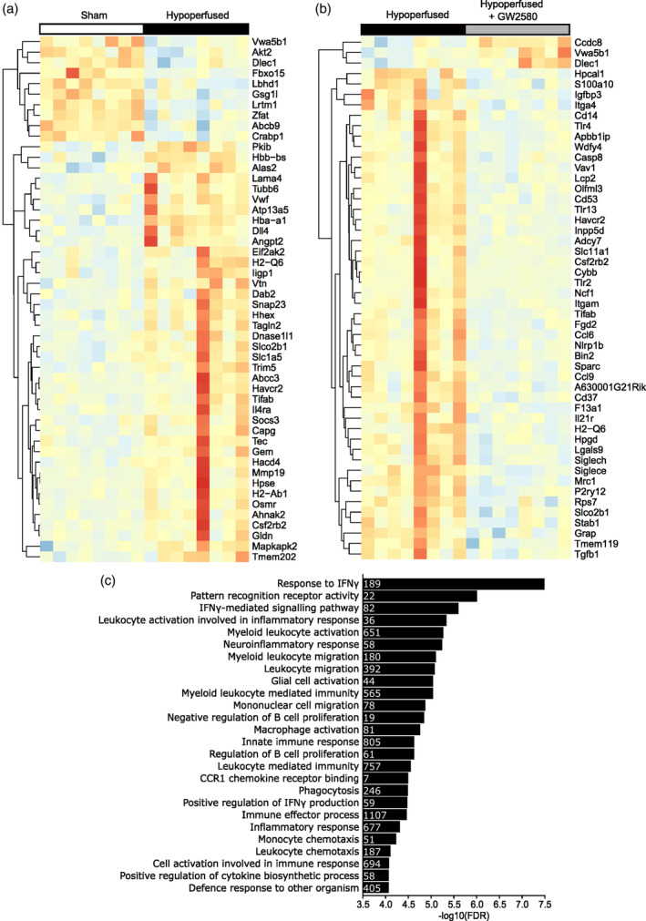
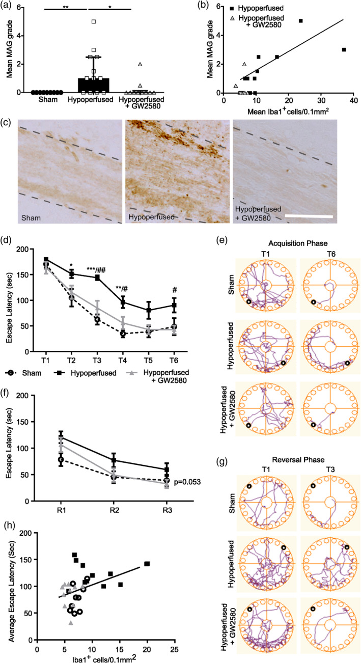
White matter abnormalities, related to poor cerebral perfusion, are a core feature of small vessel cerebrovascular disease, and critical determinants of vascular cognitive impairment and dementia. Despite this importance there is a lack of treatment options. Proliferation of microglia producing an expanded, reactive population and associated neuroinflammatory alterations have been implicated in the onset and progression of cerebrovascular white matter disease, in patients and in animal models, suggesting that targeting microglial proliferation may exert protection. Colony-stimulating factor-1 receptor (CSF1R) is a key regulator of microglial proliferation. We found that the expression of CSF1R/Csf1r and other markers indicative of increased microglial abundance are significantly elevated in damaged white matter in human cerebrovascular disease and in a clinically relevant mouse model of chronic cerebral hypoperfusion and vascular cognitive impairment. Using the mouse model, we investigated long-term pharmacological CSF1R inhibition, via GW2580, and demonstrated that the expansion of microglial numbers in chronic hypoperfused white matter is prevented. Transcriptomic analysis of hypoperfused white matter tissue showed enrichment of microglial and inflammatory gene sets, including phagocytic genes that were the predominant expression modules modified by CSF1R inhibition. Further, CSF1R inhibition attenuated hypoperfusion-induced white matter pathology and rescued spatial learning impairments and to a lesser extent cognitive flexibility. Overall, this work suggests that inhibition of CSF1R and microglial proliferation mediates protection against chronic cerebrovascular white matter pathology and cognitive deficits. Our study nominates CSF1R as a target for the treatment of vascular cognitive disorders with broader implications for treatment of other chronic white matter diseases.

Keywords: CSF1R; cerebrovascular disease; hypoperfusion; microglia; vascular cognitive impairment; white matter.


Conflict of interest statement

The authors have declared that no competing interests exist.

Figures

FIGURE 1
FIGURE 1
Human SVD is associated with increased CSF1R gene expression and microglial activation. (a–c) Hematoxylin and eosin stained representative sections of white matter from basal ganglia of human cases without SVD (a) and with mild–moderate (b) and severe (c) SVD, highlighting white matter arterioles that are (a) normal and (b) with arteriolosclerosis and loss of smooth muscle cells and (c) exhibiting vessel wall hyalinosis and complete loss of smooth muscle cells. (d–f) qPCR analysis of the relative mRNA expression of CSF1R (d), AIF1 (e) and CD68 (f) in white matter‐enriched basal ganglia tissue samples from SVD patients (n = 18) and non‐SVD control cases (n = 10). Data presented as mean ± SD and analyzed by Students' t test. *p < .05, **p < .01.
FIGURE 2
FIGURE 2
Increased microglial proliferation and Csf1r gene expression in a hypoperfusion model of white matter disease (a) Representative flow cytometry dot plots identifying neutrophil (Ly6G+), monocyte (Ly6C+), microglia (CD11b+ CD45low Ly6C Ly6G) and macrophage (CD11b+ CD45high Ly6C Ly6G) populations in the corpus callosum 7 days post‐surgery. (b) Flow cytometric quantification of the absolute numbers of microglia, macrophages, neutrophils and monocytes in the corpus callosum of sham (n = 3) and hypoperfused (n = 6) mice, based on the gating strategy shown in (a). Full gating strategy shown in Supplementary Figure 2a. (c) Representative images of Iba1+ /BrdU+ staining in the corpus callosum. Empty arrowheads indicate BrdU+ single positive cells and filled arrowheads represent Iba1+/BrdU+ cells. Scale bar = 50 μm. (d) Quantification of the number of proliferating microglial cells (Iba1+ BrdU+) and (e) Quantification of Iba1% area staining in the corpus callosum, 7 days following hypoperfusion in the corpus callosum of sham (n = 13) and hypoperfused (n = 14) mice. (f) qPCR analysis of the relative mRNA expression of Csf1r in FACS‐isolated white matter microglia from sham (n = 3) and hypoperfused (n = 6) mice. Data presented as mean ± SD and analyzed by Students' t test (b), or median ± IQR and analyzed by Mann Whitney U test (d–f). **p < .01, ***p < .001.
FIGURE 3
FIGURE 3
CSF1R inhibition following chronic hypoperfusion prevents expansion of microglia in the white matter. (a) Experimental scheme for the chronic hypoperfusion study. Final n numbers: sham, n = 9; hypoperfused + vehicle, n = 12; hypoperfused + GW2580, n = 10. (b) Quantification of the number of microglial cells (Iba1+) and (c) Iba1% area staining as a measure of microglial activation in the corpus callosum following 6 weeks of hypoperfusion and GW2580 treatment. (d) Quantification of the number of proliferating microglial cells (Iba1+ Ki67+) in the corpus callosum following chronic hypoperfusion and GW2580 treatment. (e) Representative images of Iba1 staining in the corpus callosum. Scale bar = 50 μm. Data presented as mean ± SD and analyzed by one‐way ANOVA with post hoc Bonferroni correction (b–d), *p < .05, **p < .01.
FIGURE 4
FIGURE 4
CSF1R inhibition modifies the immune‐related transcriptome profile of hypoperfused white matter (a and b) Heatmaps showing the top 50 most changed genes in white matter samples from sham versus hypoperfused mice (a) and hypoperfused versus hypoperfused + GW2580 mice (b). Data presented as log2‐transformed FPKM and scaled to average Log2 FPKM per gene, red indicating higher and blue lower expression. Sham, n = 8; hypoperfused, n = 8; hypoperfused + GW2580, n = 8. (c) Negative log10 (FDR) for the top gene sets downregulated by GW2580 treatment after chronic hypoperfusion. Inset numbers within bars represent the numbers of genes altered within that pathway.
FIGURE 5
FIGURE 5
CSF1R inhibition reduces phagocytic microglia in hypoperfused white matter (a) Representative images of Iba1+ /Lamp2+ staining in the corpus callosum following chronic hypoperfusion and GW2580 treatment. Arrowheads indicate Lamp2+ positive staining. Scale bar = 50 μm. (b) Quantification of the total number of Lamp2+/ Iba1+ microglial cells in the corpus callosum of sham (n = 9), hypoperfused (n = 12) and hypoperfused mice treated with GW2580 (n = 10). (c) Normalization of the number of Lamp2+ /Iba1+ cells to the total number of Iba1+ cells in the corpus callosum. Data presented as median ± IQR and analyzed by Kruskal‐Wallis with post hoc Dunn's test. **p < .01, ***p < .001.
FIGURE 6
FIGURE 6
CSF1R inhibition following chronic hypoperfusion improves white matter integrity and cognitive abilities. (a) Semi‐quantitative grading of white matter damage using MAG staining in the corpus callosum following chronic hypoperfusion and GW2580 treatment. Data presented as median ± IQR and analyzed by Kruskal‐Wallis with post hoc Dunn's test, *p < .05, **p < .01. (b) White matter damage evidenced by increased MAG grading positively correlates with microglial density post‐hypoperfusion and is rescued by GW2580 treatment. Data analyzed by Spearman's Rho test. (c) Representative images of MAG staining in the corpus callosum. Scale bar = 50 μm. (d) Quantification of latency to escape chamber (seconds) by sham (n = 9), hypoperfused (n = 12) and hypoperfused mice treated with GW2580 (n = 10) across the 6 training days in the acquisition phase of the Barnes maze. Each training day represents an average of two trials, maximum trial length 180 seconds. (e) Representative examples of movement traces across trial days in the acquisition phase. Black circle indicates location of escape hole. (f) Quantification of latency to escape chamber (seconds) across the 3 training days in the reversal phase of the Barnes maze. (g) Representative examples of movement traces across trial days in the reversal phase. Black circle indicates location of escape hole. Data (d, f) presented as mean ± SEM and analyzed by repeated measures two‐way ANOVA with post hoc Bonferroni correction *p < .05, **p < .01, ***p < .001, # p < .05, ## p < .01. * sham versus. hypoperfused, # hypoperfused versus hypoperfused + GW2580. (h) Impaired spatial learning evidenced by increased latency to escape chamber following chronic hypoperfusion correlates positively with increased microglial density in the corpus callosum. Data analyzed by Spearman's Rho.

References

    1. Al‐Mashhadi, S. , Simpson, J. E. , Heath, P. R., Dickman, M., Forster, G., Matthews, F. E., Brayne, C., Ince, P. G., & Wharton, S. B . (2015). Oxidative glial cell damage associated with White matter lesions in the aging human brain. Brain Pathology, 25(5), 565–574. - PMC - PubMed
    1. Armstrong, N. J. , Mather, K. A. , Sargurupremraj, M. , et al. (2020). Common genetic variation indicates separate causes for periventricular and deep White matter Hyperintensities. Stroke, 51(7), 2111–2121. - PMC - PubMed
    1. Askew, K. , Li, K. , Olmos‐Alonso, A. , Garcia‐Moreno, F. , Liang, Y. , Richardson, P. , Tipton, T. , Chapman, M. A. , Riecken, K. , Beccari, S. , Sierra, A. , Molnár, Z. , Cragg, M. S. , Garaschuk, O. , Perry, V. H. , & Gomez‐Nicola, D. (2017). Coupled proliferation and apoptosis maintain the rapid turnover of microglia in the adult brain. Cell Reports, 18, 391–405. - PMC - PubMed
    1. Barbu, M. C. , Spiliopoulou, A. , Colombo, M. , McKeigue, P. , Clarke, T. K. , Howard, D. M. , Adams, M. J. , Shen, X. , Lawrie, S. M. , McIntosh, A. M. , & Whalley, H. C. (2020). Expression quantitative trait loci‐derived scores and white matter microstructure in UK biobank: A novel approach to integrating genetics and neuroimaging. Translational Psychiatry, 10(1), 55. - PMC - PubMed
    1. Ben‐Ari, H. , Lifschytz, T. , Wolf, G. , Rigbi, A. , Blumenfeld‐Katzir, T. , Merzel, T. K. , Koroukhov, N. , Lotan, A. , & Lerer, B. (2019). White matter lesions, cerebral inflammation and cognitive function in a mouse model of cerebral hypoperfusion. Brain Research, 1711, 193–201. - PubMed

Publication types

MeSH terms

Substances