. 2023 Dec;14(6):2520-2531.
doi: 10.1002/jcsm.13265. Epub 2023 Nov 1.

Receptor interacting protein kinase-3 mediates both myopathy and cardiomyopathy in preclinical animal models of Duchenne muscular dystrophy

Affiliations

Receptor interacting protein kinase-3 mediates both myopathy and cardiomyopathy in preclinical animal models of Duchenne muscular dystrophy

Maximilien Bencze et al. J Cachexia Sarcopenia Muscle. 2023 Dec.

Abstract

Background: Duchenne muscular dystrophy (DMD) is a progressive muscle degenerative disorder, culminating in a complete loss of ambulation, hypertrophic cardiomyopathy and a fatal cardiorespiratory failure. Necroptosis is the form of necrosis that is dependent upon the receptor-interacting protein kinase (RIPK) 3; it is involved in several inflammatory and neurodegenerative conditions. We previously identified RIPK3 as a key player in the acute myonecrosis affecting the hindlimb muscles of the mdx dystrophic mouse model. Whether necroptosis also mediates respiratory and heart disorders in DMD is currently unknown.

Methods: Evidence of activation of the necroptotic axis was examined in dystrophic tissues from Golden retriever muscular dystrophy (GRMD) dogs and R-DMDdel52 rats. A functional assessment of the involvement of necroptosis in dystrophic animals was performed on mdx mice that were genetically depleted for RIPK3. Dystrophic mice aged from 12 to 18 months were analysed by histology and molecular biology to compare the phenotype of muscles from mdxRipk3+/+ and mdxRipk3-/- mice. Heart function was also examined by echocardiography in 40-week-old mice.

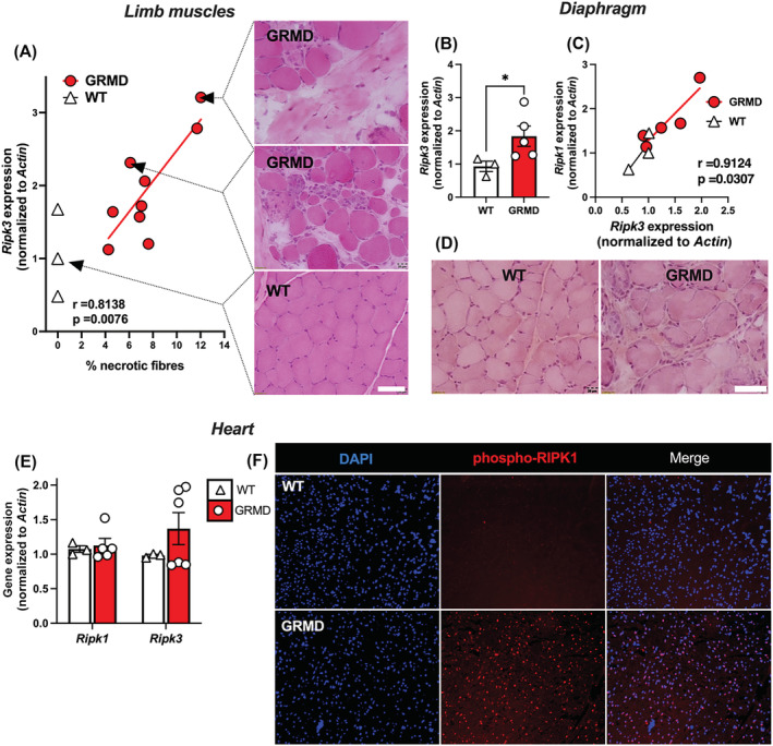
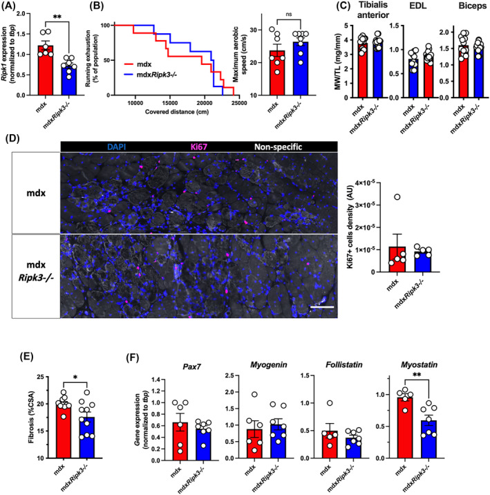
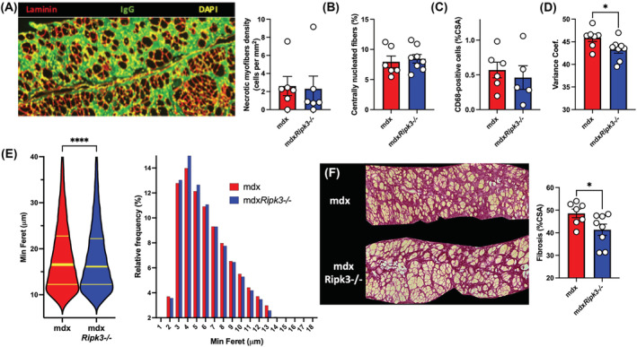
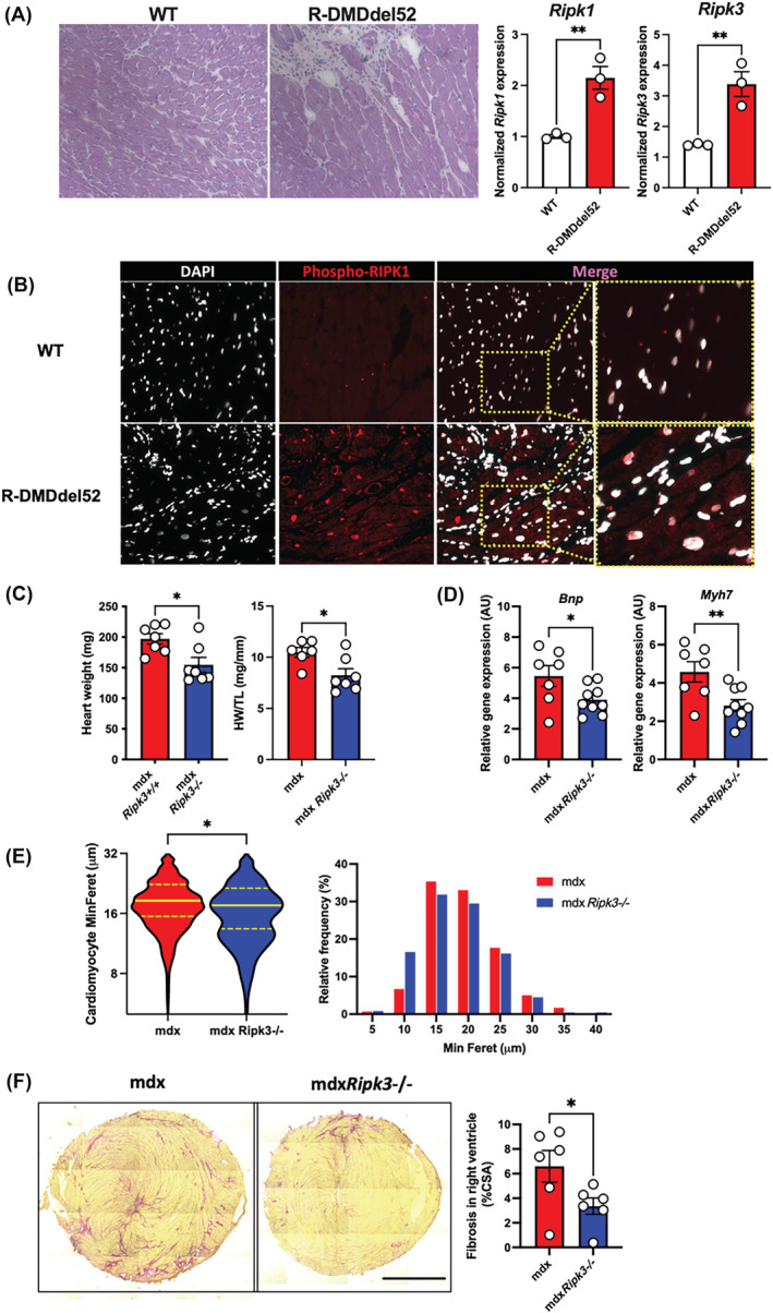
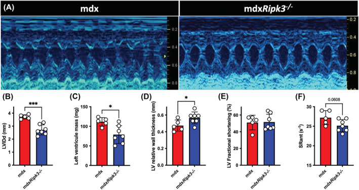
Results: RIPK3 expression in sartorius and biceps femoris muscles from GRMD dogs positively correlated to myonecrosis levels (r = 0.81; P = 0.0076). RIPK3 was also found elevated in the diaphragm (P ≤ 0.05). In the slow-progressing heart phenotype of GRMD dogs, the phosphorylated form of RIPK1 at the Serine 161 site was dramatically increased in cardiomyocytes. A similar p-RIPK1 upregulation characterized the cardiomyocytes of the severe DMDdel52 rat model, associated with a marked overexpression of Ripk1 (P = 0.007) and Ripk3 (P = 0.008), indicating primed activation of the necroptotic pathway in the dystrophic heart. MdxRipk3-/- mice displayed decreased compensatory hypertrophy of the heart (P = 0.014), and echocardiography showed a 19% increase in the relative wall thickness (P < 0.05) and 29% reduction in the left ventricle mass (P = 0.0144). Besides, mdxRipk3-/- mice presented no evidence of a regenerative default or sarcopenia in skeletal muscles, moreover around 50% less affected by fibrosis (P < 0.05).

Conclusions: Our data highlight molecular and histological evidence that the necroptotic pathway is activated in degenerative tissues from dystrophic animal models, including the diaphragm and the heart. We also provide the genetic proof of concept that selective inhibition of necroptosis in dystrophic condition improves both histological features of muscles and cardiac function, suggesting that prevention of necroptosis is susceptible to providing multiorgan beneficial effects for DMD.

Keywords: Animal model; Cardiac failure; Duchenne muscular dystrophy; Fibrosis; Myogenesis; Myonecrosis; Necroptosis; Programmed cell death.


Conflict of interest statement

The authors declare no conflict of interest.

Figures

Figure 1
Figure 1
The RIPK1‐RIPK3 axis is dysregulated in the skeletal muscles and the heart of dystrophic dogs. (A) Pearson correlation coefficient (r) and P‐value (P) between necrotic fibre percentage and RIPK3 transcripts expression normalized to ACTIN. Representative H&E staining of the sartorius muscle corresponding to related biopsied muscle. Scale bar: 20 μm. (B) Quantification of RIPK3 expression in the diaphragms of 6‐month‐old WT and GRMD dogs. (C) Pearson correlation between RIPK1 and RIPK3 expression in the diaphragm. (D) H&E staining of the diaphragm muscle. (E) in the heart left ventricle of GRMD and WT dogs, quantification of RIPK1 and RIPK3 transcripts normalized to ACTIN. (F) Representative image of a left ventricle from WT and GRMD dogs labelled with an antibody directed against the phosphorylated form of RIPK1 (S161). Data are given as means ± SEM. ns: Not significant, *P‐value < 0.05.
Figure 2
Figure 2
The genetic ablation of RIPK3 does not promote sarcopenia in 18‐month‐old mdx mice. (A) Quantification of Ripk1 transcripts in the 18‐month‐old biceps muscles. Seventeen‐month‐old mdxRipk3 +/+ (i.e., mdx) and mdxRipk3 −/− male mice were submitted to forced treadmill running with a progressive increase of speed. (B) Endurance curve of running mice against covered distance expressed in cm and maximum aerobic speed before mice exhaustion. Data are given as means ± SEM. mdxRipk3 +/+, n = 7, mdxRipk3 −/−, n = 8. Shapiro–Wilk test followed by a Mann–Whitney test. (C) Tibialis anterior (TA), EDL and biceps muscles were harvested, and muscle weight (MW) was determined (and normalized to tibial length (MW/TL). (D) TA immunolabelling using antibodies directed against Ki67 (red) and quantification of the density of Ki67‐positive cells. (E) Quantification of collagen deposition using Sirius red dye. The normal distribution of data was tested using the Kolmogorov–Smirnov test (passing normality test with α = 0.05), leading to either t‐tests or Mann–Whitney tests. (F) Quantification of the transcript levels of Pax7, myogenin, follistatin, and myostatin in the biceps, by quantitative PCR. Data are given as means ± SEM. ns: Not significant, *P‐value < 0.05, **P‐value < 0.01. Scale bar: 100 μm.
Figure 3
Figure 3
Necroptosis participates in the diaphragm fibrosis in mdx mice. (A) Representative image of IgG uptake in diaphragms of 18‐month‐old mdx (mdxRipk3 +/+) and mdxRipk3 −/− mice and quantification of myonecrosis extent. Myonecrosis is defined as the area occupied by leaky myofibres (i.e., immunoreactive for mouse IgG uptake). (B) Quantification of the percentage of centrally nucleated myofibres. (C) CD68‐positive macrophage infiltrates expressed in percentage of CSA. (D) Quantification of the variance coefficient of the minimum Feret of myofibres. (E) Violin plot of the minimum Ferets of diaphragm myofibres and relative frequency of minimum Ferets from diaphragm myofibres (mdx, n = 16 873 including seven distinct muscles, mean: 18.61 ± 0.06555; mdxRipk3 −/−, n = 31 666 including eight distinct muscles, mean: 18.21 ± 0.04554, two‐tailed Mann–Whitney test P < 0.0001). Data are expressed as a percentage. (F) Representative picture of Sirius red staining and quantification of diaphragm fibrosis. The normal distribution of data was tested using the Kolmogorov–Smirnov test (passing normality test with α = 0.05), leading to either t‐tests or Mann–Whitney tests. Data are given as means ± SEM. ns: Not significant, *P‐value < 0.05, ****P‐value < 0.0001.
Figure 4
Figure 4
RIPK3 mediates the compensatory hypertrophy of the dystrophin‐deficient heart. (A) Representative pictures of H&E labelling from 6‐month‐old wild‐type (WT) and dystrophin‐deficient R‐DMDdel52 rats and quantification of the rat Ripk1 and Ripk3 transcripts in hearts from WT and R‐DMDdel52 rats. (B) Representative pictures of heart labelling using an antibody directed against the phosphorylated form of RIPK1 at S161 from 6‐month‐old rats. (C) Heart weights of 18‐month‐old mdx and mdxRipk3−/− mice respectively without with normalization to the tibial length (TL). (D) Hearts from 18‐month‐old mdxRipk3 +/+ and −/− were analysed for Bnp and Myh7 mRNA levels by quantitative PCR. (E) Violin plot of the minimum Ferets of mouse cardiomyocytes and relative frequency of minimum Ferets from cardiomyocytes. Data are expressed as a percentage (fifty cardiomyocytes measured per mouse including n = 6 mdx and 5 mdxRipk3 −/− hearts, two‐tailed Mann–Whitney test P < 0.05). (F) Representative picture of Sirius red staining of mdx and mdxRipk3 −/−, and quantification of RV fibrosis. Scale bar: 1700 μm. The normal distribution of data was tested using the Kolmogorov–Smirnov test (passing normality test with α = 0.05), leading to either t‐tests or Mann–Whitney tests. Data are given as means ± SEM. ns: Not significant, *P‐value < 0.05, **P‐value < 0.01.
Figure 5
Figure 5
The genetic ablation of RIPK3 reduces cardiomyopathy in mdx mice. (A) Representative M‐mode echocardiogram traces in mdx and mdxRipk3 −/− 40‐week‐old mice showing a reduced left ventricle (LV) dilation in mdxRipk3 −/− mice. (B) Quantification of LVIDd, (C) LV mass, (D) LV relative wall thickness, (E) LV fractional shortening, and (F) strain rate (SR) of the anterior LV wall. The normal distribution of data was tested using the Kolmogorov–Smirnov test (passing normality test with α = 0.05), leading to t‐tests. Data are given as means ± SEM. *P‐value < 0.05, ***P‐value < 0.001.

References

    1. Relaix F, Bencze M, Borok MJ, der Vartanian A, Gattazzo F, Mademtzoglou D, et al. Perspectives on skeletal muscle stem cells. Nat Commun 2021;12:692. - PMC - PubMed
    1. Desguerre I, Mayer M, Leturcq F, Barbet JP, Gherardi RK, Christov C. Endomysial fibrosis in Duchenne muscular dystrophy: a marker of poor outcome associated with macrophage alternative activation. J Neuropathol Exp Neurol 2009;68:762–773. - PubMed
    1. Khirani S, Ramirez A, Aubertin G, Boulé M, Chemouny C, Forin V, et al. Respiratory muscle decline in Duchenne muscular dystrophy. Pediatr Pulmonol 2014;49:473–481. - PubMed
    1. Fayssoil A, Abasse S, Silverston K. Cardiac involvement classification and therapeutic management in patients with Duchenne muscular dystrophy. J Neuromuscul Dis 2017;4:17–23. - PMC - PubMed
    1. Morgan JE, Prola A, Mariot V, Pini V, Meng J, Hourde C, et al. Necroptosis mediates myofibre death in dystrophin‐deficient mice. Nat Commun 2018;9:3655. - PMC - PubMed

Publication types

Substances