

Iron status in early infancy is associated with trajectories of cognitive development up to pre-school age in rural Gambia
- PMID: 37910494
- PMCID: PMC10619872
- DOI: 10.1371/journal.pgph.0002531
Iron status in early infancy is associated with trajectories of cognitive development up to pre-school age in rural Gambia
Abstract
Introduction: Iron deficiency is among the leading risk factors for poor cognitive development. However, interventions targeting iron deficiency have had mixed results on cognitive outcomes. This may be due to previous interventions focusing on the correction of iron deficiency anaemia in late infancy and early childhood, at which point long lasting neural impacts may already be established. We hypothesise that the relationship between iron status and cognitive development will be observable in the first months of life and will not be recovered by 5 years of age.
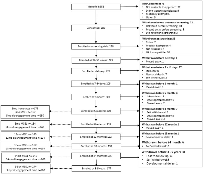
Methods: Using data from the Brain Imaging for Global Health (BRIGHT) Study in Gambia (n = 179), we conducted mixed effects modelling to assess the relationship between iron status at 5 months of age and trajectories of cognitive development from 5 months- 5 years using (i) a standardised measure of cognitive development (Mullen Scales of Early Learning) and (ii) an eye-tracking assessment of attention processing (visual disengagement time).
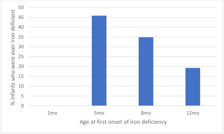
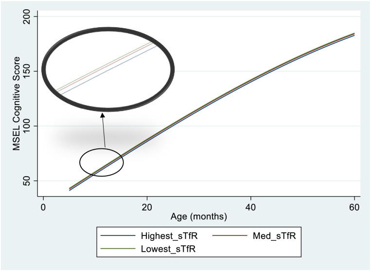
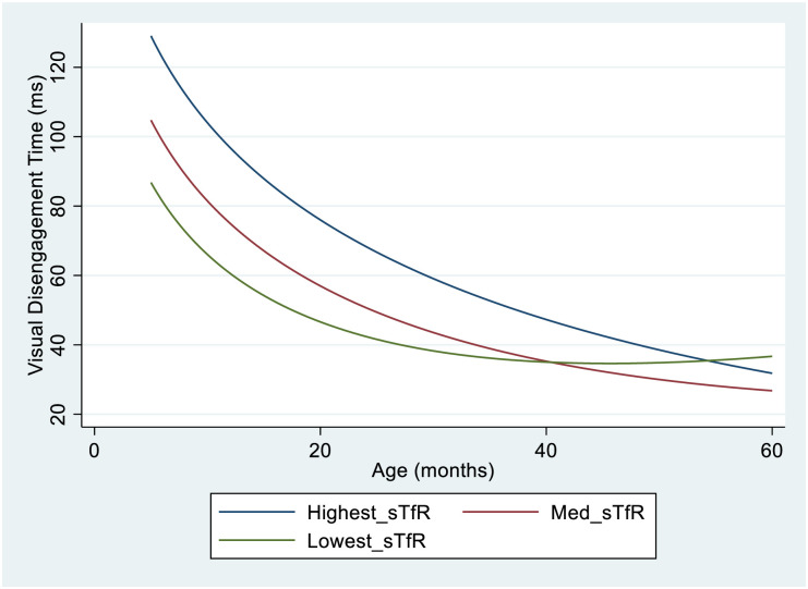
Results: All infants were iron sufficient at 1 month of age. At 5 and 12 months of age 30% and 55% of infants were iron deficient respectively. In fully adjusted analyses, infants in the lowest tercile of soluble transferrin receptor (sTfR) (best iron status) achieved MSEL Cognitive Scores on average 1.9 points higher than infants in the highest sTfR tercile (p = 0.009, effect size = 0.48). There was no evidence that this group difference was recovered by 5 years of age. Infants in the lowest sTfR tercile had visual disengagement time 57ms faster than the highest tercile (p = 0.001, effect size = 0.59). However, this difference diminished by early childhood (p = 0.024).
Conclusion: Infants are at risk of iron deficiency in early infancy. A relationship between iron status and cognitive development is apparent from 5 months of age and remains observable at 5 years of age. One mechanism by which iron availability in early infancy impacts brain development may be through effects on early attentional processing, which is rapidly developing and has substantial nutritional requirements during this period. To support neurocognitive development, prevention of iron deficiency in pre- and early postnatal life may be more effective than correcting iron deficiency once already established.
Copyright: © 2023 McCann et al. This is an open access article distributed under the terms of the Creative Commons Attribution License, which permits unrestricted use, distribution, and reproduction in any medium, provided the original author and source are credited.
Conflict of interest statement
The authors have declared that no competing interests exist.
Figures
References
-
- Stevens G., Finucane M., De-Regil L., Paciorek C., Flaxman S., Branca F. et al. Global, regional, and national trends in haemoglobin concentration and prevalence of total and severe anaemia in children and pregnant and non-pregnant women for 1995–2011: a systematic analysis of population-representative data. The Lancet Global Health. 2013;1(1):e16–e25. doi: 10.1016/S2214-109X(13)70001-9 - DOI - PMC - PubMed
Grants and funding
LinkOut - more resources
Full Text Sources