Review
. 2023 Dec:83:102804.
doi: 10.1016/j.conb.2023.102804. Epub 2023 Oct 31.

Making time and space for calcium control of neuron activity

Affiliations
Review

Making time and space for calcium control of neuron activity

Joanna Jędrzejewska-Szmek et al. Curr Opin Neurobiol. 2023 Dec.

Abstract

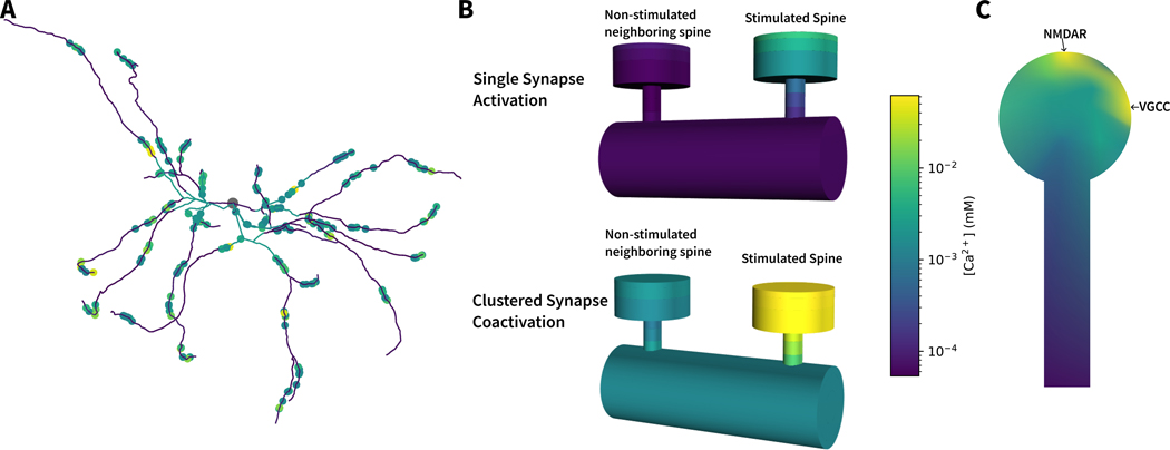
Calcium directly controls or indirectly regulates numerous functions that are critical for neuronal network activity. Intracellular calcium concentration is tightly regulated by numerous molecular mechanisms because spatial domains and temporal dynamics (not just peak amplitude) are critical for calcium control of synaptic plasticity and ion channel activation, which in turn determine neuron spiking activity. The computational models investigating calcium control are valuable because experiments achieving high spatial and temporal resolution simultaneously are technically unfeasible. Simulations of calcium nanodomains reveal that specific calcium sources can couple to specific calcium targets, providing a mechanism to determine the direction of synaptic plasticity. Cooperativity of calcium domains opposes specificity, suggesting that the dendritic branch might be the preferred computational unit of the neuron.

Keywords: Calcium release; Computational model; Nanodomains; Stochastic; Synaptic plasticity.


Conflict of interest statement

Declaration of competing interest The authors declare that they have no known competing financial interests or personal relationships that could have appeared to influence the work reported in this paper.

Figures

Figure 1:
Figure 1:. Calcium is tightly regulated in neural cells.
Calcium flows into the cell through voltage gated calcium channels (VGCC) and the N-methyl-D-aspartate receptor (NMDAR). Na+/Ca2+ exchanger (NCx) and plasma membrane Ca2+ ATPase (PMCA) are the main transmembrane proteins extruding calcium to the extracellular space. In the cytosolic, calcium binds to calcium buffers (CaB) and is transported into the Smooth Endoplasmic Reticulum Calcium ATPase (SERCA) pump to the smooth endoplasmic reticulum (SER). In the SER, calcium binds calreticulin (CRT). Calcium is released from the endoplasmic reticulum upon activation of the ryanodine receptors (RyR) by calcium or activation of the inositol trisphosphate receptors (IP3R) by inositol triphosphate (IP3). When depleted, the SER is replenished by the store operated calcium entry (SOCE), which comprises calcium channels in the cell membrane bound with the SER membrane. Calcium flows through those channels into the cytosol adjacent to SERCA pumps. Calcium from the cytosol is also transported into the mitochondria by the mitochondrial calcium uniporter (MCU) and calcium in the SER can flow into the mitochondrion after the activation of the (IP3R), which are in the vicinity of the mitochondrial voltage-dependent anion channels (vdac).
Figure 2:
Figure 2:. Calcium exhibits spatial specificity and cooperativity across multiple spatial scales.
A. Illustration of peak calcium concentration for the entire dendritic tree during a simulation of spatially distributed clusters of synaptic input trains. Each colored dot indicates a synaptically-activated dendritic spine with the color indicating the peak calcium level for that spine (dots enlarged for illustration), and the color of the dendritic branches correspond to peak dendritic calcium. Based on data from [20]. B. Illustration of simulated calcium levels in a dendritic shaft segment with a synaptically-stimulated spine and a non-stimulated neighboring spine, during either activation of a single synapse (top) or when the stimulated spine is part of a cluster of co-activated synapses (bottom—note the cluster does not include the non-stimulated neighboring spine). Specificity is illustrated by the difference in calcium levels between stimulated and non-stimulated spines; cooperativity is illustrated by the increased calcium in the stimulated spine during clustered coactivation. Heterosynaptic calcium elevations can be observed in non-stimulated neighboring spines during clustered activation due to VGCC influx evoked by the strong local depolarization, but specificity is still maintained as the stimulated spine shows about an order of magnitude higher calcium elevation. Based on data from [41]. C. Illustration of calcium nanodomains and gradients in a single spine, showing synaptically evoked calcium influx through NMDAR channels and influx through voltage gated calcium channels can generate calcium nanodomains important for synaptic signalling. Adapted from [30].

References

    1. Evans R, Morera-Herreras T, Cui Y, Du K, Sheehan T, Kotaleski J, Venance L, B. KT, The effects of nmda subunit composition on calcium influx and spike timing-dependent plasticity in striatal medium spiny neurons, PLoS Comput Biol. 8 (2012) e1002493. doi:10.1371/journal.pcbi.1002493. - DOI - PMC - PubMed
    1. Higgins D, Graupner M, Brunel N, Memory maintenance in synapses with calcium-based plasticity in the presence of background activity, PLoS Comput Biol. 10 (2014) e1003834. doi:10.1371/journal.pcbi.1003834. - DOI - PMC - PubMed
    1. Du K, Wu Y, Lindroos R, Liu Y, Rozsa B, Katona G, Ding J, Kotaleski J, Cell-type-specific inhibition of the dendritic plateau potential in striatal spiny projection neurons, Proc Natl Acad Sci USA. 114 (2017) E7612–E7621. doi:10.1073/pnas.1704893114. - DOI - PMC - PubMed
    1. Scheuss V, Yasuda R, Sobczyk A, Svoboda K, Nonlinear [ca2+] signaling in dendrites and spines caused by activity-dependent depression of ca2+ extrusion, J Neurosci 26 (2006) 8183–8194. - PMC - PubMed
    1. Bell M, Bartol T, Sejnowski T, Rangamani P, Dendritic spine geometry and spine apparatus organization govern the spatiotemporal dynamics of calcium, J. Gen. Physiol. 151 (2019) 1017–1034. - PMC - PubMed

Publication types

MeSH terms

LinkOut - more resources