. 2024 Mar;24(3):391-405.
doi: 10.1016/j.ajt.2023.10.013. Epub 2023 Oct 31.

Transplanting old organs promotes senescence in young recipients

Affiliations

Transplanting old organs promotes senescence in young recipients

Jasper Iske et al. Am J Transplant. 2024 Mar.

Abstract

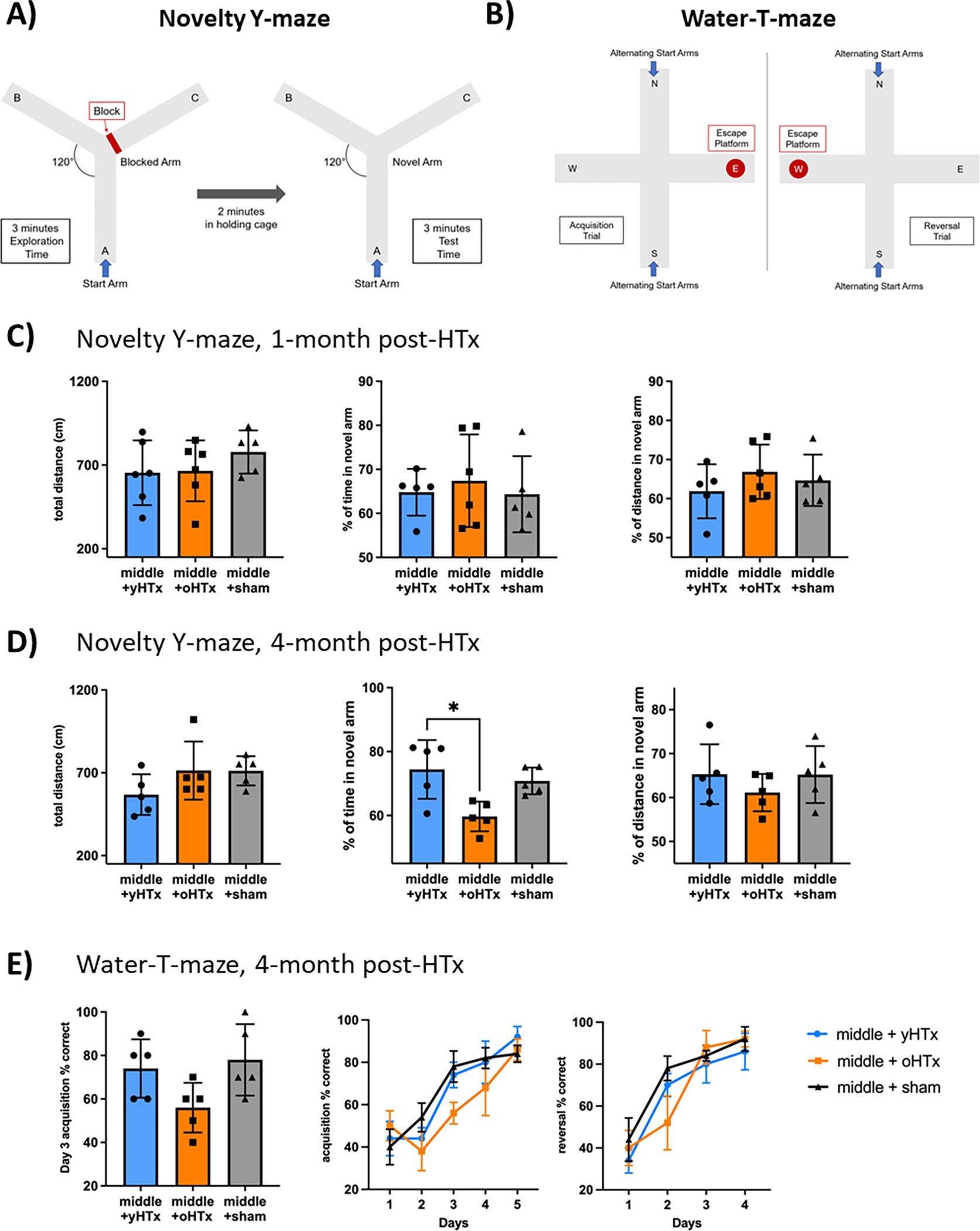
In clinical organ transplantation, donor and recipient ages may differ substantially. Old donor organs accumulate senescent cells that have the capacity to induce senescence in naïve cells. We hypothesized that the engraftment of old organs may induce senescence in younger recipients, promoting age-related pathologies. When performing isogeneic cardiac transplants between age-mismatched C57BL/6 old donor (18 months) mice and young and middle-aged C57BL/6 (3- or 12- month-old) recipients , we observed augmented frequencies of senescent cells in draining lymph nodes, adipose tissue, livers, and hindlimb muscles 30 days after transplantation. These observations went along with compromised physical performance and impaired spatial learning and memory abilities. Systemic levels of the senescence-associated secretory phenotype factors, including mitochondrial DNA (mt-DNA), were elevated in recipients. Of mechanistic relevance, injections of mt-DNA phenocopied effects of age-mismatched organ transplantation on accelerating aging. Single treatment of old donor animals with senolytics prior to transplantation attenuated mt-DNA release and improved physical capacities in young recipients. Collectively, we show that transplanting older organs induces senescence in transplant recipients, resulting in compromised physical and cognitive capacities. Depleting senescent cells with senolytics, in turn, represents a promising approach to improve outcomes of older organs.

Keywords: Aging; Senescence; age-related pathologies; cellular senescence; ischemia-reperfusion injury; organ allocation; senescence-associated secretory phenotype; senolytics.


Conflict of interest statement

Competing interests

J.L.K., T.T., and S.G.T. have a financial interest related to this research. Patents on senolytic drugs are held by Mayo Clinic and Brigham and Women’s Hospital. This research has been reviewed by the Mayo Clinic Conflict of Interest Review Board and was conducted in compliance with Mayo Clinic Conflict of Interest policies. No conflicts of interest, financial or otherwise, are declared by the other authors.

Declaration of interests

The authors declare the following financial interests/personal relationships which may be considered as potential competing interests:

Stefan G. Tullius has patent Methods And Materials For Improving Transplant Outcomes licensed to Stefan G. Tullius.

James L. Kirkland has patent Methods And Materials For Improving Transplant Outcomes licensed to Stefan G. Tullius.

Tamar Tchkonia has patent Methods And Materials For Improving Transplant Outcomes licensed to Stefan G. Tullius.

Figures

Figure 1.
Figure 1.. Transplantation of old cardiac isografts induces cellular senescence in young recipient organs
Hearts from either young or old C57BL/6 (3- and 18-months) donors were transplanted into young syngeneic recipients. (A) Liver and draining lymph nodes were collected from donors 30 days after engraftment, cut into slides, and co-stained for p16Ink4a, p21Cip1, and DAPI; cell size and percentage of senescent cells was defined as the number of p16/p21 double positive cells for lymph nodes and p16 positive cells of DAPI-stained cells for livers using a confocal microscope. (B) Expression of p16 in inguinal adipose tissue of animals receiving a cardiac transplant from either a young or old INK-ATTAC donor mouse was quantified using p16-RNA in situ hybridization. (C) Furthermore, p16 expression was quantified in femoral muscle (D) and the total brain tissue of young recipient mice by qPCR. Column plots display mean with standard deviation and individual data points are shown. Statistical significance was determined by Student’s T-test. Asterisks indicate p-values * = p≤0.05, **= p≤0.01 and *** = p≤0.001, only significant values are shown. Abbreviations: oHTx, old heart transplant; yHTx, young heart transplant.
Figure 2.
Figure 2.. Recipients of old donor hearts display inferior physical performance
(A) Hearts from either young or old C57BL/6 (3- and 18-months) were transplanted into young (3-months) and middle-aged (12-months) syngeneic recipients. Sham surgery treated recipients served as controls. Physical tests were performed over a 4-month period after transplantation. (B) RotaRod performance of young recipients. (C) Average, minimum, and maximum grip strength of middle-aged recipients are shown. (D) RotaRod performance of middle-aged recipients. Column plots display mean with standard deviation with individual data points being shown. Statistical significance was determined by One-Way ANOVA or Friedmann Test. Asterisks indicate p-values * = p≤0.05, **= p≤0.01 and *** = p≤0.001, only significant values are shown. Abbreviations: oHTx, old heart transplant; yHTx, young heart transplant; s, seconds; g, gram.
Figure 3.
Figure 3.. Recipients of old donor hearts display altered behavior
Hearts from either young or old C57BL/6 (3- and 18-months) were transplanted into middle-aged (12-months) syngeneic recipients. Sham surgery treated middle-aged mice served as controls. Behavioral tests were performed over the 4-months after transplantation. (A) Total distance of 60-minutes locomotor activity, time, and distance spent in the center are shown 1-month post HTx. (B) 30-minutes open field test, total distance, time, and distance spent in the center 1-month post HTx. (C) Distance in the first 5-minutes of the open field test. Column plots display mean with standard deviation with individual data points being shown. Statistical significance was determined by One-Way ANOVA or Friedmann Test. Asterisks indicate p-values * = p≤0.05, **= p≤0.01 and *** = p≤0.001, only significant values are shown. Abbreviations: oHTx, old heart transplant; yHTx, young heart transplant; cm, centimeter.
Figure 4.
Figure 4.. Recipients of old donor hearts display inferior cognitive performance
Hearts from either young or old C57BL/6 (3- and 18-months) were transplanted into middle-aged (12-months) syngeneic recipients. Sham surgery treated middle-aged mice served as controls. One and four months after transplant, cognitive tests were performed. (A) + (B) Schematic explanation of Novelty-Y-maze and Water-T-maze. (C) Novelty-Y-maze test after 1-month: Total distance, distance in novel arm, and time in novel arm. (D) Novelty-Y-maze after 4-months. (E) Water-T-maze: Correct responses for each day. Acquisition day three, the acquisition test and the reversal test. Column plots display mean with standard deviation with individual data points being shown. Statistical significance was determined by One-Way ANOVA or Friedmann Test. Asterisks indicate p-values * = p≤0.05, **= p≤0.01 and *** = p≤0.001, only significant values are shown. Abbreviations: oHTX, old heart transplant; yHTx, young heart transplant; cm, centimeter.
Figure 5.
Figure 5.. Recipients of old and young donor organs exhibit comparable metabolic profiles
Hearts from either young or old C57BL/6 (3- and 18-months) were transplanted into middle-aged (12-months) syngeneic recipients. Sham surgery treated middle-aged mice were included as controls. One and four months after transplant, metabolic cage analysis over 24-hours was performed. Hereby, average oxygen consumption (milliliter/hour), average carbon dioxide production (milliliter/hour), average respiratory exchange ratio (carbon dioxide production/oxygen consumption), and average food consumption were analyzed. (A) Analysis 1-month after transplantation. (B) Analysis 4-months after transplantation. (C) Locomotor activity (beam breaks) of animals while in metabolic cages one and 4-months after transplantation. Column plots display mean with standard deviation with individual data points being shown. Statistical significance was determined by One-Way ANOVA or Friedmann Test. Asterisks indicate p-values * = p≤0.05, **= p≤0.01 and *** = p≤0.001, only significant values are shown. Abbreviations: oHTx, old heart transplant; yHTx, young heart transplant.
Figure 6.
Figure 6.. Transplanting older organs promotes senescence in recipients through augmented release of SASP-factors including mt-DNA
(A) Hearts from either young or old p16Ink4a-GFP reporter mice (3- and 18-months) were transplanted into young or middle-aged syngeneic recipients. After 30 days, GFP expression was quantified in kidneys, muscle, and adipose tissue by qPCR. (B) Hearts from either young or old C57BL/6 donor mice (3- and 18-months) were transplanted into young or middle-aged syngeneic recipients (3- and 12-months). 24-hours after transplantation, mitochondrial DNA was quantified in plasma by real-time PCR according to standard curve results after DNA isolation. (C) Middle-aged mice were exposed to either isolated mt-DNA or PBS injections and physical performance was subsequently assessed using a standardized rotarod tests. (D) By two weeks, p21, p16, IL-6, and TNF-α levels were quantified in brain sections from the hippocampus by qPCR. Column plots display mean with standard deviation with individual data points being shown. Statistical significance was determined by Student’s T-test. Asterisks indicate p-values * = p≤0.05, **= p≤0.01 and *** = p≤0.001, only significant values are shown. Abbreviations: oHTX, old heart transplant; yHTx, young heart transplant.
Figure 7.
Figure 7.. Treating old donor mice with senolytics improves physical performance in recipients of organs transplanted from the old donors
Young and old (3- and 18-months) C57BL/6 mice as well as old C57BL/6 donor mice that had been treated with a single dose of Dasatinib and Quercetin prior to transplantation served as donor mice. (A) 24 hours following syngeneic transplantation of cardiac isografts into young or middle-aged recipient mice, systemic mt-DNA levels were quantified by qPCR. (B) 30 days following transplantation, physical capacity of recipient animals was assessed by a RotaRod test. Column plots display mean with standard deviation with individual data points being shown. Statistical significance was determined by Student’s T-test or Friedmann Test. Asterisks indicate p-values * = p≤0.05, **= p≤0.01 and *** = p≤0.001, only significant values are shown. Abbreviations: oHTX, old heart transplant; yHTx, young heart transplant.

References

    1. Daley MJ, Spinks WL. Exercise, mobility and aging. Sports Med 2000;29(1):1–12. - PubMed
    1. Groessl EJ, Kaplan RM, Rejeski WJ, Katula JA, Glynn NW, King AC et al. Physical Activity and Performance Impact Long-term Quality of Life in Older Adults at Risk for Major Mobility Disability. Am J Prev Med 2019;56(1):141–146. - PMC - PubMed
    1. Clark R, Freedberg M, Hazeltine E, Voss MW. Are There Age-Related Differences in the Ability to Learn Configural Responses? PLoS One 2015;10(8):e0137260. - PMC - PubMed
    1. Foster TC, Defazio RA, Bizon JL. Characterizing cognitive aging of spatial and contextual memory in animal models. Front Aging Neurosci 2012;4:12. - PMC - PubMed
    1. Hou Y, Dan X, Babbar M, Wei Y, Hasselbalch SG, Croteau DL et al. Ageing as a risk factor for neurodegenerative disease. Nat Rev Neurol 2019;15(10):565–581. - PubMed

LinkOut - more resources