. 2023 Nov 1;13(1):18864.
doi: 10.1038/s41598-023-46261-2.

Prognostic value of 18F-FDG PET in uterine cervical cancer patients with stage IIICr allocated by imaging

Affiliations

Prognostic value of 18F-FDG PET in uterine cervical cancer patients with stage IIICr allocated by imaging

Yuzu Isaji et al. Sci Rep. .

Abstract

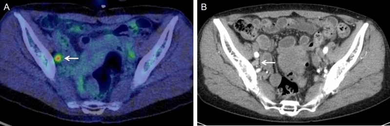
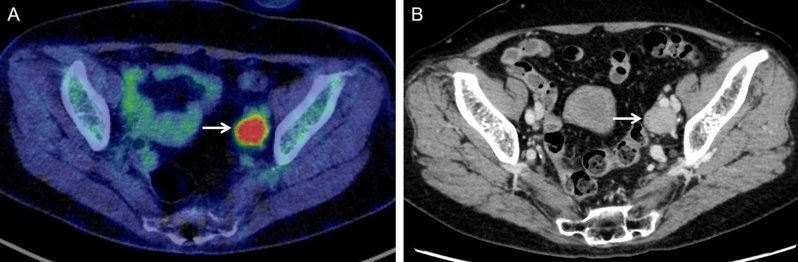
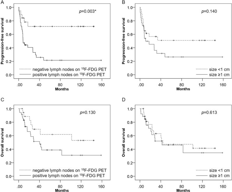
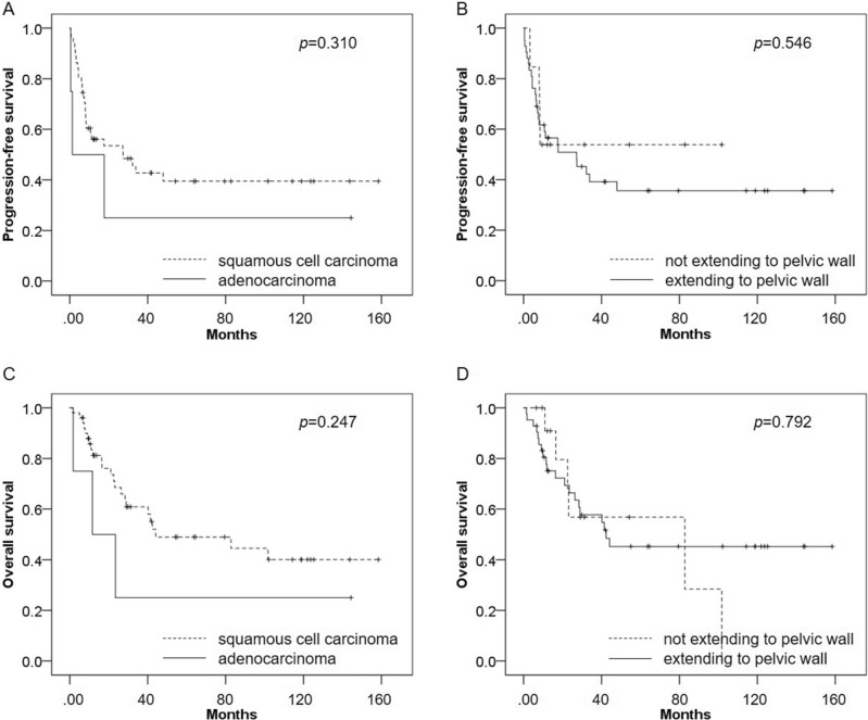
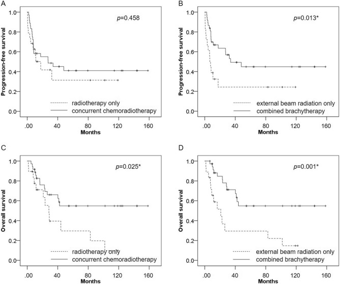
The effect on survival of radiographic lymph node metastasis in uterine cervical cancer patients is more important than before, even though its prognostic value not been well investigated. The aim of our study is to evaluate the prognostic potential of 18F-fluorodeoxyglucose Positron Emission Tomography (18F-FDG PET) compared with Computed Tomography (CT) in uterine cervical cancer patients with stage IIICr allocated by imaging. Fifty-five patients with biopsy-proven primary cervical cancer underwent definitive radiation therapy for stages IIB-IVB of The International Federation of Gynecology and Obstetrics (FIGO) 2018 classifications. The prognostic performance of pretreatment 18F-FDG PET and CT for assessing lymph node metastasis was evaluated by two experienced readers. The PET and CT findings were correlated with the risk of progression-free survival (PFS) and overall survival (OS). Kaplan-Meier survival curves showed that PFS was significantly worse in patients with positive lymph nodes on 18F-FDG PET than in those patients with negative lymph nodes on 18F-FDG PET (p = 0.003), whereas there was no significant difference in PFS between patients with lymph nodes sized ≥ 1 cm and those sized < 1 cm (p = 0.140). Univariate analysis showed that positive lymph nodes on 18F-FDG PET was significantly associated with poor PFS (p = 0.006), whereas lymph node size was not significantly associated with poor PFS (p = 0.145). In multivariate analysis, positive lymph nodes on 18F-FDG PET was significantly associated with poor PFS (p = 0.006) and was an independent prognostic factor for PFS. 18F-FDG PET offers high prognostic value for patients with stage IIICr allocated by imaging compared with CT, suggesting that 18F-FDG PET might be useful in clinical staging decisions and thus promote optimal diagnostic and therapeutic strategies.


Conflict of interest statement

The authors declare no competing interests.

Figures

Figure 1
Figure 1
A 66-year-old woman with stage IIIC1 cervical squamous cell cancer. (A) 18F-FDG PET/CT image shows positive lymph nodes on 18F-FDG PET in a right pelvic lymph node (arrow). (B) CT image shows the lymph node (arrow) of size 0.78 cm (< 1 cm).
Figure 2
Figure 2
A 72-year-old woman with stage IIIC1 cervical squamous cell cancer. (A) 18F-FDG PET/CT image shows positive lymph nodes on 18F-FDG PET in a left pelvic lymph node (arrow). (B) CT image shows the lymph node (arrow) of size 2.08 cm (≥ 1 cm).
Figure 3
Figure 3
Kaplan–Meier survival curves for PFS and OS among patients with uterine cervical cancer who underwent 18F-FDG PET/CT or PET/MRI, and CT before definitive radiation therapy. (A) PFS was significantly worse in patients with positive lymph nodes on 18F-FDG PET than in those with negative lymph nodes on 18F-FDG PET (p = 0.003). (B) There was no significant difference in PFS between patients with lymph node size ≥ 1 cm and those with size < 1 cm (p = 0.140). (C) There was no significant difference in OS between patients with positive lymph nodes on 18F-FDG PET and those with negative lymph nodes on 18F-FDG PET (p = 0.130). (D) There was no significant difference in OS between patients with lymph node size ≥ 1 cm and those with size < 1 cm (p = 0.613).
Figure 4
Figure 4
(A) In terms of clinicopathological parameters, there was no significant difference in PFS between patients with adenocarcinoma and those with squamous cell carcinoma (p = 0.310). (B) There was no significant difference in PFS between patients who had parametrial invasion extending to the pelvic wall and those who did not (p = 0.546). (C) There was no significant difference in OS (p = 0.247) between patients with adenocarcinoma and those with squamous cell carcinoma. (D) There was no significant difference in OS between patients who had parametrial invasion extending to the pelvic wall and those who did not (p = 0.792).
Figure 5
Figure 5
(A) In terms of combined treatments, there was no significant difference in PFS between patients treated with concurrent chemoradiotherapy and those treated with radiotherapy alone (p = 0.458). (B) PFS was significantly better in patients treated with external beam radiation combined with brachytherapy than in those treated with external beam radiation alone (p = 0.013). (C) OS was significantly better patients treated with concurrent chemoradiotherapy than in those treated with radiotherapy alone (p = 0.025). (D) OS was significantly better in patients treated with external beam radiation combined with brachytherapy than in those treated with external beam radiation alone (p = 0.001).

Similar articles

Cited by

References

    1. Sung H, et al. Global cancer statistics 2020: GLOBOCAN estimates of incidence and mortality worldwide for 36 cancers in 185 countries. CA Cancer J. Clin. 2021;71:209–249. doi: 10.3322/caac.21660. - DOI - PubMed
    1. Pecorelli S, Zigliani L, Odicino F. Revised FIGO staging for carcinoma of the cervix. Int. J. Gynaecol. Obstet. 2009;105:107–108. doi: 10.1016/j.ijgo.2009.02.009. - DOI - PubMed
    1. Kamura T, et al. Multivariate analysis of the histopathologic prognostic factors of cervical cancer in patients undergoing radical hysterectomy. Cancer. 1992;69:181–186. doi: 10.1002/1097-0142(19920101)69:1<181::aid-cncr2820690130>3.0.co;2-b. - DOI - PubMed
    1. Bhatla N, et al. Revised FIGO staging for carcinoma of the cervix uteri. Int. J. Gynaecol. Obstet. 2019;145:129–135. doi: 10.1002/ijgo.12749. - DOI - PubMed
    1. Corrigendum to "Revised FIGO staging for carcinoma of the cervix uteri" [Int J Gynecol Obstet 145(2019) 129–135]. Int. J. Gynaecol. Obstet.147, 279–280. 10.1002/ijgo.12969 (2019). - PubMed

MeSH terms

Substances