. 2023 Nov;623(7987):550-554.
doi: 10.1038/s41586-023-06702-4. Epub 2023 Nov 1.

Fossil evidence for a pharyngeal origin of the vertebrate pectoral girdle

Affiliations

Fossil evidence for a pharyngeal origin of the vertebrate pectoral girdle

Martin D Brazeau et al. Nature. 2023 Nov.

Abstract

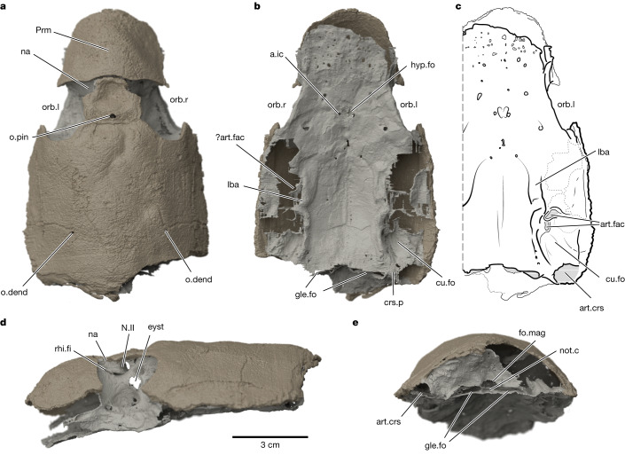
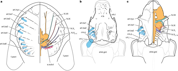
The origin of vertebrate paired appendages is one of the most investigated and debated examples of evolutionary novelty1-7. Paired appendages are widely considered as key innovations that enabled new opportunities for controlled swimming and gill ventilation and were prerequisites for the eventual transition from water to land. The past 150 years of debate8-10 has been shaped by two contentious theories4,5: the ventrolateral fin-fold hypothesis9,10 and the archipterygium hypothesis8. The latter proposes that fins and girdles evolved from an ancestral gill arch. Although studies in animal development have revived interest in this idea11-13, it is apparently unsupported by fossil evidence. Here we present palaeontological support for a pharyngeal basis for the vertebrate shoulder girdle. We use computed tomography scanning to reveal details of the braincase of Kolymaspis sibirica14, an Early Devonian placoderm fish from Siberia, that suggests a pharyngeal component of the shoulder. We combine these findings with refreshed comparative anatomy of placoderms and jawless outgroups to place the origin of the shoulder girdle on the sixth branchial arch. These findings provide a novel framework for understanding the origin of the pectoral girdle. Our evidence clarifies the location of the presumptive head-trunk interface in jawless fishes and explains the constraint on branchial arch number in gnathostomes15. The results revive a key aspect of the archipterygium hypothesis and help reconcile it with the ventrolateral fin-fold model.


Conflict of interest statement

The authors declare no competing interests.

Figures

Fig. 1
Fig. 1. The braincase and skull roof of K. sibirica Bystrow 1956 specimen TsNIGR 7656 as a virtual three-dimensional rendering.
a, Dorsal view. b, Ventral view. c, Interpretive illustration of ventral view. d, Left lateral view. e, Posterior view. a.ic, foramen for internal carotid artery; art.crs, articular facet on end of craniospinal process; art.fac, articular facets for branchial arches; crs.p, craniospinal process; cu.fo, cucullaris muscle fossa; eyst, eystalk attachment; fo.mag, foramen magnum; gle.fo, fossa for occipital glenoid facets; hyp.fo, hypophyseal fossa; lba, laterobasal angle; N.II, optic tract canal; na, naris; not.c, notochordal canal; o.dend, endolymphatic duct opening; o.pin, pineal opening; orb.l, left orbit; orb.r, right orbit; Prm, premedian plate; rhi.fi, rhinocapsular fissure. Dark beige material is dermal (exoskeletal) bone and light beige material is perichondral (endoskeletal) bone.
Fig. 2
Fig. 2. Comparative anatomy of cranial processes and branchial arch attachments in stem gnathostomes.
a, Osteostracan Nectaspis (composite based on ref. ). b, Acanthothoracid placoderm Kolymaspis. c, Acanthothoracid placoderm Romundina (original based on data from ref. and new data). Transparent blue structures represent reconstructed branchial arches. art.ba1–6, serially numbered branchial arch attachments (corresponds to art.fac in Fig. 1); art.hyo, hyoid arch articulation; a.subcl, canal for subclavian artery; crt.j, craniothoracic joint; f.pect, pectoral fin; N.VII, facial nerve canal; N.IX, glossopharyngeal canal; N.X1–4, vagus nerve canal branches (numbered 1–4); shld.grd, shoulder girdle. Not to scale.
Fig. 3
Fig. 3. Summary phylogeny of early gnathostomes with reconstructions to show comparative anatomy of pharyngeal arches and shoulder linkages.
Hypothetical intermediate is shown, for clarity of comparative anatomy; specific geometries may have varied substantially. Gill arch morphologies in osteostracan and placoderms are hypothetical and are shown to indicate location of articulations and constraints on overall pharynx architecture. Blue, branchial arches; orange, sixth branchial or thoracic arch; pink, pectoral fin attachment or scapulocoracoid. See Supplementary Information for complete phylogeny. Dashed lines indicate inferred pectoral girdle. Osteostracan is a composite based on ref. ; Romundina is based on ref. and new data; Eusthenopteron is a composite based on ref. .
Extended Data Fig. 1
Extended Data Fig. 1. Craniospinal process area of Brindabellaspis (Australian National University specimen 49493) showing the structure of the articular facet.
a, Ventral view. b, Oblique posteroventral view. Abbreviations as in Fig. 1. Original segmentation of data from (ref. ). Colour convention as in Fig. 1 (Main Text).
Extended Data Fig. 2
Extended Data Fig. 2. Posterior section of the skull and endocast of the osteostracan Mimetaspis hoeli showing the position of the subclavian artery relative to the gill compartments.
a, Skull. b, Ventral view of the skull. c, Endocast of blood vessels and ‘sel’ canals. d, Posterior view of the endocast and branchial space. e, Dorsal view of the left side arteries and branchial space. f, Dorsal view of the right side arteries and branchial space. a and c are from the same view. Additional abbreviations: a.eff.com, common efferent artery (dorsal aorta); ibr.s, interbranchial septa; k2–k10, pharyngeal cavities; sel, sensory field canals; v, canals for vessels; v.marg, marginal vein cavities. Colour scheme: dark grey, bone; red, small canals; blue, large canals and cavities; green, branchial space. Dotted lines indicate where the continuation of the specimen would be if it was complete. The arrows indicate the orientation of the specimen: A, anterior; D, dorsal; L, left; P, posterior; R, right. Top left scale bar is for a, b and c and equals 6 mm. Bottom left scale bar is for e and equals 3 mm. Bottom right scale bar is for f and equals approximately 4 mm.
Extended Data Fig. 3
Extended Data Fig. 3. Strict consensus tree from equal weights parsimony analysis.
Outgroup (jawless vertebrates) shown in light grey. See supplementary information for details of tree number and tree score. Numbers below nodes show bootstrap values 50% or above.
Extended Data Fig. 4
Extended Data Fig. 4. Strict consensus tree from implied weights (k = 12) parsimony analysis.
Outgroup (jawless vertebrates) shown in light grey. See supplementary information for details of tree number and tree score.
Extended Data Fig. 5
Extended Data Fig. 5. Romundina sp. specimen GZG 100–488A showing multiple bony elements in opercular series
.
Extended Data Fig. 6
Extended Data Fig. 6. Gnathostome phylogeny under two constraints on placoderm monophyly.
Left, strict consensus tree of equally weighted parsimony analysis.Centre, all placoderms inclusive of Entelognathus and Minjinia constrained as a clade; Right, “core placoderms” (excluding Entelognathus and Minjinia) constrained as a clade. Colour coding shows parsimony ancestral states mapping for character 180 (Endoskeletal craniothoracic [sixth branchial] facet). Black: present; white: absent; gray: ambiguous.

References

    1. Shubin N, Tabin C, Carroll S. Fossils, genes and the evolution of animal limbs. Nature. 1997;388:639–648. doi: 10.1038/41710. - DOI - PubMed
    1. Shubin N, Tabin C, Carroll S. Deep homology and the origins of evolutionary novelty. Nature. 2009;457:818–823. doi: 10.1038/nature07891. - DOI - PubMed
    1. Wagner, G. P. Homology, Genes, and Evolutionary Innovation (Princeton Univ. Press, 2014).
    1. Coates M. The evolution of paired fins. Theory Biosci. 2003;122:266–287. doi: 10.1007/s12064-003-0057-4. - DOI
    1. Wilson, M. V. H., Hanke, G. F. & Märss, T. in Major Transitions in Vertebrate Evolution (eds Anderson, J. S. & Sues, H.-D.) 122–149 (Indiana Univ. Press, 2007).

Publication types