

Deep learning algorithm performance in contouring head and neck organs at risk: a systematic review and single-arm meta-analysis
- PMID: 37915046
- PMCID: PMC10621161
- DOI: 10.1186/s12938-023-01159-y
Deep learning algorithm performance in contouring head and neck organs at risk: a systematic review and single-arm meta-analysis
Abstract
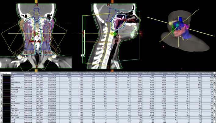
Purpose: The contouring of organs at risk (OARs) in head and neck cancer radiation treatment planning is a crucial, yet repetitive and time-consuming process. Recent studies have applied deep learning (DL) algorithms to automatically contour head and neck OARs. This study aims to conduct a systematic review and meta-analysis to summarize and analyze the performance of DL algorithms in contouring head and neck OARs. The objective is to assess the advantages and limitations of DL algorithms in contour planning of head and neck OARs.
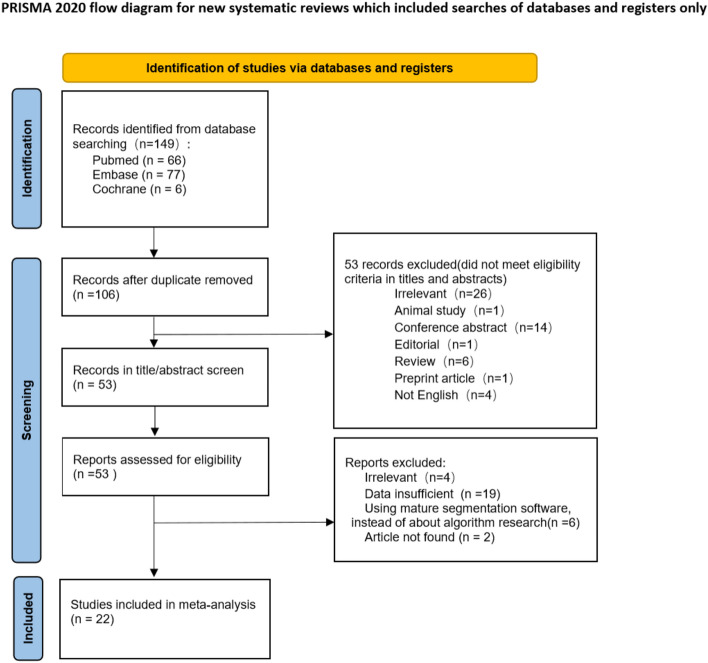
Methods: This study conducted a literature search of Pubmed, Embase and Cochrane Library databases, to include studies related to DL contouring head and neck OARs, and the dice similarity coefficient (DSC) of four categories of OARs from the results of each study are selected as effect sizes for meta-analysis. Furthermore, this study conducted a subgroup analysis of OARs characterized by image modality and image type.
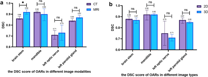
Results: 149 articles were retrieved, and 22 studies were included in the meta-analysis after excluding duplicate literature, primary screening, and re-screening. The combined effect sizes of DSC for brainstem, spinal cord, mandible, left eye, right eye, left optic nerve, right optic nerve, optic chiasm, left parotid, right parotid, left submandibular, and right submandibular are 0.87, 0.83, 0.92, 0.90, 0.90, 0.71, 0.74, 0.62, 0.85, 0.85, 0.82, and 0.82, respectively. For subgroup analysis, the combined effect sizes for segmentation of the brainstem, mandible, left optic nerve, and left parotid gland using CT and MRI images are 0.86/0.92, 0.92/0.90, 0.71/0.73, and 0.84/0.87, respectively. Pooled effect sizes using 2D and 3D images of the brainstem, mandible, left optic nerve, and left parotid gland for contouring are 0.88/0.87, 0.92/0.92, 0.75/0.71 and 0.87/0.85.
Conclusions: The use of automated contouring technology based on DL algorithms is an essential tool for contouring head and neck OARs, achieving high accuracy, reducing the workload of clinical radiation oncologists, and providing individualized, standardized, and refined treatment plans for implementing "precision radiotherapy". Improving DL performance requires the construction of high-quality data sets and enhancing algorithm optimization and innovation.
Keywords: Contouring; Deep learning; Head and neck cancer; Meta-analysis; Organs at risk; Systematic review.
© 2023. The Author(s).
Conflict of interest statement
The authors declare that they have no competing interests.
Figures


References
Publication types
MeSH terms
Grants and funding
LinkOut - more resources
Full Text Sources
Medical
