. 2023 Dec 6;31(12):3502-3519.
doi: 10.1016/j.ymthe.2023.10.018. Epub 2023 Oct 31.

Third-generation lentiviral gene therapy rescues function in a mouse model of Usher 1B

Affiliations

Third-generation lentiviral gene therapy rescues function in a mouse model of Usher 1B

Juliane W Schott et al. Mol Ther. .

Abstract

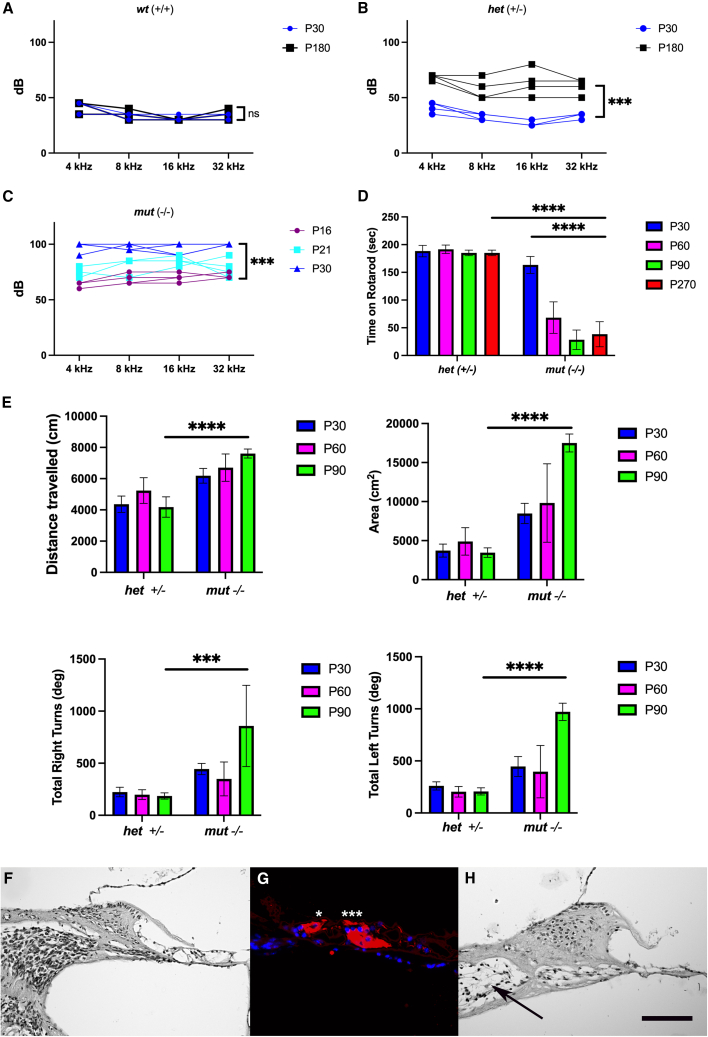
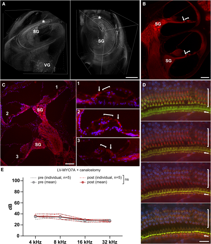
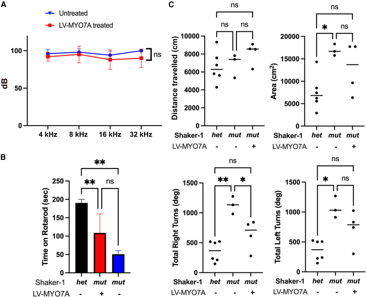
Usher syndrome 1B (USH1B) is a devastating genetic disorder with congenital deafness, loss of balance, and blindness caused by mutations in the myosin-VIIa (MYO7A) gene, for which there is currently no cure. We developed a gene therapy approach addressing the vestibulo-cochlear deficits of USH1B using a third-generation, high-capacity lentiviral vector system capable of delivering the large 6,645-bp MYO7A cDNA. Lentivirally delivered MYO7A and co-encoded dTomato were successfully expressed in the cochlear cell line HEI-OC1. In normal-hearing mice, both cochlea and the vestibular organ were efficiently transduced, and ectopic MYO7A overexpression did not show any adverse effects. In Shaker-1 mice, an USH1B disease model based on Myo7a mutation, cochlear and vestibular hair cells, the main inner ear cell types affected in USH1B, were successfully transduced. In homozygous mutant mice, delivery of MYO7A at postnatal day 16 resulted in a trend for partial recovery of auditory function and in strongly reduced balance deficits. Heterozygous mutant mice were found to develop severe hearing loss at 6 months of age without balance deficits, and lentiviral MYO7A gene therapy completely rescued hearing to wild-type hearing thresholds. In summary, this study demonstrates improved hearing and balance function through lentiviral gene therapy in the inner ear.

Keywords: MYO7A; Usher disease; Usher1B; balance loss; gene therapy; hearing loss; lentiviral vector; unconventional myosin.


Conflict of interest statement

Declaration of interests H.S. is a shareholder in Rescue Hearing Inc. and on the advisory board of MedEl GmbH. J.W.S., M.M., H.B., A.W., A.S., and H.S. have submitted patent applications for lentiviral gene therapy for the inner ear.

Figures

None
Graphical abstract
Figure 1
Figure 1
LV vector-mediated packaging, delivery, and expression of MYO7A is efficient and dose-controllable (A) Design of third-generation SIN LV vectors encoding for both human MYO7A and a dTomato reporter protein (LV-MYO7A) or encoding for a dTomato reporter protein only (LV-ctrl). An internal SFFV promoter (Prom.) was installed to drive transgene expression. ΔU3, 3′ unique region with SIN deletion; ψ, packaging signal; cPPT, central polypurine tract; PRE, post-transcriptional regulatory element; R, repeat region; RRE, Rev response element; SD, splice donor; SA, splice acceptor; U5, 5′ unique region. (B) Titers of viral vector preparations. Titer determination was based on transduction of HT1080 cells with different volumes of vector preparation and subsequent determination of the percentage of dTomato-expressing cells by flow cytometry. TU, transducing units. n = 3–5 biological replicates. (C) Percentage of HEI-OC1 cells expressing the LV vector-encoded dTomato reporter protein upon transduction with LV-MYO7A or LV-ctrl at a defined particle number per seeded cell (MOI). The percentage of dTomato-positive cells among the population of singlets within live cells was determined by flow cytometry. n = 3 biological replicates. Statistics: Multiple t-tests; discovery determined using the two-stage linear step-up procedure of Benjamini, Krieger and Yekutieli, with Q = 1%. (D) Immunofluorescence analysis of dTomato and MYO7A expression in HEI-OC1 cells transduced with LV-MYO7A or LV-ctrl at MOI of 0.2 and MOI of 2, and in non-transduced control (NTC) cells. Staining for MYO7A was performed using an anti-MYO7A primary antibody and an AF488-conjugated secondary antibody. Scale bar, 200 μm. (E) Percentage of HEI-OC1 cells expressing the vector-encoded dTomato reporter and the MYO7A protein. Flow cytometry analysis was performed upon intracellular staining for MYO7A expression in NTC cells and cells transduced with LV-ctrl or LV-MYO7A at MOI 2. Samples were either incubated with secondary antibody only (unstained) or stained with an anti-MYO7A primary and an AF488-conjugated secondary antibody. The populations shown were pre-gated for live cells using SSC-A/FSC-A characteristics, followed by gating for single cells according to FSC-A/FSC-H characteristics.
Figure 2
Figure 2
Homozygous mutant Shaker-1mut mice show early-onset hearing and balance loss, heterozygous mutant Shaker-1het mice show late-onset hearing loss and normal balance WT Shaker-1WT, heterozygous mutant Shaker-1het and homozygous mutant Shaker-1mut mice were evaluated for hearing and balance function using ABR, rotarod testing and actimeter testing. (A–C) ABR thresholds for four different frequencies (4, 8, 16, and 32 kHz) recorded at P16, P21, P30, and 6 months of age. dB, decibel; Hz, hertz. (A) WT Shaker-1WT mice demonstrated normal hearing thresholds for the first 180 days of life. (B) Mice heterozygous for the Shaker-1 mutation initially show normal ABR thresholds at P30, but develop a progressive hearing loss resulting in abnormal hearing thresholds at all frequencies by postnatal day 180. (C) Mice homozygous for the Shaker-1 mutation demonstrated testable hearing at P16 with progression to profound hearing loss by P30. (D) Balance was evaluated by testing the time thes mice could remain on a rotarod at P30, P60, P90, and P270. Heterozygotes showed consistently normal rotarod times through P270. Shaker-1mut mice initially showed normal rotarod scores at P30. These rapidly declined and remained abnormal throughout the entire test period. There is a statistically significant difference between rotarod times at P270 when comparing heterozygotes and homozygous mutant mice and when comparing P30 with P270 Shaker-1mut mice. Sec, seconds. (E) Force-plate actimeter testing was performed to determine the total distance traveled, the area covered during movement, and the total degree of right and left turns at P30, P60 and P90. Shaker-1mut mice demonstrated a progressive loss of balance function showing changes in total distance traveled, total area covered and the number of degrees of right and left turns (indicating circling behavior). Heterozygous Shaker-1het mice showed normal values throughout P90. n = 5. (F–H) Cochleae from untreated homozygous mutant Shaker-1mut mice were evaluated at P30, P90 and 1 year of age. (F) Histologic evaluation at P30 shows the presence of a normal-appearing organ of Corti (bracket). Scale bar, 95 μm. (G) By P90, immunofluorescent staining for annexin V shows labeling in inner (∗) and outer (∗∗∗) hair cells, as well as the supporting cells underneath the outer hair cell layer. Red signal: annexin V; blue signal: nuclei stained with DAPI. Scale bar, 63 μm. (H) By 1 year of age, the organ of Corti (bracket) has completely degenerated and the adjoining spiral ganglion is no longer detectable (arrow). Scale bar, 95 μm.
Figure 3
Figure 3
LV-ctrl and LV-MYO7A efficiently transduce the in vivo cochlea and do not negatively impact normal hearing Normal-hearing, WT C57BL/6 mice were injected with LV-ctrl (A and B) or LV-MYO7A (C–E) through canalostomy at 1 month of age using 1 μL of viral vector preparation. (A) Representative whole cleared cochlea from different angles. Three-dimensional views were reconstructed in Nikon software from serial images taken under a confocal microscope. dTomato signal (gray) is seen throughout the inner ear (vestibular organ and cochlea), with distribution in the cochlea from base to apex. Asterisk (∗), organ of Corti. SG, spiral ganglion; VG, vestibular ganglion. Scale bar, 500 μm. See also Figures S2A–S2C. (B) Cochlear cross-section showing multiple cochlear turns. Expression of dTomato can be seen in the spiral ganglion, inner and outer hair cells. SG, spiral ganglion. Arrow, inner hair cell; bracket, outer hair cells; red signal, dTomato. Scale bar, 140 μm. See also Figures S2D and S2E. (C) Cochlear cross-section (left) and magnification of the three regions (1, 2, and 3) from this section that contained organ of Corti tissue (right). Delivery of LV-MYO7A demonstrated transduction of both inner and outer hair cells as well as spiral ganglion cells in multiple cochlear turns. Red signal, dTomato; blue signal, nuclei stained with DAPI; SG, spiral ganglion; arrow, inner hair cell; bracket, outer hair cells. Scale bar, 100 μm (left) / 32 μm (1, 2, and 3). See also Figure S3. (D) z series of images from a representative whole mount preparation of the organ of Corti. dTomato is expressed throughout inner and outer hair cell layers. Hair cells were co-labelled with phalloidin-FITC. Arrow, inner hair cell row; bracket, outer hair cell rows; red signal, dTomato; green signal, phalloidin-FITC; blue signal, nuclei stained with DAPI. Scale bar, 28 μm. See also Figure S4. (E) ABR thresholds for four different frequencies (4, 8, 16, and 32 kHz) recorded for the same cohort of mice before (pre) and after (post) canalostomy and LV-MYO7A administration. Dotted lines show hearing thresholds for individual mice; solid lines show the mean thresholds from all mice at a given time point (pre or post). dB, decibel; Hz, hertz. n = 5. Statistics: Multiple t-tests; discovery determined using the two-stage linear step-up procedure of Benjamini, Krieger, and Yekutieli, with Q = 1%.
Figure 4
Figure 4
Vestibular hair cells are a target for LV-MYO7A gene therapy WT C57BL/6 mice were injected at 1 month of age (A–D) or homozygous mutant Shaker-1mut mice were injected at P16 (E) with 1 μL of LV-MYO7A through canalostomy. (A–C) One week after vector delivery, the utricles were removed and analyzed for dTomato expression. (A) Inner ear cross-section. Expression of dTomato was noted in the vestibular neuroepithelium. Red signal, dTomato; blue signal, nuclei stained with DAPI; s, saccule; u, utricle. Scale bar, 100 μm. (B) Utricle section. Expression of dTomato can be seen throughout the utricle. Gray signal, dTomato. Scale bar, 100 μm. (C) The number of total hair cells and of dTomato-positive (dTom+) hair cells in the utricle were counted from three mice, with five sections analyzed per mouse. Hair cells were identified as stereocilia-bearing cells through phalloidin labeling. Percentages on top of bars indicate the transduction efficiency for each mouse. Bars indicate the mean ± SD. (D) Rotarod times of the same cohort of mice before (pre) and after (post) canalostomy and LV-MYO7A administration. Sec, seconds. n = 5. Statistics: unpaired t-test. (E) Vestibular hair cells were quantified at the indicated time points in serially sectioned utricles from untreated Shaker-1het and untreated as well as LV-MYO7A-treated Shaker-1mut mice. Both type I and type II cells did not show any significant decline over time in all groups. Treatment of Shaker-1mut mice with LV-MYO7A did not alter type I or type II hair cell counts. HC, hair cell; hpf, high-power field. Shown is the mean ± SD. n = 5.
Figure 5
Figure 5
LV-MYO7A gene therapy at P16 partially improves hearing and halts balance loss in homozygous mutant Shaker-1mut mice Homozygous mutant Shaker-1mut mice were treated with 1 μL of LV-MYO7A, delivered via the PSCC at age P16. (A–D) The cochleae from treated animals were evaluated at 10 weeks after gene therapy (P90). Red signal: dTomato; blue signal: nuclei stained with DAPI. (A) Representative image from the basal turn of a cochlea from a treated animal. Expression of the dTomato marker gene can be seen in the inner hair cell (arrow). The adjoining outer hair cells already appear damaged at this point. Scale bar, 73 μm. (B) Representative image from the apical turn of a cochlea from a treated animal. Both inner and outer hair cells (arrows) clearly express the dTomato marker, showing delivery of the construct to the appropriate target cells. Scale bar, 62 μm. (C) Representative image from the vestibular organ of a treated animal. Vestibular hair cells also clearly express the dTomato marker gene (arrow). Scale bar, 27 μm. (D) Annexin V staining to localize apoptotic cells. Delivery of LV-MYO7A did not alter annexin V expression in the organ of Corti. A surviving inner hair cell and a single outer hair cell expressing dTomato are shown by arrows. Green signal, annexin V. Scale bar, 47 μm. (E) ABR thresholds for four different frequencies (4, 8, 16, and 32 kHz) in untreated and LV-MYO7A-treated Shaker-1mut mice at 2.5 months post-vector-delivery (P90). dB, decibel; Hz, hertz. n = 12. (F) Rotarod times determined at 1.5 (P60) and 2.5 months after vector delivery (P90) in LV-MYO7A-treated Shaker-1mut mice and at P90 in control Shaker-1het and untreated Shaker-1mut mice. Sec, seconds. n = 7. (G) Actimeter tracings were quantified to determine the total distance traveled, the area covered during movement, and the total degree of right and left turns at P90. There is a statistically significant improvement in actimeter scores in the LV-MYO7A-treated animals.
Figure 6
Figure 6
Actimetry demonstrates the restoration of more normal movement patterns after LV-MYO7A gene therapy Force-plate actimetry tracings for three consecutive measurement frames (left to right) of (A) control Shaker-1WT mice, (B) untreated homozygous mutant Shaker-1mut mice, and (C) LV-MYO7A-treated homozygous mutant Shaker-1mut mice. Mutant animals show only circling behavior, whereas mutants treated with LV-MYO7A demonstrate some circling, but also purposeful movement similar to Shaker-1WT animals.
Figure 7
Figure 7
LV-MYO7A gene therapy at P4 does not rescue hearing loss, but reduces balance loss in homozygous mutant Shaker-1mut mice To evaluate if better hearing outcomes could be achieved if MYO7A was delivered prior to the onset of hearing, a subset of neonatal animals was treated with LV-MYO7A at P4. (A) ABR thresholds for four different frequencies (4, 8, 16, and 32 kHz) recorded at P30 in untreated and LV-MYO7A-treated Shaker-1mut animals. dB, decibel; Hz, hertz. n = 7. (B) Rotarod times determined at P90 in control Shaker-1het mice and in untreated versus LV-MYO7A-treated Shaker-1mut animals. Sec, seconds. n = 7. (C) Force-plate actimetry tracings were quantified to determine the total distance traveled, the area covered during movement, and the total degree of right and left turns at P90 in control Shaker-1het, in untreated Shaker-1mut and in LV-MYO7A-treated Shaker-1mut animals.
Figure 8
Figure 8
LV-MYO7A gene therapy at P4 entirely prevents hearing loss in heterozygous mutant Shaker-1het mice To evaluate if late-onset hearing loss in heterozygous mutant Shaker-1het mice can be rescued by LV-MYO7A gene therapy, neonatal animals were treated with LV-MYO7A through canalostomy at P4. (A) ABR thresholds for four different frequencies (4, 8, 16, and 32 kHz) recorded at P180 in Shaker-1WT controls and in untreated versus LV-MYO7A-treated Shaker-1het mice. dB, decibel; Hz, hertz. n = 5. See also Figure S5. (B) DPOAE measured in untreated Shaker-1WT and Shaker-1het mice at one (P30) and 6 (P180) months of age, and in LV-MYO7A-treated Shaker-1het mice at 6 months of age. dB, decibel; SPL, sound pressure level; fDP, distortion product frequency. Sound floor (blue). n = 5.

References

    1. Kros C.J., Marcotti W., van Netten S.M., Self T.J., Libby R.T., Brown S.D.M., Richardson G.P., Steel K.P. Reduced climbing and increased slipping adaptation in cochlear hair cells of mice with Myo7a mutations. Nat. Neurosci. 2002;5:41–47. - PubMed
    1. Li S., Mecca A., Kim J., Caprara G.A., Wagner E.L., Du T.T., Petrov L., Xu W., Cui R., Rebustini I.T., et al. Myosin-VIIa is expressed in multiple isoforms and essential for tensioning the hair cell mechanotransduction complex. Nat. Commun. 2020;11:2066. - PMC - PubMed
    1. Weil D., Küssel P., Blanchard S., Lévy G., Levi-Acobas F., Drira M., Ayadi H., Petit C. The autosomal recessive isolated deafness, DFNB2, and the Usher 1B syndrome are allelic defects of the myosin-VIIA gene. Nat. Genet. 1997;16:191–193. - PubMed
    1. Géléoc G.G.S., El-Amraoui A. Disease mechanisms and gene therapy for Usher syndrome. Hear. Res. 2020;394 - PubMed
    1. Testa F., Melillo P., Bonnet C., Marcelli V., de Benedictis A., Colucci R., Gallo B., Kurtenbach A., Rossi S., Marciano E., et al. Clinical Presentation and Disease Course of Usher Syndrome Because of Mutations in Myo7a or Ush2a. Retina. 2017;37:1581–1590. - PubMed

LinkOut - more resources