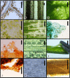
A maceration technique for soft plant tissue without hazardous chemicals
- PMID: 37915428
- PMCID: PMC10617305
- DOI: 10.1002/aps3.11543
A maceration technique for soft plant tissue without hazardous chemicals
Abstract
Premise: Current methods for maceration of plant tissue use hazardous chemicals. The new method described here improves the safety of dissection and maceration of soft plant tissues for microscopic imaging by using the harmless enzyme pectinase.
Methods and results: Leaf material from a variety of land plants was obtained from living plants and dried herbarium specimens. Concentrations of aqueous pectinase and soaking schedules were optimized, and tissues were manually dissected while submerged in fresh solution following a soaking period. Most leaves required 2-4 h of soaking; however, delicate leaves could be macerated after 30 min while tougher leaves required 12 h to 3 days of soaking. Staining techniques can also be used with this method, and permanent or semi-permanent slides can be prepared. The epidermis, vascular tissue, and individual cells were imaged at magnifications of 10× to 400×. Only basic safety precautions were needed.
Conclusions: This pectinase method is a cost-effective and safe way to obtain images of epidermal peels, separated tissues, or isolated cells from a wide range of plant taxa.
Keywords: cell isolation; dissection; epidermal peel; maceration; pectinase.
© 2023 The Authors. Applications in Plant Sciences published by Wiley Periodicals LLC on behalf of Botanical Society of America.
Figures

References
-
- Alvin, K. L. , and Boulter M. C.. 1974. A controlled method of comparative study for taxodiaceous leaf cuticles. Botanical Journal of the Linnean Society 69(4): 277–286.
-
- Bateman, D. F. 1968. The enzymatic maceration of plant tissue. Netherlands Journal of Plant Pathology 74: 67–80.
-
- Brown, R. 1951. The effects of temperature on the durations of the different stages of cell division in the root‐tip. Journal of Experimental Botany 1: 96–110.
-
- Bussotti, F. , and Grossoni P.. 1997. European and Mediterranean oaks (Quercus L.; Fagaceae): SEM characterization of the micromorphology of the abaxial leaf surface. Botanical Journal of the Linnean Society 124: 183–199.
-
- Chayen, J. 1952. Pectinase technique for isolating plant cells. Nature 170: 1070–1072. - PubMed
LinkOut - more resources
Full Text Sources

