. 2023 Oct 17:14:1250105.
doi: 10.3389/fpls.2023.1250105. eCollection 2023.

SEGS-1 a cassava genomic sequence increases the severity of African cassava mosaic virus infection in Arabidopsis thaliana

Affiliations

SEGS-1 a cassava genomic sequence increases the severity of African cassava mosaic virus infection in Arabidopsis thaliana

Cyprian A Rajabu et al. Front Plant Sci. .

Abstract

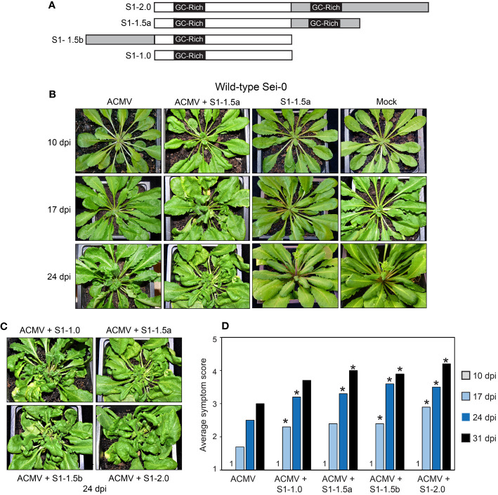
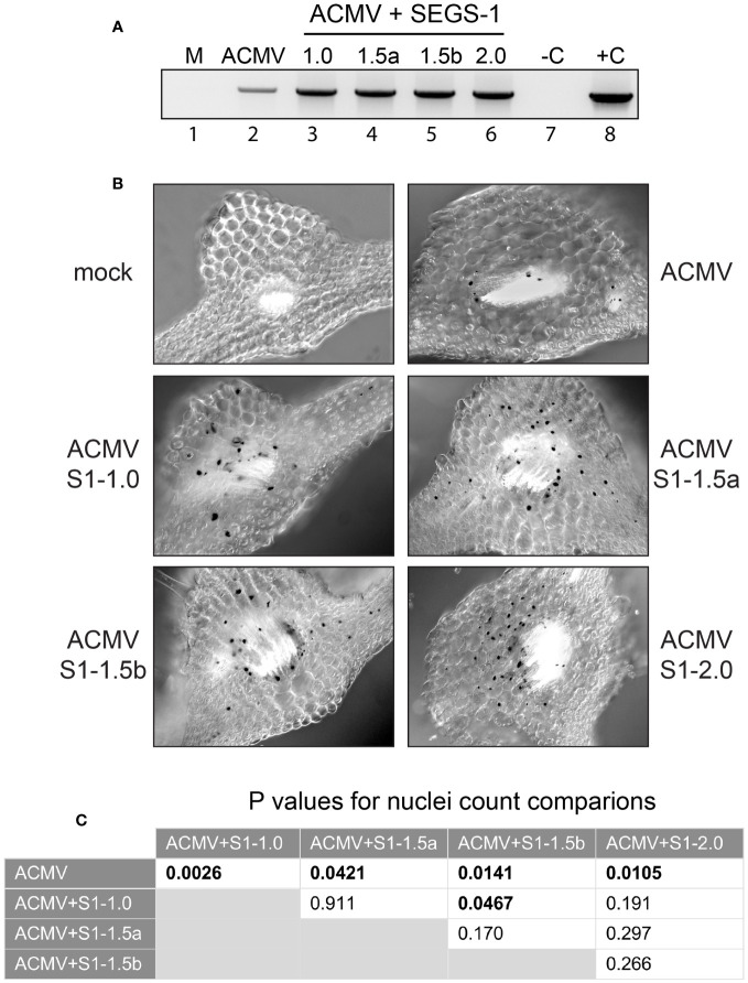
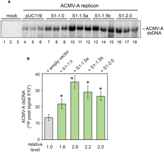
Cassava is a major crop in Sub-Saharan Africa, where it is grown primarily by smallholder farmers. Cassava production is constrained by Cassava mosaic disease (CMD), which is caused by a complex of cassava mosaic begomoviruses (CMBs). A previous study showed that SEGS-1 (sequences enhancing geminivirus symptoms), which occurs in the cassava genome and as episomes during viral infection, enhances CMD symptoms and breaks resistance in cassava. We report here that SEGS-1 also increases viral disease severity in Arabidopsis thaliana plants that are co-inoculated with African cassava mosaic virus (ACMV) and SEGS-1 sequences. Viral disease was also enhanced in Arabidopsis plants carrying a SEGS-1 transgene when inoculated with ACMV alone. Unlike cassava, no SEGS-1 episomal DNA was detected in the transgenic Arabidopsis plants during ACMV infection. Studies using Nicotiana tabacum suspension cells showed that co-transfection of SEGS-1 sequences with an ACMV replicon increases viral DNA accumulation in the absence of viral movement. Together, these results demonstrated that SEGS-1 can function in a heterologous host to increase disease severity. Moreover, SEGS-1 is active in a host genomic context, indicating that SEGS-1 episomes are not required for disease enhancement.

Keywords: ACMV; Arabidopsis thaliana; SEGS-1; begomovirus; cassava.


Conflict of interest statement

The authors declare that the research was conducted in the absence of any commercial or financial relationships that could be construed as a potential conflict of interest.

Figures

Figure 1
Figure 1
SEGS-1 enhances ACMV symptoms in Arabidopsis Sei-0. (A) SEGS-1 clones used for infection studies. The clones include a dimer (S1-2.0), a 1.5-mer with 2 GC-rich regions (S1-1.5a), a 1.5-mer with 1 GC-rich region (S1-1.5b), and a monomer (S1-1.0) that is configured like the full-length copy of SEGS-1 in the cassava genome. The gray segments and an embedded GC-rich region represent sequences that are duplicated in a construct. (B) Symptom development in plants inoculated with ACMV alone, ACMV + S1-1.5a, and S1-1.5a alone, or mock (ACMV DNA-B only). (C) Symptoms at 24 dpi in plants co-inoculated with ACMV and S1-1.0, S1-1.5a, S1-1.5b, or S1-2.0. (D) Time course of average symptom scores (1- no symptoms, 5- severe symptoms) for plants inoculated with ACMV alone or co-inoculated with ACMV + SEGS-1 clone. Values represent the mean of 10 plants per treatment. Asterisks (*) indicate significant differences between ACMV alone and ACMV+SEGS-1 treatments (p < 0.05 in a Wilcoxon ranked sum test).
Figure 2
Figure 2
SEGS-1 increases ACMV DNA accumulation in Arabidopsis Sei-0. (A) End-point PCR using the ACMV divLF/ACMV divLR primer pair to amplify ACMV DNA-A in mock (M; lane 1), ACMV alone (lane 2), or ACMV co-inoculated with S1-1.0 (lane 3), S1-1.5a (lane 4), S1-1.5b (lane 5) or S1-2.0 (lane 6) at 24 dpi. A negative no template control and a cloned positive plasmid DNA control are indicated by –C and +C (lanes 7 and 8, respectively). (B) In situ hybridization of ACMV DNA-A in plants at 24 dpi with ACMV alone or co-inoculated with ACMV and the indicated SEGS-1 clone. The 415-bp, DIG-labeled, DNA-A-specific probe forms a black precipitate over virus-positive nuclei. The leaf sections correspond to regions with vascular bundles where ACMV localizes. Mock plants were inoculated with ACMV DNA-B alone and did not contain infected cells. (C) Statistical analyses of virus-positive nuclei counts ( Supplementary Table 1 ) from in situ hybridization images using two-tailed paired Student’s t-test. Values in bold indicate significant differences (P<0.05).
Figure 3
Figure 3
SEGS-1 enhances ACMV DNA-A accumulation in tobacco protoplasts. (A) DNA gel blot showing the accumulation of nascent double-stranded ACMV DNA-A in protoplasts from NT-1 suspension cells at 48 h post transfection. The transfections are mock (pUC119: empty vector control), DNA-A + pUC119 (lanes 4-6), DNA-A + S1-1.0 (lanes 7-9), DNA-A + S1-1.5a (lanes 10-12), DNA-A + S1-1.5a (lanes 13-15) and DNA-A + S1-2.0 (lanes 16-18). The blot was hybridized to a 947-bp ACMV DNA-A fragment labeled with 32P and visualized by phosphor imaging. (B) 32P pixels were quantified using GelQuant software. Values represent the mean of 3 replicates/treatment. Bars correspond to ± 2 standard errors from the mean. Asterisks (*) indicate significant differences between the ACMV + empty vector treatment and an ACMV + SEGS-1 treatment (p < 0.05 in a two-tailed Student’s t test).
Figure 4
Figure 4
A SEGS-1 transgene enhances ACMV infection in Arabidopsis Sei-0 plants. (A) Time course (10, 17 and 24 dpi) of symptom development after inoculation with ACMV DNA-A + DNA-B or ACMV B alone (mock) in wild-type plants and in transgenic plants carrying a monomeric SEGS-1 transgene in the forward (S1-1.0F) or a reverse (S1-1.0R) orientation. (B) Time course of average symptom scores for wild-type Sei-0, S1-1.0F and S1-1.0R plants inoculated with ACMV at 10, 17, 24 and 31 dpi. Values represent the mean of 10 plants per treatment. Asterisks (*) indicate significant differences between ACMV alone and ACMV+SEGS-1 treatments (p < 0.05 in a Wilcoxon ranked sum test). (C) ACMV DNA-A copy number/ng total DNA in infected wild-type, S1-1.0F and S1-1.0R plants. The values represent the mean of 4 plants/treatment. Bars correspond to ± 2 standard errors from the mean. The ACMV DNA-A copy numbers in S1-1.0F and S1-1.0R plants were higher than in wild-type plants at 17 and 24 dpi, but no significant differences between the means were detected between the treatments by two-tailed Student’s t tests. The bars represent ± 2 standard errors from the mean. (D) Ratios of ACMV DNA-A mean copy numbers in S1-1.0F or S1-1.0R plants relative to wild-type plants. The dotted line represents the copy number in wild-type plants set to 1. (E) In situ hybridization of ACMV DNA-A in wild-type Sei-0, S1-1.0F and S1-1.0R plants at 10, 17 or 24 dpi with ACMV. The 415-bp DIG-labeled, DNA-A-specific probe forms a black precipitate over virus-positive nuclei. The leaf sections correspond to regions with vascular bundles where ACMV localizes. Mock plants were inoculated with ACMV DNA-B and did not contain infected cells. (F) Statistical analyses of virus-positive nuclei counts ( Supplementary Table 2 ) from in situ hybridization images using two-tailed paired Student’s t-test. Values in bold indicate significant differences (P<0.05). (G) SEGS-1 episome analysis. The divergent primer pair 1-4F/1-2R ( Table 1 ) was used to detect SEGS-1 episomes after RCA of total DNA. The top gel shows no PCR products amplifying across the SEGS-1 episome junction in ACMV-inoculated wild-type (wt; lane 2), S1-1.0F (lanes 3 and 4), and S1-1.0R (lanes 5 and 6) plants. C- is the water only negative PCR control (lane 7). C+ is the positive PCR control using SEGS-1 plasmid DNA as template that amplified in parallel with the Arabidopsis samples (lane 8). The bottom gel shows end-point PCR analysis using the CMAFor4/CMARev4 primer pair to amplify ACMV DNA-A in the same Arabidopsis DNA samples. Mock (lane 1) is DNA from an Arabidopsis plant inoculated with ACMV DNA-B only. In lanes 9 and 10, DNA samples from uninfected and ACMV-infected cassava plants were analyzed in parallel using the same protocol as the Arabidopsis episome assays. SEGS-1 episomes were detected in the DNA sample analyzed in lane 10 but not in the sample in lane 9.

Similar articles

Cited by

References

    1. Abbas Q., Amin I., Mansoor S., Shafiq M., Wassenegger M., Briddon R. W. (2019). The Rep proteins encoded by alphasatellites restore expression of a transcriptionally silenced green fluorescent protein transgene in Nicotiana benthamiana. Virusdisease 30, 101–105. doi: 10.1007/s13337-017-0413-5 - DOI - PMC - PubMed
    1. Aimone C. D., De Leon L., Dallas M. M., Ndunguru J., Ascencio-Ibanez J. T., Hanley-Bowdoin L. (2021. a). A new type of satellite associated with cassava mosaic begomoviruses. J. Virol. 95, e0043221. doi: 10.1128/JVI.00432-21 - DOI - PMC - PubMed
    1. Aimone C. D., Hoyer J. S., Dye A. E., Deppong D. O., Duffy S., Carbone I., et al. . (2022). An experimental strategy for preparing circular ssDNA virus genomes for next-generation sequencing. J. Virol. Methods 300, 114405. doi: 10.1016/j.jviromet.2021.114405 - DOI - PubMed
    1. Aimone C. D., Lavington E., Hoyer J. S., Deppong D. O., Mickelson-Young L., Jacobson A., et al. . (2021. b). Population diversity of cassava mosaic begomoviruses increases over the course of serial vegetative propagation. J. Gen. Virol. 102, 1622. doi: 10.1099/jgv.0.001622 - DOI - PMC - PubMed
    1. Biscotti M. A., Olmo E., Heslop-Harrison J. (2015). Repetitive DNA in eukaryotic genomes. Chromosome Res. 23, 415–420. doi: 10.1007/s10577-015-9499-z - DOI - PubMed

LinkOut - more resources