. 2023 Oct 16:14:1243480.
doi: 10.3389/fimmu.2023.1243480. eCollection 2023.

Trypanosoma cruzi P21 recombinant protein modulates Toxoplasma gondii infection in different experimental models of the human maternal-fetal interface

Affiliations

Trypanosoma cruzi P21 recombinant protein modulates Toxoplasma gondii infection in different experimental models of the human maternal-fetal interface

Guilherme de Souza et al. Front Immunol. .

Abstract

Introduction: Toxoplasma gondii is the etiologic agent of toxoplasmosis, a disease that affects about one-third of the human population. Most infected individuals are asymptomatic, but severe cases can occur such as in congenital transmission, which can be aggravated in individuals infected with other pathogens, such as HIV-positive pregnant women. However, it is unknown whether infection by other pathogens, such as Trypanosoma cruzi, the etiologic agent of Chagas disease, as well as one of its proteins, P21, could aggravate T. gondii infection.

Methods: In this sense, we aimed to investigate the impact of T. cruzi and recombinant P21 (rP21) on T. gondii infection in BeWo cells and human placental explants.

Results: Our results showed that T. cruzi infection, as well as rP21, increases invasion and decreases intracellular proliferation of T. gondii in BeWo cells. The increase in invasion promoted by rP21 is dependent on its binding to CXCR4 and the actin cytoskeleton polymerization, while the decrease in proliferation is due to an arrest in the S/M phase in the parasite cell cycle, as well as interleukin (IL)-6 upregulation and IL-8 downmodulation. On the other hand, in human placental villi, rP21 can either increase or decrease T. gondii proliferation, whereas T. cruzi infection increases T. gondii proliferation. This increase can be explained by the induction of an anti-inflammatory environment through an increase in IL-4 and a decrease in IL-6, IL-8, macrophage migration inhibitory factor (MIF), and tumor necrosis factor (TNF)-α production.

Discussion: In conclusion, in situations of coinfection, the presence of T. cruzi may favor the congenital transmission of T. gondii, highlighting the importance of neonatal screening for both diseases, as well as the importance of studies with P21 as a future therapeutic target for the treatment of Chagas disease, since it can also favor T. gondii infection.

Keywords: P21 protein; Toxoplasma gondii; Trypanosoma cruzi; coinfection; congenital toxoplasmosis; maternal-fetal interface.


Conflict of interest statement

The authors declare that the research was conducted in the absence of any commercial or financial relationships that could be construed as a potential conflict of interest.

Figures

Figure 1
Figure 1
Experimental design summarizing the T. gondii invasion and proliferation experiments carried out in this study.
Figure 2
Figure 2
T. gondii invasion and proliferation in BeWo cells infected with T. cruzi. BeWo cells were simultaneously infected with T. gondii and T. cruzi (A), or infected with T. gondii and subsequently with T. cruzi (B), or pre-infected with T. cruzi and later with T. gondii (C, D). T. gondii invasion (A, C) and intracellular proliferation (B, D) were analyzed by the β-galactosidase assay and are presented as percentage (%) of T. gondii invasion or proliferation in relation to cells only infected with T. gondii (infected medium). Data are shown as means ± SEM of three independent experiments with eight replicates. Differences between groups were analyzed with one-way ANOVA test with Bonferroni’s multiple comparisons post-test. Significant differences in relation to cells only infected with T. gondii (*).
Figure 3
Figure 3
Analysis of the role of rP21 in the viability, T. gondii invasion, and proliferation in BeWo cells. BeWo cells were treated with rP21 for 24 h and submitted to the MTT assay for cell viability analysis (A) and are presented as percentage (%) of cell viability in relation to untreated cells (untreated medium). Data are shown as means ± SEM of two independent experiments with eight replicates. In addition, BeWo cells were infected with T. gondii and treated with rP21 and/or infected with T. cruzi simultaneously (B), or infected with T. gondii and subsequently treated and/or infected with T. cruzi (C), or pre-treated and/or pre-infected with T. cruzi and subsequently with T. gondii (D, E). T. gondii invasion (B, D) and intracellular proliferation (C, E) were analyzed with the β-galactosidase assay and are presented as percentage (%) of T. gondii invasion or proliferation in relation to cells only infected with T. gondii (infected medium). Data are shown as means ± SEM of three independent experiments with eight replicates. Differences between groups were analyzed by one-way ANOVA test with Bonferroni’s multiple comparisons post-test. Significant differences in relation to cells only infected with T. gondii (*) or infected and treated with rP21 (&).
Figure 4
Figure 4
Analysis of the role of CXCR4 and actin cytoskeleton of BeWo cells in T. gondii invasion. BeWo cells were pre-treated or not with AMD3100 for 30 min and subsequently infected with T. gondii and treated simultaneously with rP21 for 3 h. T. gondii invasion (A) was analyzed with the β-galactosidase assay and are presented as percentage (%) of T. gondii invasion in relation to cells only infected with T. gondii (infected medium). Data are shown as means ± SEM of three independent experiments with eight replicates. Differences between groups were analyzed with one-way ANOVA test with Bonferroni’s multiple comparisons post-test. Significant differences in relation to cells only infected with T. gondii (*) or infected and treated with rP21 (&). In addition, BeWo cells were seeded on glass coverslips and infected with T. gondii and treated with rP21 simultaneously for 3 h. Then, the cells were fixed and labeled for the actin cytoskeleton (red), T. gondii (green), and nuclei (blue). The actin cytoskeleton was analyzed under confocal fluorescence microscopy (B). The mean actin fluorescence intensity was evaluated in 100 cells in triplicate of each condition in ImageJ software (National Institutes of Health, USA). Differences between groups were analyzed by Kruskall–Wallis test with Dunn’s multiple comparison post-test. Representative images are shown of uninfected and untreated BeWo cells (C), uninfected and treated with rP21 (D), infected and untreated (E), and infected and treated with rP21 (F). Significant differences in relation to uninfected and untreated cells (#) or infected and untreated (*). Scale bar: 20 µm.
Figure 5
Figure 5
Cell cycle analysis and reinfection capacity of T. gondii. BeWo cells were infected with T. gondii for 3 h and then treated with rP21 for 24 h. The intracellular parasites were isolated through syringes with 21- and 26-gauge needles, and submitted to cell cycle analysis (A, B) or reinfection assay (C). Cell cycle data are shown as percentage (%) of parasites in G1 and S/M phase, and are presented as means ± SEM of one experiment with five replicates. For the reinfection assay, the data are presented as percentage (%) of T. gondii invasion in relation to the parasites untreated (infected medium). Data are shown as means ± SEM of three independent experiments with eight replicates. Differences between groups were analyzed with one-way ANOVA test with Bonferroni’s multiple comparisons post-test. Significant differences in relation to parasites untreated (*).
Figure 6
Figure 6
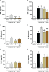
Cytokine production in BeWo cells. The supernatants of BeWo cells infected or not with T. gondii and/or T. cruzi and treated or not with rP21 were submitted to ELISA assay for measurement of MIF (A, D), IL-6 (B, E), and IL-8 (C, F). Data are presented in pg/mL according to the standard curve for each cytokine, and are shown as means ± SEM of three independent experiments with three replicates. Differences between groups were analyzed by means of one-way ANOVA test with Bonferroni’s multiple comparisons post-test or by means of Kruskall–Wallis test with Dunn’s multiple comparisons post-test. Significant differences in relation to uninfected and untreated cells (#) or infected or not with T. gondii and treated with rP21 (&) or only infected with T. gondii (*).
Figure 7
Figure 7
Analysis of rP21 treatment and T. cruzi infection in T. gondii proliferation in human placental explants. Human placental explants were treated with rP21 for 24 h and submitted to the MTT assay (A). Data are presented as percentage (%) viability of villous in relation to untreated villous (untreated medium). Data are shown as means ± SEM of two independent experiments with five replicates. Representative photomicrographs are shown of untreated villous (B) or treated with rP21 (C). Histological sections stained by Harris hematoxylin and eosin. Scale bar: 200 µm. In addition, the villi were pre-treated and/or infected with T. cruzi and subsequently with T. gondii (D) or infected with T. gondii and subsequently treated and/or infected with T. cruzi (E). T. gondii intracellular proliferation was analyzed by means of the β-galactosidase assay and are presented as percentage (%) of T. gondii proliferation in relation to villi only infected with T. gondii (infected medium). Data are shown as means ± SEM of three independent experiments with eight replicates. Differences between groups were analyzed by means of one-way ANOVA test with Bonferroni’s multiple comparisons post-test. Significant differences in relation to villi only infected with T. gondii (*) or infected and treated with rP21 (&).
Figure 8
Figure 8
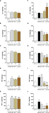
Cytokine production in human placental explants. The supernatants of villous infected or not with T. gondii and/or T. cruzi and treated or not with rP21 were submitted to ELISA assay for measurement of IL-4 (A, F), IL-6 (B, G), IL-8 (C, H), MIF (D, I), and TNF-α (E, J). Data are presented in pg/mg according to the standard curve for each cytokine, and are shown as means ± SEM of three independent experiments with three replicates. Differences between groups were analyzed by means of one-way ANOVA test with Bonferroni’s multiple comparisons post-test or by means of Kruskall–Wallis test with Dunn’s multiple comparisons post-test. Significant differences in relation to uninfected and untreated villous (#), only infected with T. gondii (*) and treated with rP21 (&) or only infected with T. gondii (*), infected with T. gondii and treated with rP21 (&), or infected with T. gondii and T. cruzi ($).
Figure 9
Figure 9
Proposed model of the role of rP21 and T. cruzi during T. gondii infection in human trophoblast cells and human villous explants. BeWo cells treated and/or infected with T. cruzi increase T. gondii invasion and decrease its proliferation. In addition, rP21 binds to CXCR4 and promotes actin polymerization, increasing T. gondii invasion, as well as arresting the T. gondii cell cycle in the S/M phase, upregulating IL-6, and downmodulating IL-8 production (A). In human placental explants, the pre-treatment with rP21 decreases T. gondii proliferation and downmodulates TNF-α production, whereas post-treatment as well as T. cruzi infection increases T. gondii proliferation, in addition to the upregulation of IL-4 and downmodulation of IL-6, IL-8, MIF, and TNF-α (B).

References

    1. Tenter AM, Heckeroth AR, Weiss LM. Toxoplasma gondii: from animals to humans. Int J Parasitol (2000) 30(12-13):1217–58. doi: 10.1016/s0020-7519(00)00124-7 - DOI - PMC - PubMed
    1. Djurkovic-Djakovic O, Dupouy-Camet J, van der Giessen J, Dubey JP. Toxoplasmosis: overview from a one health perspective. Food Waterborne Parasitol (2019) 15:e00054. doi: 10.1016/j.fawpar.2019.e00054 - DOI - PMC - PubMed
    1. Molan A, Nosaka K, Hunter M, Wang W. Global status of Toxoplasma gondii infection: systematic review and prevalence snapshots. Trop BioMed (2019) 36(4):898–925. - PubMed
    1. Bollani L, Auriti C, Achille C, Garofoli F, De Rose DU, Meroni V, et al. . Congenital toxoplasmosis: the state of the art. Front Pediatr (2022) 10:894573. doi: 10.3389/fped.2022.894573 - DOI - PMC - PubMed
    1. Sana M, Rashid M, Rashid I, Akbar H, Gomez-Marin JE, Dimier-Poisson I. Immune response against toxoplasmosis-some recent updates RH: Toxoplasma gondii immune response. Int J Immunopathol Pharmacol (2022) 36:3946320221078436. doi: 10.1177/03946320221078436 - DOI - PMC - PubMed

Publication types