. 2023 Oct 14;26(11):108170.
doi: 10.1016/j.isci.2023.108170. eCollection 2023 Nov 17.

Elevated high-mannose N-glycans hamper endometrial decidualization

Affiliations

Elevated high-mannose N-glycans hamper endometrial decidualization

Siyi Chen et al. iScience. .

Abstract

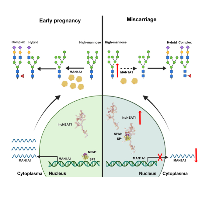
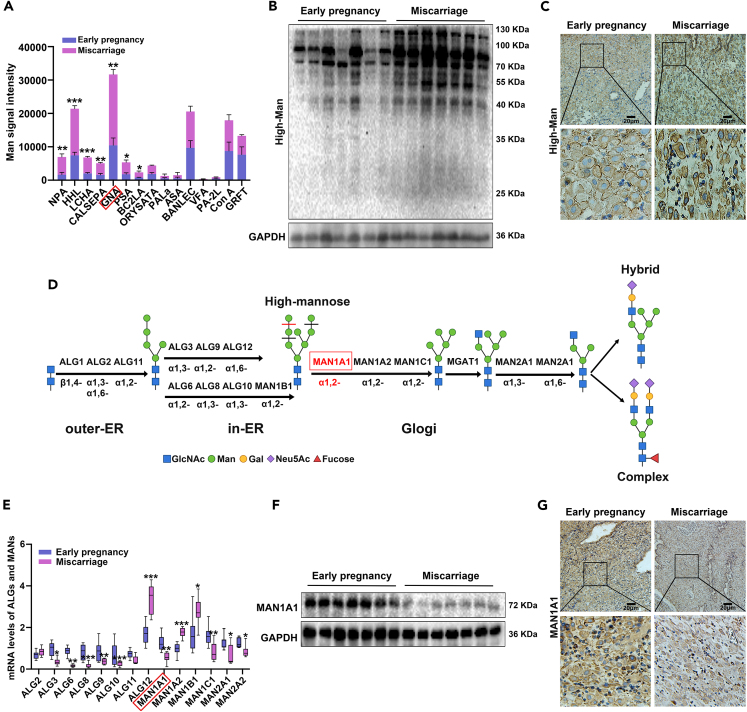
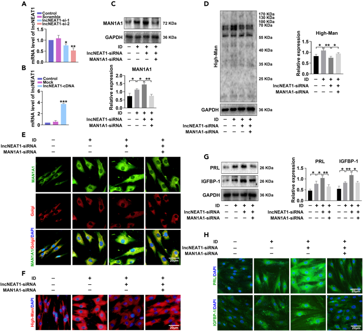
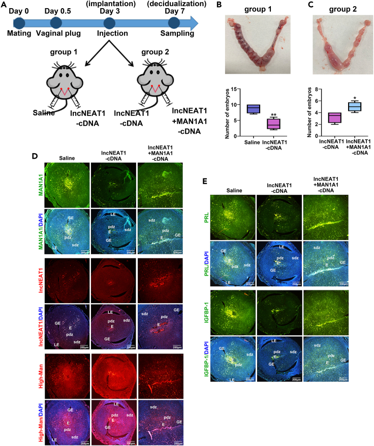
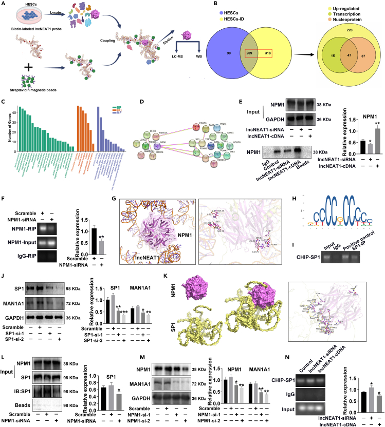
Decidualization of endometrial stromal cells is a hallmark of endometrial receptivity for embryo implantation, and dysfunctional decidualization is associated with pregnancy failure. Protein glycosylation is an important posttranslational modification that affects the structure and function of glycoproteins. Our results showed that high-mannose epitopes were elevated in the decidual tissues of miscarriage patients compared with early pregnant women by Lectin microarray. Furthermore, the level of mannosyl-oligosaccharide α-1,2 mannosidase IA (MAN1A1), a key enzyme for high-mannose glycan biosynthesis, was decreased in the decidual tissues of miscarriage patients. Screening of lncRNAs showed that lncNEAT1 level was increased in the serum and decidua of miscarriage patients, and negatively correlated with MAN1A1 expression. The results also revealed that specific binding of lncNEAT1 with nucleophosmin (NPM1)-SP1 transcription complex inhibited MAN1A1 expression and hampered endometrial decidualization and embryo implantation potential. The study suggests the new insights into the function of high-mannose glycans/MAN1A1 modification during endometrial decidualization.

Keywords: Biochemistry; Molecular biology; Physiology.


Conflict of interest statement

The authors declare no competing interests.

Figures

None
Graphical abstract
Figure 1
Figure 1
Increased high-mannose glycans and decreased MAN1A1 in the decidual tissues of miscarriage patients (A) Analysis of differential mannose-specific lectins in the bar graph. GNA: Galanthus nivalis lectin. (B and C) Lectin blot and immunohistochemical staining analysis of high-mannose glycans in the decidual tissues of early pregnant women (n = 7) and miscarriage patients (n = 7). (D) Schematic diagram of mannosyltransferases (ALGs) and mannosidases (MANs) catalyzing N-glycosylation synthesis by BioRender. (E) qPCR analysis of ALGs and MANs in the decidual tissues of early pregnancy and miscarriage. (F and G) Western blot and immunohistochemical staining analysis of MAN1A1 in the decidual tissues of early pregnant women and miscarriage patients. Scale bars, 20 μm ∗p < 0.05, ∗∗p < 0.01, ∗∗∗p < 0.001.
Figure 2
Figure 2
Silencing MAN1A1 increases high-mannose synthesis and inhibits decidualization of endometrial stromal cells (A–D) HESCs were transfected with MAN1A1-siRNA and MAN1A1-cDNA, and mRNA and protein levels of MAN1A1 were detected by qPCR and western blot. (E–G) Western blot analysis of MAN1A1, PRL (prolactin), and insulin-like growth factor-binding protein 1 (IGFBP-1) expression and Lectin blot analysis of high-mannose expression in HESCs after artificial decidua induction (ID) with MPA and dbcAMP, followed by transfection with MAN1A1-siRNA and MAN1A1-cDNA. (H–J) Immunofluorescent staining of MAN1A1 (green), high-mannose (red), decidualization markers PRL (green), and IGFBP-1 (green) in HESCs after ID or uninduced control, followed by transfection with MAN1A1-siRNA or cotransfection with MAN1A1-cDNA. Golgi apparatus was labeled by Golgi-Tracker Red. Nuclei were stained with DAPI (blue). Scale bars, 20 μm ∗p < 0.05, ∗∗p < 0.01, ∗∗∗p < 0.001.
Figure 3
Figure 3
Increased IncNEAT1 level in the decidual tissues of miscarriage patients (A) Integrative analysis of upregulated and downregulated lncRNAs in the serum of infertile patients compared with healthy women, and MAN1A1-correlated lncRNAs by Venn diagram. (B) Intersection of lncRNAs (fold change >2) in correlation with MAN1A1 by volcano plot. lncNEAT1: para-nuclear assembled transcript 1. (C) GO enrichment graph for upregulated lncRNAs by LncSEA database. (D) Expression and localization of lncNEAT1 in human tissues by LncSEA database. (E) qPCR analysis of lncNEAT1 level in the decidual tissues of early pregnancy and miscarriage. (F) Pearson correlation analysis of lncNEAT1 and MAN1A1. Pearson R = 0.52; p = 0.04; n = 16. Linear regression: y = −0.2274x + 1.633; r2 = 0.2604. (G) lncNEAT1 level in the nucleus and cytoplasm of HESCs by PCR. (H and I) Fluorescence in situ hybridization (FISH) analysis of lncNEAT1 (red) in HESCs (H) and endometrial tissues (I). Nuclei are stained with DAPI (blue). Scale bars, 20 μm ∗p < 0.05.
Figure 4
Figure 4
lncNEAT1 hampered decidualization of endometrial stromal cells by decreasing MAN1A1 expression (A and B) qPCR analysis of lncNEAT1 level in HESCs after transfection with lncNEAT1-siRNA or lncNEAT1-cDNA. (C, D, and G) Western blot analysis of MAN1A1, PRL, and IGFBP-1 expression and Lectin blot analysis of high-mannose in HESCs after ID, and transfection with lncNEAT1-siRNA or cotransfection with MAN1A1-siRNA. (E, F, and H) Immunofluorescent staining of MAN1A1 (green), high-mannose (red), PRL (green), and IGFBP-1 (green) in HESCs after ID, and transfection with lncNEAT1-siRNA or cotransfection with MAN1A1-siRNA. Golgi apparatus was labeled by Golgi-Tracker Red. Nuclei are stained with DAPI (blue). Scale bars, 20 μm ∗p < 0.05, ∗∗p < 0.01.
Figure 5
Figure 5
lncNEAT1 and MAN1A1 affect embryo implantation by decreasing decidualization in vivo (A) Schematic diagram of mouse pregnant model plan. (B and C) Number of embryos implanted at GD 7 in group 1 injected normal saline (control) and lncNEAT1-cDNA, and group 2 injected lncNEAT1-cDNA and lncNEAT1-cDNA + MAN1A1-cDNA. (D and E) Immunofluorescent staining of MAN1A1 (green), lncNEAT1 (red), high-mannose (red), PRL (green), and IGFBP-1 (green) in the uterus of GD 7. Nuclei are stained with DAPI (blue). Scale bars, 200 μm. LE: luminal epithelium; GE: glandular epithelium; E: embryo; pdz: primary decidual zone; sdz: secondary decidual zone. ∗p < 0.05, ∗∗p < 0.01.
Figure 6
Figure 6
Binding of lncNEAT1 and NPM1-SP1 complex inhibits MAN1A1 transcription (A) Schematic summary of biotin-labeled lncNEAT1 pull-down protocol. (B) LC-MS/MS analysis of lncNEAT1 binding proteins by Venn diagram: lncNEAT1 pull-down protein in HESCs and HESCs-ID (left); transcription-associated proteins and nuclear proteins in upregulated HESCs-ID (right). (C) GO analysis of 47 intersecting proteins. (D) Protein-protein interaction network analysis of lncNEAT1-binding proteins associated with decidualization and transcription factors of MAN1A1 by STRING database. (E) lncNEAT1 pull-down analysis of NPM1 (nucleophosmin) expression in HESCs after transfection with lncNEAT1-siRNA or lncNEAT1-cDNA. (F) RIP (RNA-binding protein immunoprecipitation) analysis of lncNEAT1 levels in HESCs after transfection with NPM1-siRNA. (G) Demonstration of molecular docking of lncNEAT1 and NPM1. Yellow dashed line: hydrogen bonds. (H) SP1 binding motif sequence logo from JASPAR database. (I) ChIP (Chromatin immunoprecipitation) analysis of the binding site between SP1 and the promoter of MAN1A1. (J and M) Western blot analysis of MAN1A1 expression in HESCs after transfection with SP1-siRNA or NPM1-siRNA. (K) Molecular docking of NPM1 and SP1. Yellow dashed line: hydrogen bonds; pink dashed line: salt bridges. (L) Immunoprecipitation analysis of NPM1 and SP1 in HESCs transfected with scramble or NPM1 siRNA. IP: protein pulled down with an anti-NPM1 antibody. IB: detection of SP1 expression by antibody. (N) ChIP analysis of SP1 and the MAN1A1 promoter binding in HESCs after transfection with lncNEAT1-siRNA and lncNEAT1-cDNA. ∗p < 0.05, ∗∗p < 0.01, ∗∗∗p < 0.001.

Similar articles

Cited by

References

    1. Lidegaard Ø., Mikkelsen A.P., Egerup P., Kolte A.M., Rasmussen S.C., Nielsen H.S. Pregnancy loss: A 40-year nationwide assessment. Acta Obstet. Gynecol. Scand. 2020;99:1492–1496. - PubMed
    1. Ramhorst R., Grasso E., Vota D., Gori S., Hauk V., Paparini D., Calo G., Pérez Leirós C. From decidualization to pregnancy progression: An overview of immune and metabolic effects of VIP. Am. J. Reprod. Immunol. 2022;88:e13601. - PubMed
    1. Ochoa-Bernal M.A., Fazleabas A.T. Physiologic events of embryo implantation and decidualization in human and non-human primates. Int. J. Mol. Sci. 2020;21:1973. - PMC - PubMed
    1. Wu J., Lin S., Huang P., Qiu L., Jiang Y., Zhang Y., Meng N., Meng M., Wang L., Deng W., et al. Maternal anxiety affects embryo implantation via impairing adrenergic receptor signaling in decidual cells. Commun. Biol. 2022;5:840. - PMC - PubMed
    1. Yoshie M., Kusama K., Tanaka R., Okubo T., Kojima J., Takaesu Y., Isaka K., Nishi H., Tamura K. Possible roles of calreticulin in uterine decidualization and receptivity in rats and humans. Int. J. Mol. Sci. 2021;22:10505. - PMC - PubMed

LinkOut - more resources