. 2023 Oct 31:6:0256.
doi: 10.34133/research.0256. eCollection 2023.

Hepatocellular SETDB1 Regulates Hepatic Ischemia-Reperfusion Injury through Targeting Lysine Methylation of ASK1 Signal

Affiliations

Hepatocellular SETDB1 Regulates Hepatic Ischemia-Reperfusion Injury through Targeting Lysine Methylation of ASK1 Signal

Kang Xia et al. Research (Wash D C). .

Abstract

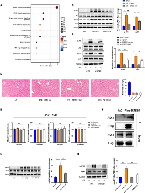
Background: Hepatic ischemia-reperfusion injury (HIRI) stands as an unavoidable complication arising from liver surgery, profoundly intertwined with its prognosis. The role of lysine methyltransferase SET domain bifurcated 1 (SETDB1) in HIRI remains elusive, despite its confirmation as a potential therapeutic target for diverse diseases. Here, we investigated the mechanism by which SETDB1 regulated HIRI. Methods: RNA sequencing data were used to identify the expression and potential targets of SETDB1 through bioinformatics analysis. To elucidate the impact of SETDB1 on HIRI, both an in vivo model of HIRI in mice and an in vitro model of hepatocyte hypoxia/reoxygenation were established. Biochemical and histological analyses were used to investigate the influence of SETDB1 on liver damage mediated by HIRI. Chromatin immunoprecipitation and coimmunoprecipitation were implemented to explore the in-depth mechanism of SETDB1 regulating HIRI. Results: We confirmed that hepatocellular SETDB1 was up-regulated during HIRI and had a close correlation with HIRI-related inflammation and apoptosis. Moreover, inhibition of SETDB1 could mitigate HIRI-induced liver damage, inflammation, and apoptosis. Through our comprehensive mechanistic investigation, we revealed that SETDB1 interacts with apoptosis-signal-regulating kinase 1 (ASK1) and facilitates the methylation of its lysine residues. Inhibition of SETDB1 resulted in reduced phosphorylation of ASK1, leading to a marked suppression of downstream c-Jun N-terminal kinase (JNK)/p38 signaling pathway activation. The therapeutic effect on inflammation and apoptosis achieved through SETDB1 inhibition was nullified by the restoration of JNK/p38 signaling activation through ASK1 overexpression. Conclusions: The findings from our study indicate that SETDB1 mediates lysine methylation of ASK1 and modulates the activation of the ASK1-JNK/p38 pathway, thus involved in HIRI-induced inflammation and apoptosis. These results suggest that SETDB1 holds promise as a potential therapeutic target for mitigating HIRI.


Conflict of interest statement

Competing interests: The authors declare that they have no competing interests.

Figures

Fig. 1.
Fig. 1.
In vivo HIRI models of mice demonstrated up-regulation of SETDB1 expression. (A) Volcano plots were used to illustrate DEGs in the HIRI group compared to the sham group (red, up-regulated genes; green, down-regulated genes). The horizontal dashed gray lines represent log2-normalized fold changes between −0.585 and 0.585. The vertical dashed gray line represents a P value of 0.05. (B) The heatmap illustrates the top 10 up-regulated and down-regulated DEGs (red, up-regulated; blue, down-regulated). (C) SETDB1 protein expression in human liver tissue before reperfusion (pre) and after reperfusion (post) (n = 4 for each group). (D) The mRNA level of SETDB1 in human liver tissue before reperfusion and after reperfusion (n = 4 for each group). (E) SETDB1 protein expression was examined in liver samples from the sham group and HIRI groups at 1, 3, and 6 h (n = 3 mice for each group, relative to Sham group). (F) The mRNA level of Setdb1 was assessed in liver tissue samples from the indicated groups (n = 3 mice for each group, relative to Sham group). (G and H) The serum levels of alanine aminotransferase (ALT) and aspartate aminotransferase (AST) were measured in the indicated groups (n = 5 mice for each group, relative to Sham group). (I) Representative H&E staining images of liver tissue slices from the indicated groups (n = 4 mice for each group, relative to Sham group). Scale bars, 100 μm. (J) IF staining was performed to detect the expression of SETDB1 and HNF4 in liver tissues from the indicated groups (n = 4 mice for each group). Scale bars, 50 or 100μm. All data are presented as means ± SDs. Statistical significance was denoted as *P < 0.05 and **P < 0.01.
Fig. 2.
Fig. 2.
In vivo, the administration of (R,R)-59 exhibited a mitigating effect on inflammation and apoptosis during HIRI. Mice were subjected to peritoneal injection of (R,R)-59 (at doses of 10, 30, and 50 mg/kg) or DMSO 5 days before the establishment of the animal model. (A) On the left, representative H&E staining images of liver tissue are displayed, while on the right, the statistical analysis of necrotic areas is presented [n = 4 mice for each group, relative to Sham group or I/R + group (50 mg/kg)]. Scale bars, 100μm. (B and C) ALT and AST were measured in the specified groups [n = 5 mice for each group, relative to Sham group or I/R + group (50 mg/kg)]. (D) Correlation analysis was established to identify the relationship between SETDB1 and molecules associated with HIRI events in mouse models. (E) The protein expression of H3K9me3 of the specified groups was evaluated (n = 3 mice for each group). (F) On the left, representative IF staining images of CD11b are displayed, while on the right, the statistical analysis of the positive area is presented (n = 4 mice for each group). Scale bars, 10 or 50 μm. (G) The mRNA levels of TNF, IL6, IL1b, and C-C motif chemokine ligand 2 (CCL2) were assessed in liver tissue of the designated groups (n = 3 mice for each group). (H) The protein levels of the NF-κB signaling pathway were assessed in liver tissues of the designated groups (n = 3 mice for each group). (I) On the left, representative images of TUNEL in liver tissue slices from the specified mouse samples are shown. On the right, the statistical analysis of the TUNEL-positive area is presented (n = 4 mice for each group). Scale bars, 50 μm. (J) The mRNA levels of genes associated with apoptosis were assessed in the specified groups (n = 3 mice for each group). (K) The levels of apoptosis-associated proteins in liver tissues of the specified groups were analyzed (n = 3 mice for each group). All data are presented as means ± SDs. Statistical significance was denoted as *P < 0.05 and **P < 0.01.
Fig. 3.
Fig. 3.
In vitro, the inhibition of SETDB1 suppressed inflammation and apoptosis in hepatocytes treated with H/R. (A) The mRNA level of Setdb1 was assessed in H/R-treated hepatocytes (n = 3 for each group). (B) The protein level of SETDB1 was assessed in H/R-treated hepatocytes (n = 3 for each group). (C) The mRNA level of Setdb1 was assessed in H/R-treated KCs (n = 3 for each group). (D) The protein level of SETDB1 was assessed in H/R-treated KCs (n = 3 for each group). (E) The mRNA level of Setdb1 was assessed in H/R-treated LSECs (n = 3 for each group). (F) The protein level of SETDB1 was assessed in H/R-treated LSECs (n = 3 for each group). (G) The mRNA level of Setdb1 was assessed in H/R-treated NPCs (n = 3 for each group). (H) The protein level of SETDB1 was assessed in H/R-treated NPCs (n = 3 for each group). L02 hepatocytes were transfected with si-SETDB1 in vitro. (I) The mRNA levels of inflammatory factors were assessed in the specified groups (n = 3 for each group). (J) The protein levels of the NF-κB signaling pathway were examined in the specified groups (n = 3 for each group). (K) The mRNA levels of genes associated with apoptosis were assessed in the specified groups (n = 3 for each group). (L) The levels of proteins associated with apoptosis were assessed in the specified groups (n = 3 for each group). All data are presented as means ± SDs. “ns” indicates no significance, while *P < 0.05 and **P < 0.01 denote statistical significance.
Fig. 4.
Fig. 4.
SETDB1 regulated ASK1–JNK/p38 signaling during HIRI in vivo and in vitro. (A) Key biological pathways associated with SETDB1 function were identified through KEGG enrichment analysis. (B and C) The protein levels of total and phosphorylated JNK and p38 were assessed in the specified groups (n = 3 for each group). (D) Representative images of H&E staining in mice liver tissues (left) and related quantitative analysis (right) (n = 4 for each group). Scale bar, 100 μm. (E) ChIP enrichment levels of SETDB1, H3K9me1, H3K9me2, and H3K9me3 on the promoter region of ASK1 were assessed in the specified groups (n = 3 for each group). (F) L02 cells were transfected with vectors containing Flag-tagged SETDB1. IP was conducted using a Flag or immunoglobulin G (IgG) antibody, followed by Western blot analysis using either a Flag or ASK1 antibody (n = 3 for each group). (G and H) The protein levels of total and phosphorylated ASK1 were examined in the indicated groups (n = 3 for each group). All data are presented as means ± SDs. The abbreviation **P < 0.01 indicates a statistically significant difference.
Fig. 5.
Fig. 5.
There was a direct interaction between SETDB1 and ASK1. (A) Representative images of IF to detect SETDB1 (red), p-ASK1 (green), and DAPI (blue) in L02 cells (n = 4 for each group). Scale bars, 20μm. (B and C) Shown here were representative Co-IP results of SETDB1 and ASK1 in L02 cells transfected with either SETDB1 tagged with Flag or ASK1 tagged with HA (n = 3 for each group). (D) L02 hepatocytes were transfected with the designated plasmids, followed by IP using anti-methyl lysine antibodies. The associated proteins were subsequently eluted and probed with related antibodies. Methyl lysine (Me-K) levels were normalized using the input of HA-ASK1 (n = 3 for each group). (E) IP detection of the lysine methylation of ASK1. The levels of methyl lysine were standardized by the input of ASK1 (n = 3 for each group).
Fig. 6.
Fig. 6.
The therapeutic effect of SETDB1 knockdown on H/R-treated hepatocytes was reversed upon overexpression of ASK1. (A) The protein levels of total and phosphorylated ASK1, JNK, and p38 were assessed in si-SETDB1-treated and si-NC-treated cell lines infected with adenovirus (ad)-ASK1 or ad-Vector (n = 3 for each group). (B) The mRNA levels of inflammatory factors were measured in the indicated groups (n = 3 for each group). (C) The protein levels of NF-κB signaling pathway components were analyzed in the indicated groups (n = 3 for each group). (D) The mRNA levels of apoptosis-associated genes were determined in the indicated groups (n = 3 for each group). (E) The protein levels of apoptosis-associated markers were examined in the indicated groups (n = 3 for each group). All data are presented as the means ± SD. **P < 0.01 indicates a statistically significant difference.
Fig. 7.
Fig. 7.
In vivo, the overexpression of ASK1 counteracted the liver damage observed in the HIRI model treated with (R,R)-59. (A) The protein levels of total and phosphorylated ASK1, JNK, and p38 were assessed in the livers of mice treated with (R,R)-59 or DMSO and transfected with AAV9-ASK1 or AAV9. (B and C) ALT and AST were measured in the specified groups (n = 3 for each group). (D) On the left, representative H&E-stained images from the specified mouse samples are displayed, while on the right, the statistical analysis of necrotic areas is presented (n = 4 for each group). Scale bar, 100 μm. (E) On the left, representative IF staining CD11b images from the specified mouse samples are presented, and on the right, the statistical analysis of the positive area is provided (n = 4 for each group). Scale bars, 10 or 50 μm. (F) The mRNA levels of inflammatory factors were assessed from the designated groups (n = 3 for each group). (G) The protein levels of the NF-κB signaling pathway were assessed of the specified groups (n = 3 for each group). (H) On the left, representative TUNEL staining from the specified mouse samples are presented, while on the right, the statistical analysis of the TUNEL-positive area is provided (n = 4 for each group). Scale bars, 50μm. (I) The mRNA levels of genes associated with apoptosis were measured in the specified groups (n = 3 for each group). (J) The levels of apoptosis-associated proteins were assessed in liver tissues of the specified groups (n = 3 for each group). All data are presented as the means ± SD. **P < 0.01 indicates a statistically significant difference.

References

    1. Liu Y, Lu T, Zhang C, Xu J, Xue Z, Busuttil RW, Xu N, Xia Q, Kupiec-Weglinski JW, Ji H. Activation of YAP attenuates hepatic damage and fibrosis in liver ischemia-reperfusion injury. J Hepatol. 2019;71(4):719–730. - PMC - PubMed
    1. Huang M, Cai H, Han B, Xia Y, Kong X, Gu J. Natural killer cells in hepatic ischemia-reperfusion injury. Front Immunol. 2022;13: Article 870038. - PMC - PubMed
    1. Hirao H, Nakamura K, Kupiec-Weglinski JW. Liver ischaemia-reperfusion injury: A new understanding of the role of innate immunity. Nat Rev Gastroenterol Hepatol. 2022;19(4):239–256. - PubMed
    1. Liu H, Man K. New insights in mechanisms and therapeutics for short- and Long-term impacts of hepatic ischemia reperfusion injury post liver transplantation. Int J Mol Sci. 2021;22(15). - PMC - PubMed
    1. Lin H, Caroll KS. Introduction: Posttranslational protein modification. Chem Rev. 2018;118(3):887–888. - PubMed