

White matter microstructural integrity continues to develop from adolescence to young adulthood in mice and humans: Same phenotype, different mechanism
- PMID: 37916059
- PMCID: PMC10619509
- DOI: 10.1016/j.ynirp.2023.100179
White matter microstructural integrity continues to develop from adolescence to young adulthood in mice and humans: Same phenotype, different mechanism
Abstract
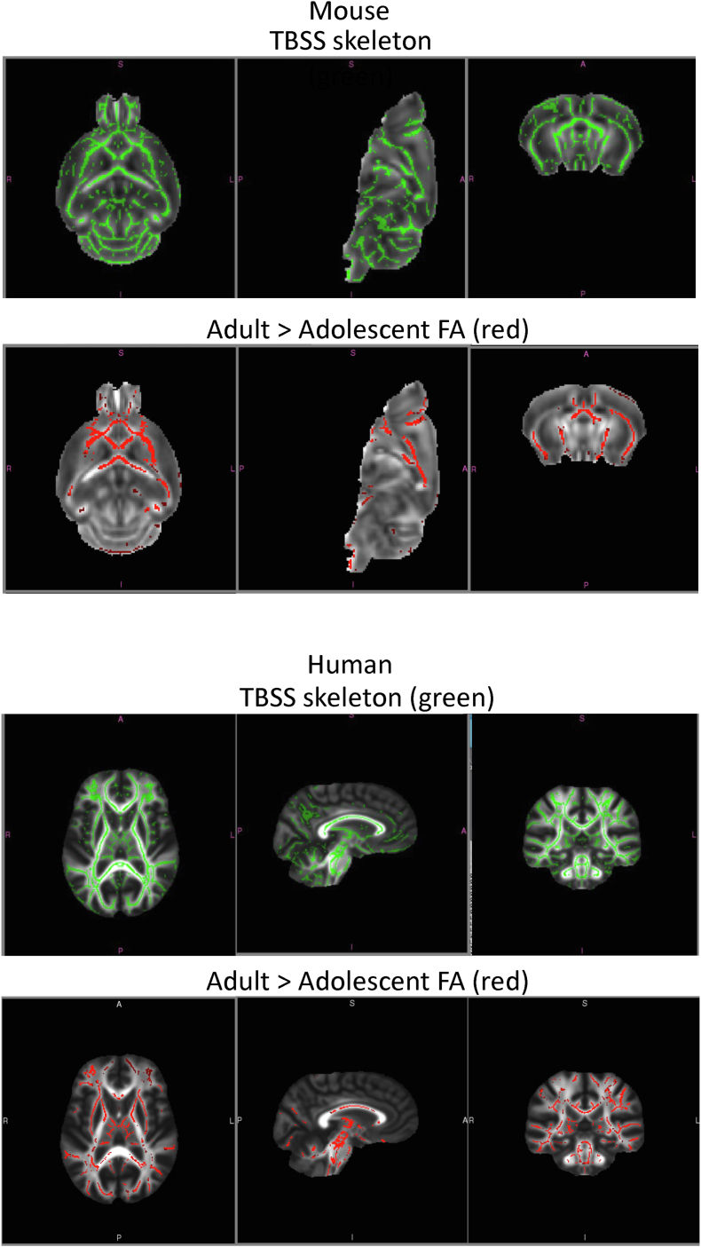
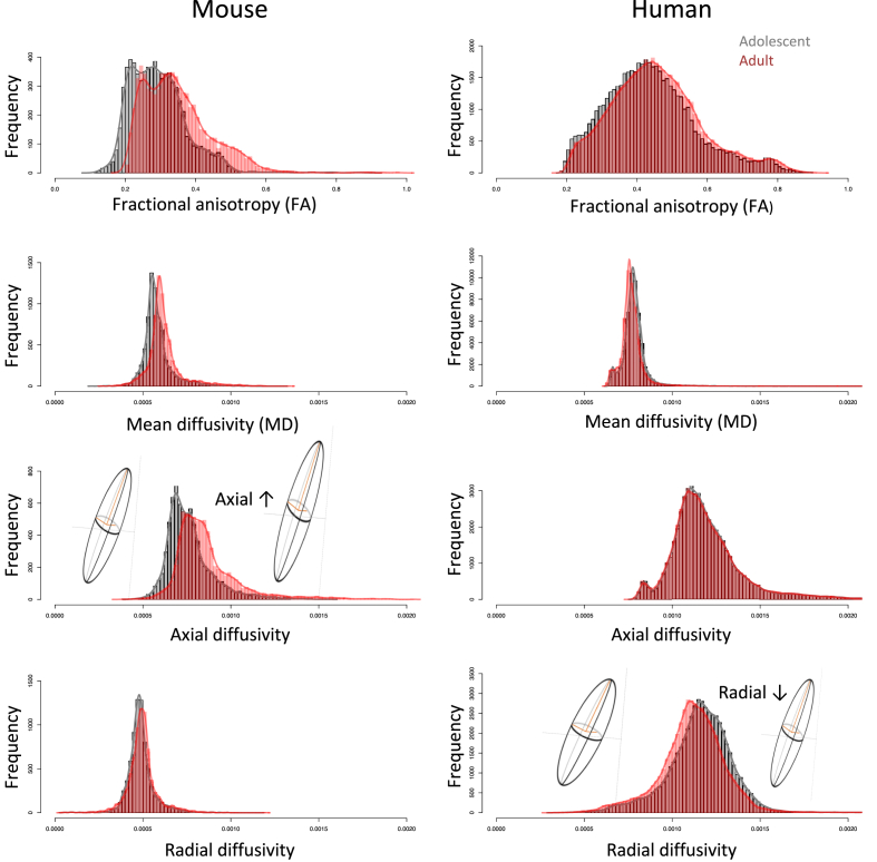
As direct evaluation of a mouse model of human neurodevelopment, adolescent and young adult mice and humans underwent MR diffusion tensor imaging to quantify age-related differences in microstructural integrity of brain white matter fibers. Fractional anisotropy (FA) was greater in older than younger mice and humans. Despite the cross-species commonality, the underlying developmental mechanism differed: whereas evidence for greater axonal extension contributed to higher FA in older mice, evidence for continuing myelination contributed to higher FA in human adolescent development. These differences occurred in the context of species distinctions in overall brain growth: whereas the continued growth of the brain and skull in the murine model can accommodate volume expansion into adulthood, human white matter volume and myelination continue growth into adulthood within a fixed intracranial volume. Appreciation of the similarities and differences in developmental mechanism can enhance the utility of animal models of brain white matter structure, function, and response to exogenous manipulation.
Keywords: Adolescence; Development; Diffusion tensor imaging; Human; Mouse; Translational.
Conflict of interest statement
Declaration of competing interest The authors declare that they have no known competing financial interests or personal relationships that could have appeared to influence the work reported in this paper. The authors have no competing interests to disclose.
Figures

Similar articles
-
Harmonizing DTI measurements across scanners to examine the development of white matter microstructure in 803 adolescents of the NCANDA study.Neuroimage. 2016 Apr 15;130:194-213. doi: 10.1016/j.neuroimage.2016.01.061. Epub 2016 Feb 10. Neuroimage. 2016. PMID: 26872408 Free PMC article.
-
Association of Heavy Drinking With Deviant Fiber Tract Development in Frontal Brain Systems in Adolescents.JAMA Psychiatry. 2021 Apr 1;78(4):407-415. doi: 10.1001/jamapsychiatry.2020.4064. JAMA Psychiatry. 2021. PMID: 33377940 Free PMC article.
-
Voxel-based analysis of white matter during adolescence and young adulthood.Brain Dev. 2010 Aug;32(7):531-7. doi: 10.1016/j.braindev.2009.08.006. Epub 2009 Sep 8. Brain Dev. 2010. PMID: 19740616
-
A review of diffusion MRI of typical white matter development from early childhood to young adulthood.NMR Biomed. 2019 Apr;32(4):e3778. doi: 10.1002/nbm.3778. Epub 2017 Sep 8. NMR Biomed. 2019. PMID: 28886240 Review.
-
Developmental trajectory of the prefrontal cortex: a systematic review of diffusion tensor imaging studies.Brain Imaging Behav. 2018 Aug;12(4):1197-1210. doi: 10.1007/s11682-017-9761-4. Brain Imaging Behav. 2018. PMID: 28913594
Cited by
-
Critical analysis of translational potential of rodent models of white matter pathology across a wide spectrum of human diseases.Cell Death Dis. 2025 Jul 31;16(1):580. doi: 10.1038/s41419-025-07893-6. Cell Death Dis. 2025. PMID: 40744926 Free PMC article. Review.
-
Direct 3-D printing of complex optical phantoms using dynamic filament mixing.Sci Rep. 2025 Mar 21;15(1):9705. doi: 10.1038/s41598-025-94390-7. Sci Rep. 2025. PMID: 40113981 Free PMC article.
-
Fabrication of complex optical phantoms using on-the-fly multi-filament mixing 3-D printing.Res Sq [Preprint]. 2024 Dec 11:rs.3.rs-5500473. doi: 10.21203/rs.3.rs-5500473/v1. Res Sq. 2024. Update in: Sci Rep. 2025 Mar 21;15(1):9705. doi: 10.1038/s41598-025-94390-7. PMID: 39711566 Free PMC article. Updated. Preprint.
-
Deep Learning-Based Pediatric Brain Region Segmentation and Volumetric Analysis for General Growth Pattern in Healthy Children.J Imaging Inform Med. 2025 Aug;38(4):1999-2011. doi: 10.1007/s10278-024-01305-5. Epub 2024 Nov 13. J Imaging Inform Med. 2025. PMID: 39538049 Free PMC article.
-
Obesity accelerates age-related memory deficits and alters white matter tract integrity in Ldlr-/-.Leiden mice.Brain Behav Immun Health. 2025 Apr 15;45:100991. doi: 10.1016/j.bbih.2025.100991. eCollection 2025 May. Brain Behav Immun Health. 2025. PMID: 40291340 Free PMC article.
References
Grants and funding
- R01 AA005965/AA/NIAAA NIH HHS/United States
- U24 AA021697/AA/NIAAA NIH HHS/United States
- U01 AA021696/AA/NIAAA NIH HHS/United States
- U01 AA021691/AA/NIAAA NIH HHS/United States
- U01 AA021681/AA/NIAAA NIH HHS/United States
- U01 AA021695/AA/NIAAA NIH HHS/United States
- U01 AA021692/AA/NIAAA NIH HHS/United States
- K99 AA028840/AA/NIAAA NIH HHS/United States
- R00 AA028840/AA/NIAAA NIH HHS/United States
- U24 AA021695/AA/NIAAA NIH HHS/United States
- R37 AA005965/AA/NIAAA NIH HHS/United States
- R37 AA010723/AA/NIAAA NIH HHS/United States
- U01 AA021690/AA/NIAAA NIH HHS/United States
- R01 DA057567/DA/NIDA NIH HHS/United States
- U01 AA021697/AA/NIAAA NIH HHS/United States
- R01 AA010723/AA/NIAAA NIH HHS/United States
LinkOut - more resources
Full Text Sources
Research Materials