Review
. 2024 Jan;38(1):95-119.
doi: 10.1007/s40259-023-00632-3. Epub 2023 Nov 2.

Dystrophin- and Utrophin-Based Therapeutic Approaches for Treatment of Duchenne Muscular Dystrophy: A Comparative Review

Affiliations
Review

Dystrophin- and Utrophin-Based Therapeutic Approaches for Treatment of Duchenne Muscular Dystrophy: A Comparative Review

Sylwia Szwec et al. BioDrugs. 2024 Jan.

Erratum in

Abstract

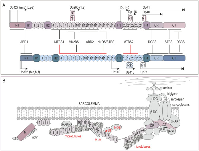
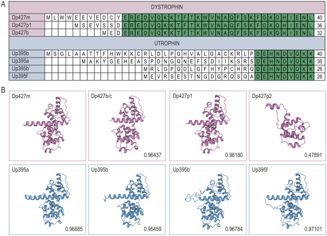
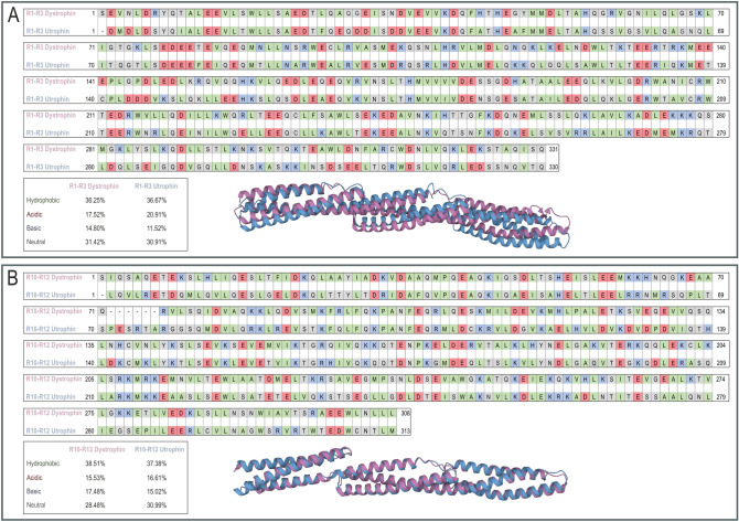
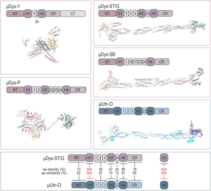
Duchenne muscular dystrophy is a devastating disease that leads to progressive muscle loss and premature death. While medical management focuses mostly on symptomatic treatment, decades of research have resulted in first therapeutics able to restore the affected reading frame of dystrophin transcripts or induce synthesis of a truncated dystrophin protein from a vector, with other strategies based on gene therapy and cell signaling in preclinical or clinical development. Nevertheless, recent reports show that potentially therapeutic dystrophins can be immunogenic in patients. This raises the question of whether a dystrophin paralog, utrophin, could be a more suitable therapeutic protein. Here, we compare dystrophin and utrophin amino acid sequences and structures, combining published data with our extended in silico analyses. We then discuss these results in the context of therapeutic approaches for Duchenne muscular dystrophy. Specifically, we focus on strategies based on delivery of micro-dystrophin and micro-utrophin genes with recombinant adeno-associated viral vectors, exon skipping of the mutated dystrophin pre-mRNAs, reading through termination codons with small molecules that mask premature stop codons, dystrophin gene repair by clustered regularly interspaced short palindromic repeats (CRISPR)/CRISPR-associated protein 9 (CRISPR/Cas9)-mediated genetic engineering, and increasing utrophin levels. Our analyses highlight the importance of various dystrophin and utrophin domains in Duchenne muscular dystrophy treatment, providing insights into designing novel therapeutic compounds with improved efficacy and decreased immunoreactivity. While the necessary actin and β-dystroglycan binding sites are present in both proteins, important functional distinctions can be identified in these domains and some other parts of truncated dystrophins might need redesigning due to their potentially immunogenic qualities. Alternatively, therapies based on utrophins might provide a safer and more effective approach.


Conflict of interest statement

JSC is one of the inventors on patents covering muscle-specific gene editing and patents covering muscle-specific gene regulatory cassettes, micro-dystrophins, and systemic delivery of AAV. JSC holds equity in and is a member of the scientific advisory board of Solid Biosciences. PK, SS, and ZK have no competing interests to declare.

Figures

Fig. 1
Fig. 1
Comparison of binding properties of dystrophin and utrophin. A Both the full length Dp427 dystrophin and Up395 utrophin have four main regions: the N-terminal domain (NT), the central rod domain composed of four hinges (H1–H4) and 24/22 spectrin repeats, the cysteine-rich domain (CR), and the C-terminal domain (CT). Red lines note that the binding properties of dystrophin are not retained in utrophin. B Dystrophin assembles the dystrophin glycoprotein complex that includes dystroglycans, sarcoglycans, sarcospan, syntrophins, and dystrobrevins, and associate with other proteins as indicated in the figure. Utrophin also assembles the complex but loses the ability to interact with microtubules, actin through ABD2, and nNOS (marked red). ABD1/2 actin biding domain 1/2, DBBS α-dystrobrevin binding site, DGBS β-dystroglycan binding site, MK2BS Ser/Thr kinase MAP/Microtubule affinity-regulating kinase 2 (MARK2) binding site, nNOS/STBS nNOS/α-syntrophin binding site, MTBS1/2 microtubules binding site 1/2, STBS α/β-syntrophins binding site
Fig. 2
Fig. 2
Comparison of ABD1 of the full-length human dystrophin and utrophin variants. Amino acid alignments (A) and 3D structures of ABD1 (I-TASSER software) (B) were based on the following fragments: Dp427m, 1–246 (NP_003997.2); Dp427p1, 1–242 (NP_004000.1); Dp427p2, 1–123 (NP_004001.1); Dp427b (Dp427c), 1–238 (NP_000100.3), Up395a, 1–261 (NP_009055.2); Up395b, 1–266 (XP_005267184.1); Up395b’, 1–252 (XP_024302304), and Up395f, 1–252 (XP_005267190.1). The structures were compared with Dp427m with the TM-align software. Note, very high TM-scores (above 0.95), except Dp427p2 (0.47891). Note also that transcription of Dp427p1 and p2 begins from the same promoter but the coding sequence of p2 starts from the 124th amino acid (methionine) of Dp427m. The Up395b’ amino acid sequence is marked as putative [64]. Up395a and Up395a’ transcription starts from different promoters but have the same coding sequence
Fig. 3
Fig. 3
Comparison of 3D structures of protein binding motifs of dystrophin and utrophin. The images represent superimposed 3D structures (TM-align software) of distinct domains and binding sites of dystrophin (pink) and utrophin (blue). The microtubules binding site 2 (MTBS2) is shown as the repeat sequence R20–R23 (the sequence that directly binds microtubules) and R20–R24 (includes the whole region between two hinges). ABD1/2 actin biding domain 1/2, DBBS α-dystrobrevin binding site, DGBS β-dystroglycan binding site, EF-hands a region composed of two EF-hands motifs, 1 and 2, MK2BS Ser/Thr kinase MAP/Microtubule affinity-regulating kinase 2 (MARK2) binding site, nNOS/STBS, nNOS/α-syntrophin binding site, MTBS1/2 microtubules binding site 1/2, STBS, α/β-syntrophins binding site, WW WW motif, ZZ ZZ-type zinc finger motif
Fig. 4
Fig. 4
Comparison of dystrophin and utrophin spectrin repeats R1–R3 and R10–R12. Amino acid alignments and 3D structures of R1–R3 (A) and R10–R12 (B) of dystrophin and utrophin are shown. Green indicates hydrophobic amino acids (A, alanine; F, phenylalanine; I, isoleucine; L, leucine; M, methionine; P, proline; V, valine; W, tryptophan); red, acidic amino acids (D, aspartic acid; E, glutamic acid); blue, basic amino acids (H, histidine; K, lysine; R, arginine); gray, other amino acids (C, cysteine; G, glycine; N, asparagine; S, serine; T, threonine; Q, glutamine; Y, tyrosine). The superimposed 3D structures of spectrin repeats R1–R3 and R10–R12 of dystrophin (pink) and utrophin (blue) were generated with the TM-align software
Fig. 5
Fig. 5
Graphical representation, 3D structures, and sequence alignment of μDys and μUtr proteins. 3D structures were predicted with the I-TASSER software. Note that μDys–Y and μDys–P show a more condensed structure, while μDys–ST/G, μDys–SB, and μUtr–O are linear, being more comparable to the full-length dystrophin. Sequence alignment of μDys and μUtr proteins revealed that H1 and H3 regions are dissimilar (lower panel, marked red). μDys–Y micro-dystrophin from [118], μDys–P micro-dystrophin manufactured and tested by Pfizer [102], μDys–ST/G micro-dystrophins manufactured and tested by Sarepta Therapeutics (μDys–ST) and by Genethon and Sarepta Therapeutics (μDys–G) [90], μDys–SB micro-dystrophin manufactured and tested by Solid Biosciences [113], μUtr–O micro-utrophin designed by Odom et al. [112, 187]. N/A not applicable
Fig. 6
Fig. 6
Restorative repair of the DMD gene expression with antisense oligonucleotides (AONs) and CRISPR/Cas9 technology. A Expression of the DMD gene in control samples based on a DMD fragment that encompasses exons 50–54 that is transcribed and translated into a protein region composed of H3 and spectrin repeats R20 and R21. B, C Deletion of DMD exons 50, 51, 52, or 55 (Δ50, Δ51, Δ52, Δ55) causes DMD as it changes the dystrophin reading frame and the protein cannot be synthesized. In (B) is shown an example where the reading frame can be restored in patients carrying Δ52 mutation via the use of AONs that induce skipping of exons 51 and 53 in pre-mRNA. Note that although the truncated dystrophin is missing part of hinge 3 (H3) and R20 or part of R20 and R21, the synthesized protein fragments have largely unaffected 3D structures. As presented in C, the dystrophin reading frame in patients with distinct mutations, including Δ50, Δ51, Δ52, Δ55, can be restored by deleting a relatively large fragment of the DMD gene with the CRISPR/Cas9 technology. Note that the gRNAs are designed to cut within exons 47 and 58 to remove a relatively large region within the rod domain (Δ47–58) so that the perfect spectrin repeat structure is recreated from the remaining R18 and R23 fragments [161]
Fig. 7
Fig. 7
Schematic representation of utrophin-based therapies for DMD. Utrophin increased levels could be achieved through rAAV-mediated delivery of genes encoding μUtr (A), activation of the endogenous UTRN gene promoter directly through ezutromid/SMT022357, or indirectly via heregulin that induces distinct signaling events (B), stabilization of the utrophin-glycoprotein complex (UGC) through biglycan or GALGT2 (C), or counteracting UTRN mRNA degradation by blocking microRNAs (miRNAs; (D) with AONs (1) or by the CRISPR/Cas9-directed excision of the DNA sequence, which upon transcription serves as a binding site for miRNAs (2)

References

    1. Gao QQ, McNally EM. The dystrophin complex: structure, function, and implications for therapy. Compr Physiol. 2015. 10.1002/cphy.c140048. - PMC - PubMed
    1. Muntoni F, Torelli S, Ferlini A. Dystrophin and mutations: one gene, several proteins, multiple phenotypes. Lancet Neurol. 2003. 10.1016/s1474-4422(03)00585-4. - PubMed
    1. Crisafulli S, Sultana J, Fontana A, Salvo F, Messina S, Trifirò G. Global epidemiology of Duchenne muscular dystrophy: an updated systematic review and meta-analysis. Orphanet J Rare Dis. 2020. 10.1186/s13023-020-01430-8. - PMC - PubMed
    1. Broomfield J, Hill M, Guglieri M, Crowther M, Abrams K. Life expectancy in Duchenne muscular dystrophy. Neurology. 2021. 10.1212/WNL.0000000000012910. - PMC - PubMed
    1. Dumont NA, Rudnicki MA. Targeting muscle stem cell intrinsic defects to treat Duchenne muscular dystrophy. NPJ Regen Med. 2016. 10.1038/npjregenmed.2016.6. - PMC - PubMed

MeSH terms

LinkOut - more resources