Review
. 2023 Nov 2;24(1):55.
doi: 10.1186/s10195-023-00734-2.

Bernese periacetabular osteotomy (PAO): from its local inception to its worldwide adoption

Affiliations
Review

Bernese periacetabular osteotomy (PAO): from its local inception to its worldwide adoption

Reinhold Ganz et al. J Orthop Traumatol. .

Abstract

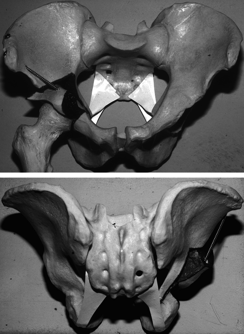
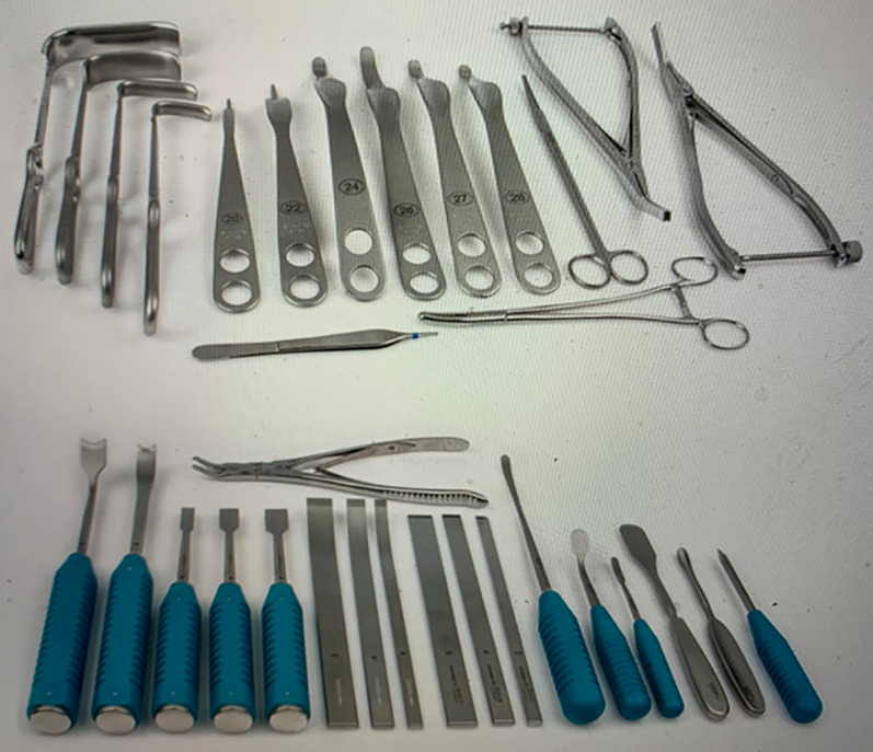
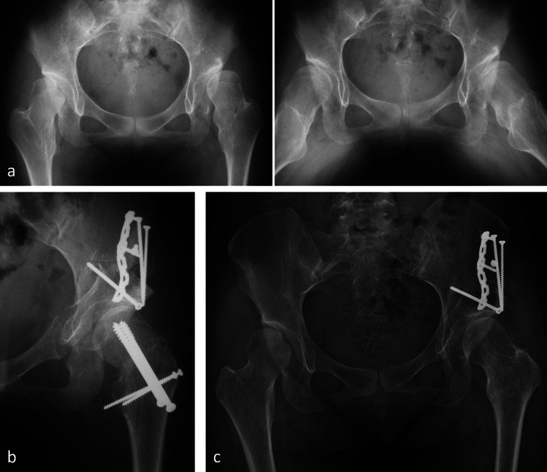
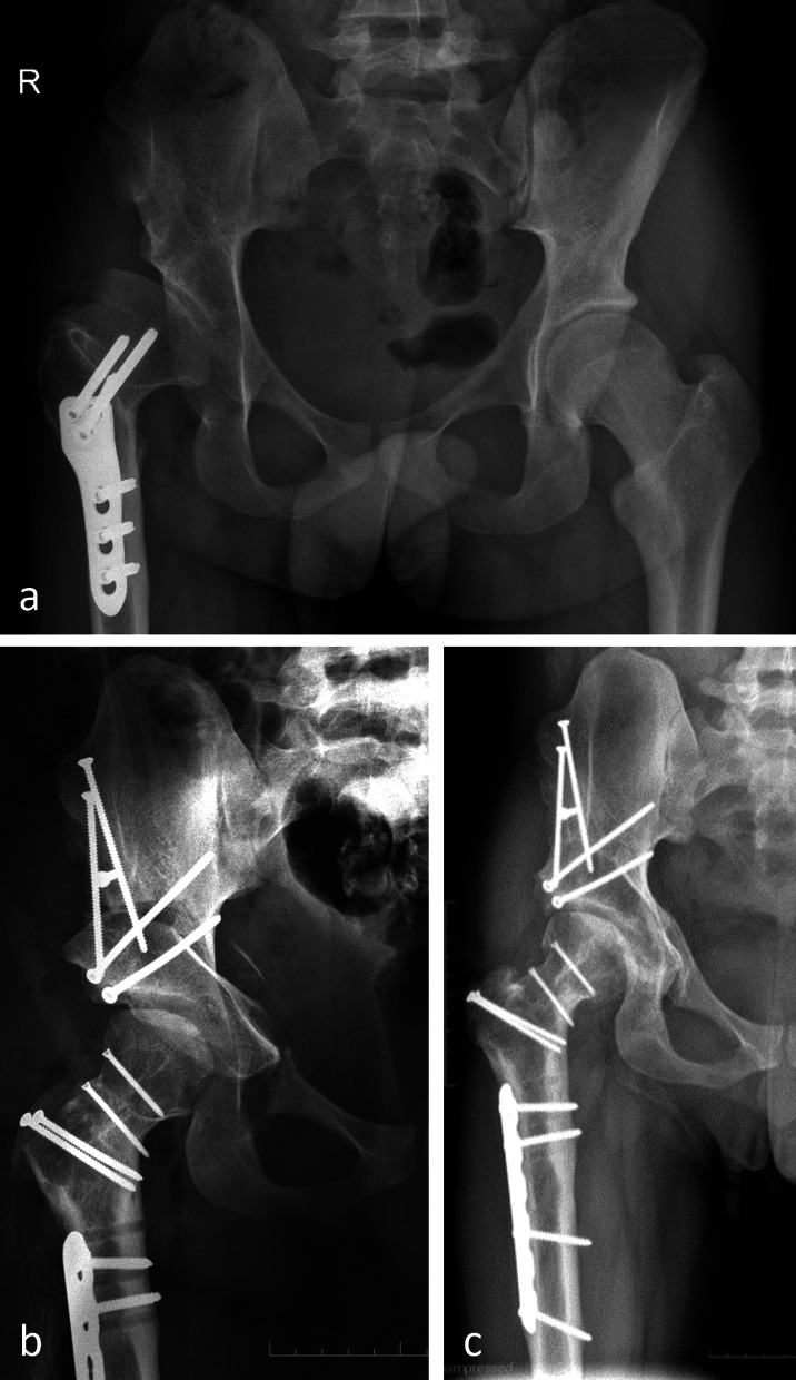
The development of the Bernese periacetabular osteotomy (PAO) is based on a structured approach starting with an analysis of the preexisting procedures to improve the coverage of the femoral head and was followed by a list of additional goals and improvements. Cadaveric dissections with a detailed description of the vascular supply of acetabulum and periacetabular bone set the stage for an intrapelvic approach, which offered the largest acetabular correction possible combined with safe intracapsular access. The final composition of osteotomies required the development of several instruments and cutting devices before the feasibility could be tested on a series of cadaveric hips.While the sequence of the osteotomies remained largely unchanged over time (except for the pubic and ischial osteotomies), several propositions for an easier/less invasive approach have been discussed; some made it into standard practice. Efforts were undertaken to optimize the learning curve and minimize failures using video-clips, hands-on courses, fellowships, publications, and ongoing mentoring programs. In retrospect, with almost 40 years of experience, such efforts have promoted a worldwide adoption of the Bernese periacetabular osteotomy.

Keywords: Bernese PAO; Hip joint preservation surgery; PAO; Pelvic osteotomy history; Periacetabular osteotomy.


Conflict of interest statement

All the authors declare that they have no affiliations with or involvement in any organization or entity with any financial interest, or non-financial interest, in the subject matter or materials discussed in this manuscript.

Figures

Fig. 1
Fig. 1
Periacetabular vascular supply cadaver study on vascular supply of the periacetabular bone. Right hip, osteotomies from pelvic inside. The branches from superior and inferior gluteal artery can be preserved during osteotomy (left) and fragment correction (right)
Fig. 2
Fig. 2
PAO osteotomy cuts arrangement of the five osteotomy cuts, visible on the left lateral view, in the middle anterior view, and on the right medial view of a right hemipelvis, namely incomplete ischial cut (first), pubis cut (second), supraacetabular cut (third), retroacetabular cut (fourth), and infraacetabular cut completing the ischial cut (fifth)
Fig. 3
Fig. 3
Pelvis plastic model: plastic model showing separation of the acetabular fragment from posterior column including sacrospinal and sacrotuberal ligaments. Top: anterior view; bottom: posterior view
Fig. 4
Fig. 4
PAO instrumentation set of instruments with special osteotomes and retractors
Fig. 5
Fig. 5
1984: first operated case—13-year-old female with PFFD of the left hip. a Varus proximal femur after intertrochanteric osteotomy 3 years earlier. b Periacetabular osteotomy followed by intertrochanteric revalgisation for better joint congruity. Good lateral coverage of the head. Increased retroversion of the acetabulum led to posterior subluxation 3 months after surgery, which was treated with a posterior shelf. c Progressive joint degeneration after 36 years. Plate for shelf fixation still in place. d Follow-up radiography of THR 2 years later. The patient regained a normal gait pattern
Fig. 6
Fig. 6
Ischial cut via femoral approach in hips with combined acetabular and femoral surgery. On top, access to the infracotyloid groove between obturator internus and gemellus inferior muscles. Bottom: it allows optimal protection of the ischial nerve during execution of the cut
Fig. 7
Fig. 7
Bilateral severe acetabular dysplasia of 16-year-old female; more pain on the right side. a Several subchondral bone cysts, best visible in a lateral view (black arrow). Displaced rim fracture with some reduction in abduction (white arrow). b PAO combined with femoral varus osteotomy. A 6-month interval between surgery of the hips was observed (right side first). Slight overcorrection of the PAO on the right side for better unloading of the damaged rim area was performed. Partial metal removal after 2 years was performed. Radiological result observed after 5 years. c Closer look to the critical joint area of the right hip before surgery and after sugery (d) showing reduced and healed rim fragment (double arrow)
Fig. 8
Fig. 8
Perthes disease in the right hip of a 13-year-old male, operated on with Salter osteotomy. a Persistent subluxation and adduction with extrusion of the healthy lateral pillar and loading of the necrotic area. Adaptive widening of the acetabular cavity. On the right, computer simulation showing relocated head after resection of the necrotic area (red portion) and optimal coverage with PAO. b Intraoperative pictures showing the resection of the necrotic central part of the head with the template in place and the final size of the head after screw fixation of the mobile lateral part of the head with two screws. c Postoperative sequences with the new head healed without necrosis
Fig. 9
Fig. 9
Acetabular dysplasia, multiple exostoses, and subluxation multiple exostoses near both hips with acetabular dysplasia in a very gracile 21-year-old female. a Pain and discomfort of the left hip during subluxation, palpable with rotation in full extension. Lateral view (right) showing the causative exostosis at the posterior neck. b Postoperative result after surgical dislocation, femoral neck varus osteotomy, and removal of the exostosis through the neck osteotomy, followed by PAO. c The 10-year-result. Removal of the femoral screws for local pain soon after surgery. Both hips are pain-free
Fig. 10
Fig. 10
Posttraumatic dysplasia in a 19-year-old male. a Typical posttraumatic dysplasia of the right hip after a car accident at age 3. Large acetabular rim fragment. Laborious execution of the osteotomy due to the thick supra- and retroacetabular bone. Screw fixation of the large rim fragment. PAO with maximal possible medial displacement of the acetabular fragment. b Radiological result after 2 years with healed fracture and osteotomies
Fig.11
Fig.11
Acetabular protrusio was observed, with a bilateral protrusion in a 21-year-old lady with osteogenesis imperfecta. Constant pain on the right side while the left side was pain-free during most daily activities. Patient refused to get THR. On the top, preoperative radiography showing fatigue fracture through the bottom of the joint (white arrow). b Result 1 year after excessive rim trimming and PAO decovering the head. Fatigue fracture healed (black arrow) with substantially reduced pain
Fig. 12
Fig. 12
Posttraumatic protrusio observed in patient. a Posttraumatic protrusio and deformation of the hemipelvis in a 23-year-old female. Her gynecologist discussed the risk of abortion due to deviation of the uterus, which was the reason why she was asking for correction. A modified PAO was first tested on a plastic model. Correction of protrusio und pelvic deformation was assisted by a distractor. b The new position was stabilized with a long recoplate placed along the pelvic brim. Ultrasound was performed just before natural delivery. c At 16 years after surgery, she is now the mother of five children and is very active in outdoor activities. The straight leg is the operated one
Fig. 13
Fig. 13
Severe deformity (courtesy Prof. P. Rego, Hospital da Luz, Lisbon, Portugal) of the right hip in a 16-year-old male after open reduction at the age of 1.5 years, followed by avascular necrosis. a Varus osteotomy at the age of 14 years, which did not improve limping and pain. b Postoperative radiography after complex surgery with relative lengthening of the neck, intracapital osteotomy to reduce the size of the head, and subtrochanteric derotation osteotomy followed by PAO. c The 4-year follow-up result with good and pain-free function

References

    1. Codivilla A. Operative treatment of congenital dislocation of the hip. Z Orthop Chir. 1901;9:124–137.
    1. Codivilla A. The operative treatment of congenital dislocations. New York Medical J. 1902;73:741.
    1. Colonna PC. Congenital dislocation of the hips in older subjects: based on a study of sixty-six open operations. J Bone Joint Surg. 1932;14:277–298.
    1. Bordman DL, Moseley CF. Finding patients after 40 years: a very long-term follow-up study of the Colonna arthroplasty. J Pediatr Orthop. 1999;19:169–176. doi: 10.1097/01241398-199903000-00007. - DOI - PubMed
    1. Ganz R, Slongo Th, Siebenrock KA, Turchetto L, Leunig M. Clin Orthop Relat Res. 2012;470:2957–2967. doi: 10.1007/s11999-012-2444-y. - DOI - PMC - PubMed

LinkOut - more resources