Comparative Study
. 2024 Sep 17;16(10):1060-1065.
doi: 10.1136/jnis-2023-020789.

Direct comparison of digital subtraction myelography versus CT myelography in lateral decubitus position: evaluation of diagnostic yield for cerebrospinal fluid-venous fistulas

Affiliations
Comparative Study

Direct comparison of digital subtraction myelography versus CT myelography in lateral decubitus position: evaluation of diagnostic yield for cerebrospinal fluid-venous fistulas

Niklas Lützen et al. J Neurointerv Surg. .

Abstract

Background: Cerebrospinal fluid (CSF)-venous fistulas (CVFs) are increasingly identified as a cause of spontaneous intracranial hypotension (SIH). Lateral decubitus digital subtraction myelography (LD-DSM) and CT myelography (LD-CTM) are mainly used for detection, but the most sensitive method is yet unknown.

Objective: To compare LD-DSM with LD-CTM for diagnostic yield of CVFs.

Methods: Patients with SIH diagnosed with a CVF between January 2021 and December 2022 in which the area of CVF(s) was covered by both diagnostic modalities were included. LD-CTM immediately followed LD-DSM without repositioning the spinal needle, and the second half of the contrast agent was injected at the CT scanner. Patients were awake or mildly sedated. Retrospectively, two neuroradiologists evaluated data independently and blinded for the presence of CVF.

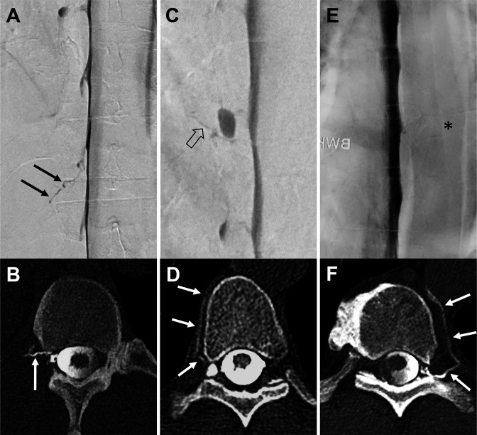
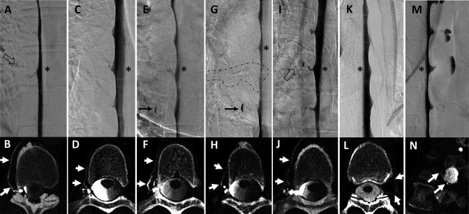
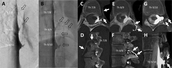
Results: Twenty patients underwent a total of 27 combined LD-DSM/LD-CTM examinations (4/20 with follow-up and 3/20 with bilateral examinations). Both raters identified significantly more CVFs with LD-CTM than with LD-DSM (rater 1: 39 vs 9, P<0.001; rater 2: 42 vs 12, P<0.001). Inter-rater agreement was substantial for LD-DSM (κ=0.732) and LD-CTM (κ=0.655). The results remained significant after considering the senior rating for cases of disagreement (39 vs 10; P<0.001), and no CVF detected on LD-DSM was missed on LD-CTM.

Conclusion: In this study, LD-CTM has a higher diagnostic yield for the detection of CVFs than LD-DSM and should supplement LD-DSM, but further studies are needed. LD-CTM can be easily acquired in awake or mildly sedated patients with the second half of contrast injected just before CT scanning, or it may be considered as a stand-alone investigation.

Keywords: CT; Intervention; Spine.


Conflict of interest statement

Competing interests: None declared.

Figures

Figure 1
Figure 1
Three exemplary patients with a combined examination (A+B, C+D, and E+F): lateral decubitus digital subtraction myelography (LD-DSM; A, C, E) in anterior-posterior view followed in each case by lateral decubitus CT myelography (LD-CTM; B, D, F) with a second half of contrast application in axial view. In the first example, a contrasted paraspinal vein at the Th 12/L1 level on the right side is visible in both examinations, LD-DSM (black arrows in A) and LD-CTM (white arrow in B): rated as positive for a CSF-venous fistula (CVF) by raters 1 and 2 in each modality. The second example shows a tiny hyperdense line on LD-DSM (open black arrow in C) adjacent to a nerve root diverticulum at the level of Th 8/9 on the right side (rater 1: CVF-negative, rater 2: CVF-positive, senior rater adjudicated: CVF-positive). On LD-CTM of the same patient, a contrasted paraspinal vein (white arrows in D) is visible at the same level (raters 1 and 2: CVF-positive). The third example does not show a contrasted vein on LD-DSM (asterisk in E) at the level Th 9/10 left (raters 1 and 2: CVF-negative). The LD-CTM of the same patient clearly demonstrates a contrasted paravertebral vein (white arrows in F) at the same level (raters 1 and 2: CVF-positive).
Figure 2
Figure 2
Seven examples of combined lateral decubitus digital subtraction myelography (LD-DSM) and lateral decubitus CT myelography (LD-CTM) studies (A+B, C+D, E+F, G+H, I+J, K+L, M+N), in which DSM was rated negative but CTM was rated positive for CSF-venous fistulas (CVFs) after senior rating in cases of disagreement between raters 1 and 2. The asterisk in each LD-DSM (A, C, E, G, I, K, M) indicates the spinal level at which a CVF was found at LD-CTM (white arrows in B, D, F, H, J, L, N). Retrospectively, the DSM shows a small lesion as hint of a potential CVF in two examples: a tiny tubular and a dot-like structure in (A) and (I; open black arrows). In example E+F, a patient detected with a CVF only at CTM (F) was treated by transvenous embolization (onyx cast circled by dashed line in G) and was evaluated at follow-up with a de novo CVF at the spinal level above (asterisk in G), also seen only at CTM (white arrows in H). The black arrow in (E) and (G) shows a spinal diverticulum filled with contrast agent only in the lower aspect.
Figure 3
Figure 3
Example of a patient evaluated as having multiple synchronous CSF-venous fistulas (CVFs; n=3) at the initial examination on the left side (Th 7/8, 8/9, and 9/10). Lateral decubitus digital subtraction myelography (LD-DSM) (A) and lateral decubitus fluoroscopy (B) indicated a CVF at level Th 8/9 by rater 1 and at Th 7/8, 8/9, and 9/10 by rater 2 and the senior rater (open black arrows in A and B). At the level Th 7/8 was a dot-like structure only visible at DSM (open black arrow in A at level Th 7/8). Raters 1 and 2, both identified three CVFs at lateral decubitus CT myelography (LD-CTM) at adjacent spinal levels, shown on axial (C, E, G) and sagittal (D, F) and oblique sagittal (H) CTM by white arrows. As the contrasted veins shown here each have contact with the nerve root sleeve, it is more likely that three CVFs are present than that they originate from one point of fistula only.

References

    1. Urbach H. Intracranial hypotension: clinical presentation, imaging findings, and imaging-guided therapy. Curr Opin Neurol 2014;27:414–24. 10.1097/WCO.0000000000000105 - DOI - PubMed
    1. Schievink WI, Moser FG, Maya MM. CSF-venous fistula in spontaneous intracranial hypotension. Neurology 2014;83:472–3. 10.1212/WNL.0000000000000639 - DOI - PubMed
    1. Roytman M, Salama G, Robbins MS, et al. CSF-venous fistula. Curr Pain Headache Rep 2021;25:5. 10.1007/s11916-020-00921-4 - DOI - PubMed
    1. Schievink WI, Maya MM, Moser FG, et al. Lateral decubitus digital subtraction myelography to identify spinal CSF-venous fistulas in spontaneous intracranial hypotension. J Neurosurg Spine 2019:1–4. 10.3171/2019.6.SPINE19487 - DOI - PubMed
    1. Mamlouk MD, Ochi RP, Jun P, et al. Decubitus CT myelography for CSF-venous fistulas: a procedural approach. AJNR Am J Neuroradiol 2021;42:32–6. 10.3174/ajnr.A6844 - DOI - PMC - PubMed

Publication types