. 2023 Dec;72(12):4441-4456.
doi: 10.1007/s00262-023-03555-8. Epub 2023 Nov 3.

Cholesterol induction in CD8+ T cell exhaustion in colorectal cancer via the regulation of endoplasmic reticulum-mitochondria contact sites

Affiliations

Cholesterol induction in CD8+ T cell exhaustion in colorectal cancer via the regulation of endoplasmic reticulum-mitochondria contact sites

Han Shuwen et al. Cancer Immunol Immunother. 2023 Dec.

Abstract

Background: Hypercholesterolemia is one of the risk factors for colorectal cancer (CRC). Cholesterol can participate in the regulation of human T cell function and affect the occurrence and development of CRC.

Objective: To elucidate the pathogenesis of CRC immune escape mediated by CD8+ T cell exhaustion induced by cholesterol.

Methods: CRC samples (n = 217) and healthy individuals (n = 98) were recruited to analyze the relationship between peripheral blood cholesterol levels and the clinical features of CRC. An animal model of CRC with hypercholesterolemia was established. Intraperitoneal intervention with endoplasmic reticulum stress (ERS) inhibitors in hypercholesterolemic CRC mice was performed. CD69, PD1, TIM-3, and CTLA-4 on CD8+ T cells of spleens from C57BL/6 J mice were detected by flow cytometry. CD8+ T cells were cocultured with MC38 cells (mouse colon cancer cell line). The proliferation, apoptosis, migration and invasive ability of MC38 cells were detected by CCK-8 assay, Annexin-V APC/7-AAD double staining, scratch assay and transwell assay, respectively. Transmission electron microscopy was used to observe the ER structure of CD8+ T cells. Western blotting was used to detect the expression of ERS and mitophagy-related proteins. Mitochondrial function and energy metabolism were measured. Immunoprecipitation was used to detect the interaction of endoplasmic reticulum-mitochondria contact site (ERMC) proteins. Immunofluorescence colocalization was used to detect the expression and intracellular localization of ERMC-related molecules.

Results: Peripheral blood cholesterol-related indices, including Tc, low density lipoproteins (LDL) and Apo(a), were all increased, and high density lipoprotein (HDL) was decreased in CRCs. The proliferation, migration and invasion abilities of MC38 cells were enhanced, and the proportion of tumor cell apoptosis was decreased in the high cholesterol group. The expression of IL-2 and TNF-α was decreased, while IFN-γ was increased in the high cholesterol group. It indicated high cholesterol could induce exhaustion of CD8+ T cells, leading to CRC immune escape. Hypercholesterolemia damaged the ER structure of CD8+ T cells and increased the expression of ER stress molecules (CHOP and GRP78), lead to CD8+ T cell exhaustion. The expression of mitophagy-related proteins (BNIP3, PINK and Parkin) in exhausted CD8+ T cells increased at high cholesterol levels, causing mitochondrial energy disturbance. High cholesterol enhanced the colocalization of Fis1/Bap31, MFN2/cox4/HSP90B1, VAPB/PTPIP51, VDAC1/IPR3/GRP75 in ERMCs, indicated that high cholesterol promoted the intermolecular interaction between ER and mitochondrial membranes in CD8+ T cells.

Conclusion: High cholesterol regulated the ERS-ERMC-mitophagy axis to induce the exhaustion of CD8+ T cells in CRC.

Keywords: CD8+ T-cell exhaustion; Cholesterol; Colorectal cancer (CRC); ER-mitochondria contact sites (ERMCs); Endoplasmic reticulum stress (ER stress); Mitochondrial energy metabolism.


Conflict of interest statement

The authors declare that no conflicts of interest exist.

Figures
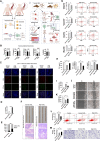
Fig. 1
Fig. 1
High cholesterol promotes CRC by inducing exhaustion in CD8+ T cells. A Workflow of this part. B histogram of cholesterol (Tc), HDL, LDL, Apo(a) and ApoB contents in CRC patients and healthy individuals (ncontrol = 98; nCRC = 217). C IF assay was used to detect the expression of CTLA-4, PD1 and TIM-3 in CD8+ T cells from peripheral blood in CRC patients with healthy serum cholesterol levels and hypercholesterolemia. D comparison of the peripheral blood cholesterol level between CRC mice model with high cholesterol diets and normal cholesterol diets. E, F Pathological morphology and HE staining of colon tissues from CRC mice model of inflammation induced by AOM/DSS. G Expression of CD69, CTLA-4, PD1 and TIM-3 in CD8+ T cells from non-CRC mice with hypercholesterolemia (high CHOL) and CD8+ T cells from non-CRC mice with normal cholesterol level (Normal CHOL). The red box shows the surface of receptor-positive CD8+ T cells. H Histogram of the contents of the cytokines IFN-γ, TNF-α and IL-2 in peripheral blood from mice in the Normal CHOL group and High CHOL group. IM MC38 cells were used as the control group, Normal CHOL CD8+ T cells were used as the positive control group, and High CHOL CD8+ T cells were used as the experimental group. The CCK8 method (I) was used to detect the proliferation activity of MC38 cells, a scratch test (J, K) was used to detect the migration ability of MC38 cells, flow cytometry L) was used to detect the percentage of apoptosis in MC38 cells, and Transwell assays (M) were used to detect the invasion ability of MC38 cells. * indicating P < 0.05, ** indicating P < 0.01, *** indicating P < 0.001
Fig. 2
Fig. 2
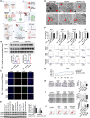
ERS in exhausted CD8+ T cells induced by high cholesterol. A Workflow of this part. B The expression of the CD8+ T cell ERS proteins CHOP and GRP78 in the peripheral blood of CRC patients with normal cholesterol and high cholesterol was detected by WB. C IF assay was used to detect the expression of CHOP and GRP78 in CD8+ T cells from cancerous tissues in CRC patients with normal cholesterol and hypercholesteremia. D Expression of the ERS-related proteins CHOP and GPR78 in mice from each group detected by WB. E Endoplasmic reticulum morphology of different groups of CD8+ T cells observed by transmission electron microscopy. In the normal CHOL group, the rough endoplasmic reticulum distribution was also reduced. In the High CHOL group (CD8+ T cells from mice in the High cholesterol group), the endoplasmic reticulum was rare. In the MC-38/CD8+ T-WT group (MC-38 cells were cocultured with CD8+ T cells from wild-type mice), endoplasmic reticulum disintegration was observed in some parts. In the MC-38/CD8+ T-high CHOL group (coculture of MC-38 cells with CD8 + T cells from mice in the high cholesterol group), the ER was disintegrated. In the CD8+ T-4-PBA group (mice CD8+ T cells treated with the ERS inhibitor 4-PBA), ER structures were less common in the cytoplasm. In the MC-38/CD8+ T-4-PBA group (intervention with the ERS inhibitor 4-PBA in the coculture system of MC-38 cells and mice CD8+T cells), the arrow shows ERS disintegration. F Histogram and flow chart of PD1, TIM-3, CTLA-4 and CD69 expression in spleen T cells from mice in the 3 groups. G Cell proliferation was detected by the CCK-8 method, and differences were observed among the 3 groups of tumor cells (P < 0.05). H Cell invasion ability detected by the Transwell method. I Cell migration detected by a scratch test. J apoptosis was detected by Annexin-V APC/7-AAD double staining. In the bar chart, * indicates P < 0.05, ** indicates P < 0.01, *** indicates P < 0.001
Fig. 3
Fig. 3
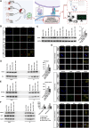
Contact between the endoplasmic reticulum and mitochondria occurs in CRCs at different cholesterol levels. A Workflow of this part. B The expression of CD8+ T cell ERMC proteins (Fis1 and Bap31, MFN2, VAPB and PTPIP51, VDAC1 and IPR3 and GRP75) in the peripheral blood of CRC patients with normal cholesterol and high cholesterol was detected by WB. CF IF assay was used to detect the expression and location of Fis1 and Bap31, CoX4 and HSP90B1, VAPB and PTPIP51, VDAC1 and IPR3 and GRP75 in CD8+ T cells from CRC patients with normal cholesterol and high cholesterol levels. In the bar chart, * indicates P < 0.05, ** indicates P < 0.01
Fig. 4
Fig. 4
Exhaustion of CD8+ T cells induced by high cholesterol showed structural and functional changes in ERMCs. A Workflow of this part. B Confocal immunofluorescence microscopy of mitochondria and ER of CD8+ T cells in the normal cholesterol group, high cholesterol group, normal CHOL CRC group, and high CHOL CRC group. Among these groups, the nucleus was blue, mitochondrial probe was green, and the ER probe was red. The higher the yellow overlap in the merged diagram is, the greater the colocalization of mitochondria and endoplasmic reticulum. C The expression of the mitochondrial fusion protein MFN2 in CD8+ T cells from the 4 groups was detected by Western blot. DF Co-immunoprecipitation was performed to clarify the interaction between Fis1 and Bap31, VAPB and PTPIP51, and VDAC1 and IPR3 and GRP75 proteins of ERMCs in CD8+ T cells. Input refers to the protein content in cells, and IP refers to the protein content measured by the antigen antibody response. G, Immunofluorescence was performed to clarify the expression and location of MFN2 and CoX4 and HSP90B1, VAPB and PTPIP51, and VDAC1 and IPR3 and GRP75 in CD8+ T cells. The nucleus (blue) and other colors correspond to the probe colors of each molecule. The more orange parts in the combined figure, the more molecules are located in the cell

References

    1. Siegel RL, Miller KD, Fuchs HE, Jemal A (2021) Cancer statistics, 2021. CA Cancer J Clin 71(1):7–33 - PubMed
    1. Zhang L, Cao F, Zhang G, Shi L, Chen S, Zhang Z, Zhi W, Ma T (2019) Trends in and predictions of colorectal cancer incidence and mortality in China From 1990 to 2025. Front Oncol 9:98 - PMC - PubMed
    1. Jun SY, Brown AJ, Chua NK, Yoon JY, Lee JJ, Yang JO, Jang I, Jeon SJ, Choi TI, Kim CH et al (2021) Reduction of squalene epoxidase by cholesterol accumulation accelerates colorectal cancer progression and metastasis. Gastroenterology 160(4):1194-1207.e1128 - PubMed
    1. Cornish AJ, Law PJ, Timofeeva M, Palin K, Farrington SM, Palles C, Jenkins MA, Casey G, Brenner H, Chang-Claude J et al (2020) Modifiable pathways for colorectal cancer: a mendelian randomisation analysis. Lancet Gastroenterol Hepatol 5(1):55–62 - PMC - PubMed
    1. Wang Y, Sun XQ, Lin HC, Wang DS, Wang ZQ, Shao Q, Wang FH, Yan SM, Liang JY, Zeng ZL et al (2019) Correlation between immune signature and high-density lipoprotein cholesterol level in stage II/III colorectal cancer. Cancer Med 8(3):1209–1217 - PMC - PubMed