. 2024 Feb;20(2):170-179.
doi: 10.1038/s41589-023-01458-4. Epub 2023 Nov 2.

DNA-encoded library-enabled discovery of proximity-inducing small molecules

Affiliations

DNA-encoded library-enabled discovery of proximity-inducing small molecules

Jeremy W Mason et al. Nat Chem Biol. 2024 Feb.

Abstract

Small molecules that induce protein-protein associations represent powerful tools to modulate cell circuitry. We sought to develop a platform for the direct discovery of compounds able to induce association of any two preselected proteins, using the E3 ligase von Hippel-Lindau (VHL) and bromodomains as test systems. Leveraging the screening power of DNA-encoded libraries (DELs), we synthesized ~1 million DNA-encoded compounds that possess a VHL-targeting ligand, a variety of connectors and a diversity element generated by split-and-pool combinatorial chemistry. By screening our DEL against bromodomains in the presence and absence of VHL, we could identify VHL-bound molecules that simultaneously bind bromodomains. For highly barcode-enriched library members, ternary complex formation leading to bromodomain degradation was confirmed in cells. Furthermore, a ternary complex crystal structure was obtained for our most enriched library member with BRD4BD1 and a VHL complex. Our work provides a foundation for adapting DEL screening to the discovery of proximity-inducing small molecules.


Conflict of interest statement

Competing interests

The authors declare the following competing financial interests: X.M. is a shareholder of Terremoto Biosciences. C.W.C. is an advisor to Anagenex. P.A.C. is an advisor to nference, Inc., Pfizer, Inc., and Belharra Therapeutics. S.L.S. is a shareholder and serves on the Board of Directors of Jnana Therapeutics and Kojin Therapeutics; is a shareholder and advises Kisbee Therapeutics, Belharra Therapeutics, Magnet Biomedicine, Exo Therapeutics, and Eikonizo Therapeutics; advises Vividian Therapeutics, Eisai Co., Ltd., Ono Pharma Foundation, F-Prime Capital Partners, and the Genomics Institute of the Novartis Research Foundation; and is a Novartis Faculty Scholar. The remaining authors declare no competing interests.

Figures

Extended Data Fig. 1.
Distribution of barcode counts for each CIP-DEL library member across each screening condition. (a) Barcode counts for the beads-only screen. (b) Barcode counts for the BRD4 (-) VHL screen. (c) Barcode counts for the BRD4 (+) VHL screen.
Extended Data Fig. 2.
Individual building block barcode counts for each screening condition.
Extended Data Fig. 3.
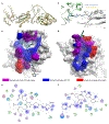
CIP-1 ternary complex structural analysis. (a) Six copies of the ternary complex are present in each asymmetric unit. An overlay of the six ternary complexes shows the structures are highly similar with RMS deviations of 1.06 – 1.37 Å. (b) The electron density for CIP-1 is well defined in 4 of the 6 complexes of the asymmetric unit. 2Fo – Fc maps contoured at 2.5σ and 1σ are shown for CIP-1 in a ternary complex with well-defined density. (c) BRD4BD1 protein surface with residues colored based on interactions with CIP-1 (blue), VHL (red), or both (purple). CIP-1 is shown in sticks. (d) VHL protein surface with residues colored based on interactions with CIP-1 (blue), BRD4BD1 (red), or both (purple). (e) 2D diagram of CIP-1 interactions with BRD4BD1. (f) 2D diagram of CIP-1 interactions with VHL.
Extended Data Fig. 4.
Comparison of the small molecule components of VHL PROTAC ternary complex structures. (a) Overlaying the VHL binding moieties of ternary complex assemblies, the diverse projection vectors of the connectors and target protein ligands are shown. The initial point of divergence for the bifunctional molecules is the carbon atom following the terminal amide of the VH032 ligand. (b) Comparison of the buried surface area for four VHL-targeting CIPs.
Extended Data Fig. 5.
Comparison of the ternary complex assemblies for four VHL-targeting bifunctional degraders. The first three structures show bromodomains targets (BRD4BD1, BRD4BD2, and SMARCA4BD) and the fourth complex is a non-bromodomain targeting degrader (Bcl-XL). VHL proteins are aligned at the bottom of each structure and the target protein orientation is shown at the top. Key details for each complex are provided in the table. Inset: the three bromodomain containing complexes are overlaid with VHL aligned at the bottom of the structures, highlighting the diverse target protein orientations.
Figure 1.
Figure 1.. Library design and synthesis.
(a) Using the ternary complex crystal structure of MZ1 with BRD4BD2 and VHL (PDB: 5T35), the thiazole-methyl group of MZ1 was selected as the site for DNA attachment as it projects towards a solvent exposed channel, potentially minimizing disruption of the ternary complex. (b) MZ1 was used as a template for CIP-DEL library design. The VHL ligand was functionalized with a connector library and appended to DNA. Each connector terminated with an amine that was functionalized using split-and-pool DEL synthesis methods. (c) Affinity screening strategy for identifying complex-inducing library members. Parallel screens with the protein of interest +/− the E3 ubiquitin ligase are performed and enrichment signals that remain high in both screens suggest library members that are capable of stable ternary complex induction. (d) The VHL ligand was modified with an alkyne to enable DNA attachment and functionalized with 22 Fmoc-amino acid connectors. The Fmoc groups were replaced by acetyl groups, and each compound was assayed for binding to VHL. We selected 15 connectors that retained binding to VHL to include in the CIP-DEL library. (e) Overview of the CIP-DEL library synthesis.
Figure 2.
Figure 2.. CIP-DEL screening analysis and evaluation of compounds synthesized off-DNA.
(a - c) Cube plots of enriched features in (a) the BRD4 (−) VHL screen (enrichment cutoff ≥ 7), (b) the BRD4 (+) VHL screen (enrichment cutoff ≥ 80), and (c) both the BRD4 (−) VHL and BRD4 (+) VHL screens (enrichment cutoff ≥ 5 in both). Points are sized by (−) VHL enrichment values in (a) and (+) VHL enrichment values in (b) and (c). Coloring: red = MZ1-DEL (shown at 1,1,1); grey = meets enrichment cutoff; green = contains C15 and BB2–109 or BB3–114; blue = synthesized off-DNA. Compounds selected for synthesis are shown in all plots and may not meet indicated enrichment cutoffs. (d) Structures of select library members synthesized off-DNA with binary and ternary KD values from SPR (see Supplementary Table 1 for additional details). (e) Ternary complex induction by select CIP compounds using the NanoBiT assay. (f) Compound induced BRD4BD1 degradation measured by the HiBiT assay. (g) BRD4BD1 degradation with CIP compounds is reversed in the presence of a Nedd8 inhibitor. Data in e - g represent the mean ± s.d. of n = 2 independent experiments. (h) Western blot analysis of endogenous BRD4 degradation with CIP compounds and controls (n = 1).
Figure 3.
Figure 3.. Correlations between CIP-DEL screening results and off-DNA validation data.
(a) Correlation between BRD4 (−) VHL enrichment values (lower-bound of 95% confidence interval) and binary KD values obtain by SPR. (b) Relationship between BRD4 (+) VHL enrichments and ternary KD values obtained by SPR. (c) BRD4 (+) VHL enrichment values compared to NanoBiT EC50 and HiBiT DC50 values. (d) BRD4 (+) VHL enrichments compared to NanoBiT Emax and HiBiT Dmax results. (e) Relationship between NanoBiT EC50 and HiBiT DC50 values, and the ternary KD values obtained from SPR. (f) Correlation of NanoBiT Emax and HiBiT Dmax with the ternary KD results obtained by SPR. (g) Association between the NanoBiT EC50 and HiBiT DC50 results obtained for each active off-DNA compound. (h) Correlation between NanoBiT Emax and HiBiT Dmax values for active DNA-free compounds. (i) Comparison of CIP-DEL enrichment ratios (+/−) VHL and the alpha values calculated from binary and ternary SPR KD values. In all panels, data points for MZ1 were excluded from the line fitting due to potential ternary complex disruptions caused by attachment of the DNA tag (see text).
Figure 4.
Figure 4.
Ternary complex crystal structure of CIP-1 with BRD4 and VHL-Elongin C-Elongin B. (a) Overall architecture of complex assembly. Protein components are shown as ribbons with semi-transparent surfaces and labeled by protein color. CIP-1 is shown as sticks. (b) Key interactions of CIP-1 with BRD4BD1. An amide carbonyl from CIP-1 forms an H-bond with Asn140 of BRD4BD1 and the pyrazole substituent of CIP-1 forms a pi-stacking interaction with Trp81. An intramolecular H-bond between an amide NH and a triazine nitrogen of CIP-1 reinforces the bound conformation of the BRD4 binding component. The ternary complex is stabilized by two H-bonds formed between Asp145 of BRD4BD1 and Tyr112 and His110 of VHL. (c) Key interactions of CIP-1 with VHL. The VHL binding component of CIP-1 maintains equivalent interactions with the E3 ligase as the parent VH032 ligand.
Figure 5.
Figure 5.. CIP-DEL screening against BRD2BD1 and BRDTBD1 with off-DNA profiling of two shared hits.
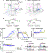
(a) Selection analysis of the BRD2BD1 (+) VHL screen (enrichment cutoff ≥ 7), and (b) the BRDTBD1 (+) VHL screen (enrichment cutoff ≥ 5). Points are sized by (+) VHL enrichment values and the two shared hits are labeled and colored blue. (c) Structures of hit compounds synthesized off-DNA with enrichment values observed in each screen. (d) Ternary complex induction between VHL and BRD2BD1 (filled) and BRDTBD1 (hollow) for CIP-22 (blue) and CIP-23 (green) measured using the NanoBiT assay. (e) CIP mediated degradation of BRD2BD1 and BRDTBD1 measured using the HiBiT assay. (f) The bromodomain degradation with CIPs is reversed in the presence of a Neddylation inhibitor. (g) Cell viability is not affected by the CIP compounds, shown by the CellTiter-Glo assay. Data in d - g represent the mean ± s.d. of n = 2 independent experiments. (h) Western blot analysis of endogenous BRD2 degradation with CIP compounds (n = 3 independent experiments).

References

    1. Schreiber SL The Rise of Molecular Glues. Cell 184, 3–9 (2021). - PubMed
    1. Liu J et al. Calcineurin is a common target of cyclophilin-cyclosporin A and FKBP-FK506 complexes. Cell 66, 807–815 (1991). - PubMed
    1. Brown EJ et al. A mammalian protein targeted by G1-arresting rapamycin–receptor complex. Nature 369, 756–758 (1994). - PubMed
    1. Sabatini DM, Erdjument-Bromage H, Lui M, Tempst P & Snyder SH RAFT1: A mammalian protein that binds to FKBP12 in a rapamycin-dependent fashion and is homologous to yeast TORs. Cell 78, 35–43 (1994). - PubMed
    1. Haggarty SJ et al. Dissecting cellular processes using small molecules: identification of colchicine-like, taxol-like and other small molecules that perturb mitosis. Chemistry & Biology 7, 275–286 (2000). - PubMed

MeSH terms

LinkOut - more resources