Review
. 2023 Nov 3;10(1):45.
doi: 10.1186/s40662-023-00363-0.

Multimodal diagnostics for keratoconus and ectatic corneal diseases: a paradigm shift

Affiliations
Review

Multimodal diagnostics for keratoconus and ectatic corneal diseases: a paradigm shift

Renato Ambrósio Jr et al. Eye Vis (Lond). .

Abstract

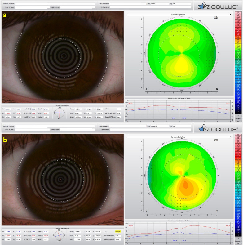
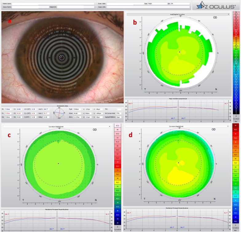
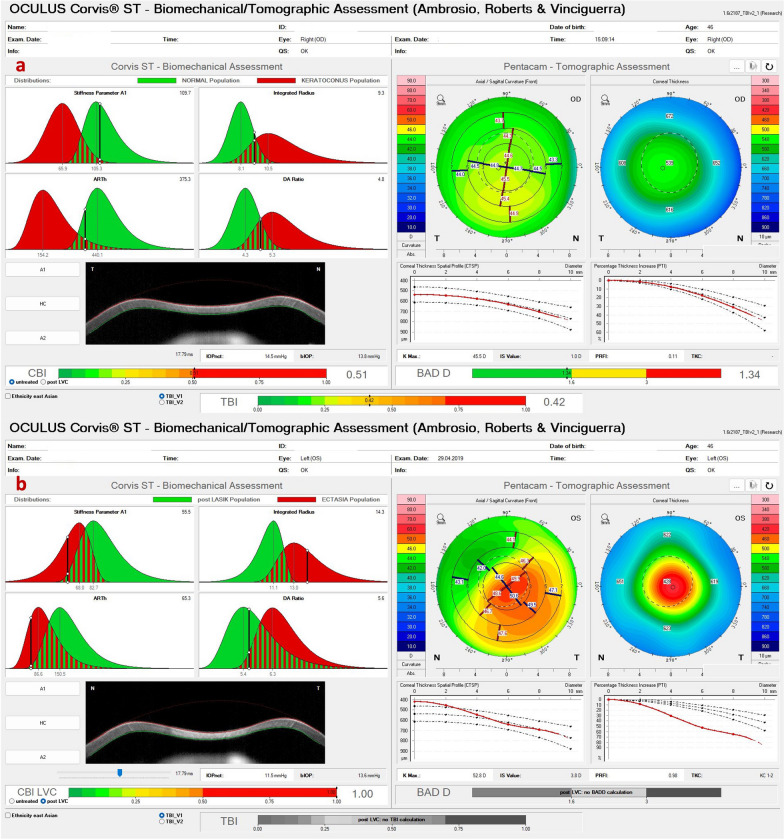
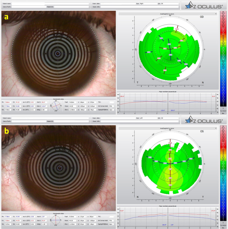
Different diagnostic approaches for ectatic corneal diseases (ECD) include screening, diagnosis confirmation, classification of the ECD type, severity staging, prognostic evaluation, and clinical follow-up. The comprehensive assessment must start with a directed clinical history. However, multimodal imaging tools, including Placido-disk topography, Scheimpflug three-dimensional (3D) tomography, corneal biomechanical evaluations, and layered (or segmental) tomography with epithelial thickness by optical coherence tomography (OCT), or digital very high-frequency ultrasound (dVHF-US) serve as fundamental complementary exams for measuring different characteristics of the cornea. Also, ocular wavefront analysis, axial length measurements, corneal specular or confocal microscopy, and genetic or molecular biology tests are relevant for clinical decisions. Artificial intelligence enhances interpretation and enables combining such a plethora of data, boosting accuracy and facilitating clinical decisions. The applications of diagnostic information for individualized treatments became relevant concerning the therapeutic refractive procedures that emerged as alternatives to keratoplasty. The first paradigm shift concerns the surgical management of patients with ECD with different techniques, such as crosslinking and intrastromal corneal ring segments. A second paradigm shift involved the quest for identifying patients at higher risk of progressive iatrogenic ectasia after elective refractive corrections on the cornea. Beyond augmenting the sensitivity to detect very mild (subclinical or fruste) forms of ECD, ectasia risk assessment evolved to characterize the inherent susceptibility for ectasia development and progression. Furthermore, ectasia risk is also related to environmental factors, including eye rubbing and the relational impact of the surgical procedure on the cornea.

Keywords: Corneal biomechanics; Corneal ectasia; Corneal tomography; Keratoconus; Multimodal corneal imaging; Refractive surgery; Susceptibility.


Conflict of interest statement

Dr. Ambrósio is a consultant for Oculus, Alcon, Zeiss and Mediphacos.

Figures

Fig. 1
Fig. 1
The Scheimpflug image (Pentacam AXL) (a), the Tomey Cassia 2 (b) and Anterion OCT (Heidelberg Engineering GmbH) (c) of the cornea and anterior chamber of the right eye of a 45-year-old patient with traumatic ectasia with a pellucid marginal degeneration thinning pattern
Fig. 2
Fig. 2
Pentacam Quad maps of a pellucid marginal degeneration (PMD) after trauma in a right eye (a) and subclinical PMD-like pattern in a left eye (b)
Fig. 3
Fig. 3
Mild, subclinical or fruste pellucid marginal corneal degeneration. a Keratograph 5M shows the Placido rings and the axial curvature topography of a subclinical pellucid marginal degeneration-like pattern in a left eye (OS). b Tomographic-Biomechanical Display shows borderline Corvis biomechanical index (CBI) and abnormal tomographic biomechanical index version 2 (TBIv2)
Fig. 4
Fig. 4
Keratograph 5M shows the topography of a low keratometry keratoconus (KC) with a maximum keratometry (Kmax) of (a) 43.3 D in the right eye (OD) and (b) 44.0 D in the left eye (OS)
Fig. 5
Fig. 5
Tomographic-Biomechanical Display of the patient with low keratometry KC shows tomographic biomechanical index (TBI) of (a) 1.0 in the right eye (OD) and (b) 0.96 in the left eye (OS) and Belin/Ambrósio enhanced ectasia display deviation (BAD-D) of (a) 4.04 in OD and (b) 3.89 in OS
Fig. 6
Fig. 6
Tomographic ABCD ectasia/keratoconus (KC) staging shows the patient with low keratometry KC demonstrating stability over ten years in all parameters
Fig. 7
Fig. 7
Keratograph 5M shows the Placido rings and the topography of (a) a very asymmetric ectasia (VAE) with a moderate keratoconus (KC) in the right eye (OD) and (b) advanced disease in the left eye (OS) of twin 1
Fig. 8
Fig. 8
The Pentacam Belin/Ambrósio enhanced ectasia display deviation (BAD-D) shows (a) a value of 3.28 in the right eye (OD) and (b) 8.95 in the left eye (OS) of twin 1
Fig. 9
Fig. 9
Slit-lamp biomicroscopy of intrastromal corneal ring segments (ICRS, AF 320/200) in the right eye of twin 1
Fig. 10
Fig. 10
Pentacam anterior curvature differential maps shows anterior curvature maps from the right eye (OD) in March 2023 and September 2022, respectively (a and c); anterior curvature maps from the left eye (OS) in March 2023 and September 2022, respectively (b and d). Note that there was no evident progression of the ectatic disease, even a mild keratometric reduction in OD (ac) with clinical treatment with oral supplementation of vitamin B2 and a decrease in curvature in OS (bd) with the intrastromal corneal ring segments
Fig. 11
Fig. 11
Keratograph 5M shows the Placido rings and the topography of a typical topography in the right eye (a) and an forme fruste keratoconus (FFKC) in the left eye (b) of the twin 2
Fig. 12
Fig. 12
Cornea ectasia report from optical coherence tomography (OCT) of the right eye (OD) of twin 2, demonstrating a Gatinel score of − 0.3 in the OD (a) and an abnormal score of 1.9 in the left eye (OS) (b)
Fig. 13
Fig. 13
Corvis ST Tomographic-Biomechanical Display shows a forme fruste keratoconus (FFKC) in the right eye (OD) and (b) subclinical keratoconus (KC) in the left eye (OS) of twin 2. Despite a relatively regular anterior tomographic assessment (top right), note the abnormal tomographic biomechanical index version 2 (TBIv2) values of 0.28 in OD
Fig. 14
Fig. 14
Keratograph 5M shows the unoperated right eye (OD) of a patient that developed ectasia in the contralateral left eye (OS) after unilateral laser in situ keratomileusis (LASIK). a The Placido rings; bd The axial curvature topography with the Ambrósio-2 absolute scale (b), Klyce/Smolek absolute 1.5 D scale (c) and the absolute 0.5 D Atlas scale (d)
Fig. 15
Fig. 15
Corvis ST Tomographic-Biomechanical Display of the same patient of Fig. 14 shows (a) abnormal Corvis biomechanical index (CBI, 0.51) and tomographic biomechanical index (TBI, 0.49) despite borderline BAD-D (v3) 1.34 in the unoperated right eye (OD) and (b) high CBI post-LVC in the left eye (OS) with post-LASIK ectasia. BAD-D (v3), Belin/Ambrósio enhanced ectasia deviation (third version); LVC, laser vision correction; LASIK, laser in situ keratomileusis
Fig. 16
Fig. 16
Optical coherence tomography (OCT) of the cornea shows the epithelial map and pachymetry of the contralateral eye (the right eye) of the same patient in Figs. 14 and 15. Note mild but relevant epithelial thinning
Fig. 17
Fig. 17
Keratograph 5M shows the Placido rings and the relatively normal axial curvature topography of a 27-year-old female with a posterior polymorphous corneal dystrophy (PPCD) and considered with (a) forme fruste keratoconus (FFKC) in the right eye (OD) and (b) a mild inferior steepening in the left eye (OS)
Fig. 18
Fig. 18
Slit-lamp biomicroscopy of the right eye (OD) with the “snail track” sign (a) and specular microscopy with endothelial alterations in OD (b)
Fig. 19
Fig. 19
The optical coherence tomography (OCT) of the right eye shows the pachymetry and epithelium maps
Fig. 20
Fig. 20
Tomographic-Biomechanical Display of the patient with posterior polymorphous corneal dystrophy (PPCD) and forme fruste keratoconus (FFKC) with (a) relatively high tomographic biomechanical index (TBI) in the right eye (OD) and (b) relatively normal TBI in the left eye (OS)

References

    1. Seiler T. The paradigm change in keratoconus therapy. Indian J Ophthalmol. 2013;61(8):381. doi: 10.4103/0301-4738.116050. - DOI - PMC - PubMed
    1. McGhee CN, Kim BZ, Wilson PJ. Contemporary treatment paradigms in keratoconus. Cornea. 2015;34(Suppl 10):S16–23. doi: 10.1097/ICO.0000000000000504. - DOI - PubMed
    1. Ambrósio R, Jr, Lopes B, Amaral J, Faria-Correia F, Canedo ALC, Salomão MQ, et al. Keratoconus: breaking paradigms and contradictions of a new subspecialty. Rev Bras Oftalmol. 2019;78(2):81–85.
    1. Esporcatte LPG, Salomão MQ, Neto ABDC, Machado AP, Lopes BT, Ambrósio R., Jr Enhanced diagnostics for corneal ectatic diseases: the whats, the whys, and the hows. Diagnostics. 2022;12(12):3027. doi: 10.3390/diagnostics12123027. - DOI - PMC - PubMed
    1. Ambrósio R., Jr Cirurgia refrativa terapêutica: por que diferenciar? Rev Bras Oftalmol. 2013;72(2):85–86. doi: 10.1590/S0034-72802013000200002. - DOI

LinkOut - more resources