

Multiple Foci of Basal Cell Carcinoma Arising in Rhinophyma: A Case Report and Literature Review
- PMID: 37920281
- PMCID: PMC10618623
- DOI: 10.1016/j.jpra.2023.10.001
Multiple Foci of Basal Cell Carcinoma Arising in Rhinophyma: A Case Report and Literature Review
Abstract
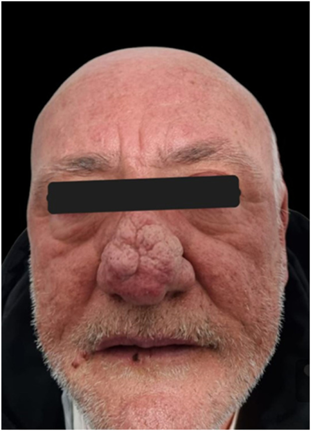
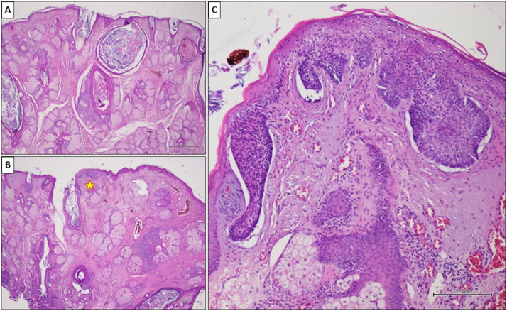
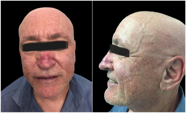
Background: Rhinophyma is a benign condition caused by the excessive growth of sebaceous glands in the nasal tissue, presenting with symptoms such as nasal hypertrophy, erythema, and papules. Cases of basal cell carcinoma in rhinophyma have been reported in literature, but its etiological role remains unclear. It is uncertain whether rhinophyma is predisposed to neoplasm development or if their coexistence is coincidental.
Material and method: We conducted a literature survey to identify such cases reported over the years.
Results: We identified 22 studies reporting a total of 47 cases in the literature, all involving male patients. The most common pattern of occurrence was the rapid growth of a nodular formation within the context of rhinophyma.
Discussions and conclusion: The elucidation of the association between basal cell carcinoma and rhinophyma remains challenging. The presence of multiple foci supports the theory that rhinophyma may play a role in their development, but larger studies are needed to establish a causal relationship.
Keywords: Basal cell carcinoma; Basocellular cancer; Case report; Rhinophyma.
© 2023 The Author(s).
Figures
Similar articles
-
Adenoid Cystic Basal Cell Carcinoma Arising in Rhinophyma.Curr Health Sci J. 2020 Jul-Sep;46(3):309-314. doi: 10.12865/CHSJ.46.03.15. Epub 2020 Sep 30. Curr Health Sci J. 2020. PMID: 33304635 Free PMC article.
-
[A systematic review and current recommendation for treatment of rhinophyma].Laryngorhinootologie. 2020 Nov;99(11):772-778. doi: 10.1055/a-1208-5284. Epub 2020 Oct 27. Laryngorhinootologie. 2020. PMID: 33111293 German.
-
[Rhinophyma and skin carcinoma: a case report and literature review].Ann Chir Plast Esthet. 2012 Apr;57(2):169-72. doi: 10.1016/j.anplas.2011.10.003. Epub 2011 Dec 28. Ann Chir Plast Esthet. 2012. PMID: 22209650 Review. French.
-
Malignancies within rhinophyma: report of three new cases and review of the literature.Aesthetic Plast Surg. 2012 Apr;36(2):396-405. doi: 10.1007/s00266-011-9802-0. Epub 2011 Aug 20. Aesthetic Plast Surg. 2012. PMID: 21858597 Review.
-
Basal cell carcinoma and rhinophyma.Ann Plast Surg. 2008 Oct;61(4):410-2. doi: 10.1097/SAP.0b013e31816cad18. Ann Plast Surg. 2008. PMID: 18812712
References
LinkOut - more resources
Full Text Sources
