. 2023 Aug 11:4:100124.
doi: 10.1016/j.jvssci.2023.100124. eCollection 2023.

Vascular smooth muscle cell mechanotransduction through serum and glucocorticoid inducible kinase-1 promotes interleukin-6 production and macrophage accumulation in murine hypertension

Affiliations

Vascular smooth muscle cell mechanotransduction through serum and glucocorticoid inducible kinase-1 promotes interleukin-6 production and macrophage accumulation in murine hypertension

Mario Figueroa et al. JVS Vasc Sci. .

Abstract

Objective: The objective of this investigation was to demonstrate that in vivo induction of hypertension (HTN) and in vitro cyclic stretch of aortic vascular smooth muscle cells (VSMCs) can cause serum and glucocorticoid-inducible kinase (SGK-1)-dependent production of cytokines to promote macrophage accumulation that may promote vascular pathology.

Methods: HTN was induced in C57Bl/6 mice with angiotensin II infusion (1.46 mg/kg/day × 21 days) with or without systemic infusion of EMD638683 (2.5 mg/kg/day × 21 days), a selective SGK-1 inhibitor. Systolic blood pressure was recorded. Abdominal aortas were harvested to quantify SGK-1 activity (pSGK-1/SGK-1) by immunoblot. Flow cytometry quantified the abundance of CD11b+/F480+ cells (macrophages). Plasma interleukin (IL)-6 and monocyte chemoattractant protein-1 (MCP-1) was assessed by enzyme-linked immunosorbent assay. Aortic VSMCs from wild-type mice were subjected to 12% biaxial cyclic stretch (Stretch) for 3 or 12 hours with or without EMD638683 (10 μM) and with or without SGK-1 small interfering RNA with subsequent quantitative polymerase chain reaction for IL-6 and MCP-1 expression. IL-6 and MCP-1 in culture media were analyzed by enzyme-linked immunosorbent assay. Aortic VSMCs from SGK-1flox+/+ mice were transfected with Cre-Adenovirus to knockdown SGK-1 (SGK-1KD VSMCs) and underwent parallel tension experimentation. Computational modeling was used to simulate VSMC signaling. Statistical analysis included analysis of variance with significance at a P value of <.05.

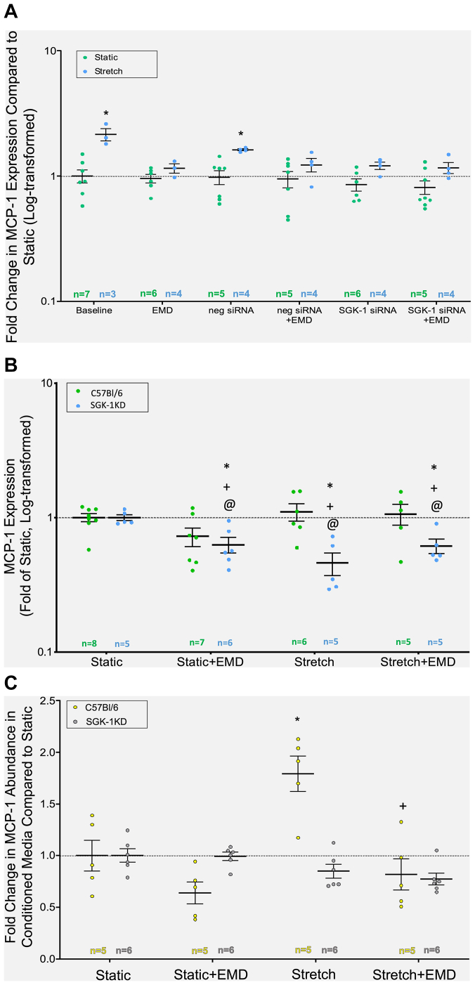
Results: SGK-1 activity, abundance of CD11b+/F4-80+ cells, and plasma IL-6 were increased in the abdominal aorta of mice with HTN and significantly reduced by treatment with EMD638683. This outcome mirrored the increased abundance of IL-6 in media from Stretch C57Bl/6 VSMCs and attenuation of the effect with EMD638683 or SGK-1 small interfering RNA. C57Bl/6 VSMCs also responded to Stretch with increased MCP-1 expression and secretion into the culture media. Further supporting the integral role of mechanical signaling through SGK-1, target gene expression and cytokine secretion was unchanged in SGK-1KD VSMCs with Stretch, and computer modeling confirmed SGK-1 as an intersecting node of signaling owing to mechanical strain and angiotensin II.

Conclusions: Mechanical activation of SGK-1 in aortic VSMCs can promote inflammatory signaling and increased macrophage abundance, therefore this kinase warrants further exploration as a pharmacotherapeutic target to abrogate hypertensive vascular pathology.

Keywords: Hypertension; Interleukin-6; Serum and glucocorticoid-inducible kinase-1 (SGK-1).


Conflict of interest statement

J.M.R. is a surgical proctor for CVRx, outside the scope of the submitted work.

Figures

Fig 1
Fig 1
Systolic blood pressure as measured by tail cuff. ∗P < .05 vs day 0. AngII, Angiotensin II.
Fig 2
Fig 2
(A) SGK-1 activity (pSGK-1/SGK-1) in aortic homogenate at day 21, represented as a fold change from the value in C57Bl/6 control mice (baseline normal physiology). (B) Aortic digestion and cell extraction for flow cytometry quantification of CD11b+/F480+ cells (macrophages) in abdominal aorta on day 21, presented as a percentage of the total cell complement. (C) Quantification of interleukin (IL)-6 in plasma of experimental mice at day 21 using ELISA to quantify the protein from the standard curve. Values have been represented as a fold change from the C57Bl/6 control (baseline normal physiology). ∗ P < .05 vs C57Bl/6; #P < .05 vs C57Bl/6+ angiotensin II (AngII).
Fig 3
Fig 3
(A) Interleukin (IL)-6 expression in C57Bl/6 vascular smooth muscle cells (VSMCs) treated with or without Stretch, with or without small interfering RNA (siRNA), and with or without EMD for 3 hours, represented as a fold change from the C57Bl/6 Static VSMCs (baseline). (B) IL-6 expression in C57Bl/6 and SGK-1KD VSMCs treated for 12 hours with or without Stretch and with or without EMD, represented as a fold change from the C57Bl/6 Static VSMCs (baseline). Data in (A) and (B) graphed and statistically analyzed after log10 transformation. (C) IL-6 abundance in conditioned culture media from C57Bl/6 and SGK-1KD VSMCs treated for 12 hours with or without Stretch and with or without EMD. Abundance values were obtained from the enzyme-linked immunosorbent assay (ELISA) standard curve and represented as a fold change from the C57Bl/6 Static VSMC culture media (baseline). ∗ P < .05 vs wild-type (WT) Static, +P < .05 vs WT Stretch.
Fig 4
Fig 4
(A) MCP-1 expression in C57Bl/6 vascular smooth muscle cells (VSMCs) treated with or without Stretch, with or without small interfering RNA (siRNA), and with or without EMD for 3 hours, represented as a fold change from the C57Bl/6 Static VSMCs (baseline). (B) MCP-1 expression in C57Bl/6 and SGK-1KD VSMCs treated for 12 hours with or without Stretch and with or without EMD, represented as a fold change from the C57Bl/6 Static VSMCs (Baseline). [Data in A and B graphed and statistically analyzed following log10 transformation.] (C) Monocyte chemoattractant protein-1 (MCP-1) abundance in conditioned culture media from C57Bl/6 and SGK-1KD VSMCs treated for 12 hours with or without Stretch and with or without EMD. Abundance values were obtained from the enzyme-linked immunosorbent assay (ELISA) standard curve and represented as a fold change from the C57Bl/6 Static VSMC culture media (baseline). ∗P < .05 vs wild-type (WT) Static; +P < .05 vs WT Stretch; @P < .05 vs SGK-1KO Static.
Fig 5
Fig 5
(A) A network model of smooth muscle cell (SMC) signaling enabling quantitative, differential equation-based predictions of cell expression responses to angiotensin II (AngII) and mechanical strain. (B, C) Further, a simplified model topology identified negative feedback loops (B) and positive feedback loops (C) that contribute to the complexity of interleukin (IL)-6 regulation. (D) Model prediction that inhibiting SGK-1 activity with EMD638683 will result in a substantial reduction of IL-6 expression in vascular SMCs (VSMCs) under mechanical and AngII treatments.
Supplementary Fig 1
Supplementary Fig 1
Representative flow cytometry gating to quantify CD11b+/F480+ cells. AngII, angiotensin II.
Supplementary Fig 2
Supplementary Fig 2
Verification of cell lysate as vascular smooth muscle cells (VSMCs).
Supplementary Fig 3
Supplementary Fig 3
Relative abundance of SGK-1 in C57Bl/6 vs SGK-1KD vascular smooth muscle cells (VSMCs).

References

    1. Haga J.H., Li Y.S., Chien S. Molecular basis of the effects of mechanical stretch on vascular smooth muscle cells. J Biomech. 2007;40:947–960. - PubMed
    1. Anwar M.A., Shalhoub J., Lim C.S., Gohel M.S., Davies A.H. The effect of pressure-induced mechanical stretch on vascular wall differential gene expression. J Vasc Res. 2012;49:463–478. - PubMed
    1. Harrison D.G., Marvar P.J., Titze J.M. Vascular inflammatory cells in hypertension. Front Physiol. 2012;3:128. - PMC - PubMed
    1. Akerman A.W., Stroud R.E., Barrs R.W., et al. Elevated wall tension initiates interleukin-6 expression and abdominal aortic dilation. Ann Vasc Surg. 2018;46:193–204. - PMC - PubMed
    1. Cheng J., Wang Y., Ma Y., et al. The mechanical stress-activated serum-, glucocorticoid-regulated kinase 1 contributes to neointima formation in vein grafts. Circ Res. 2010;107:1265–1274. - PubMed