. 2024 Apr;20(4):830-846.
doi: 10.1080/15548627.2023.2274251. Epub 2023 Nov 3.

Distinct roles of core autophagy-related genes in zebrafish definitive hematopoiesis

Affiliations

Distinct roles of core autophagy-related genes in zebrafish definitive hematopoiesis

Xiang-Ke Chen et al. Autophagy. 2024 Apr.

Abstract

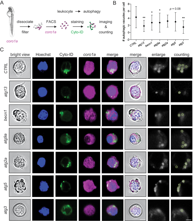
Despite the well-described discrepancy between ATG (macroautophagy/autophagy-related) genes in the regulation of hematopoiesis, varying essentiality of core ATG proteins in vertebrate definitive hematopoiesis remains largely unclear. Here, we employed zebrafish (Danio rerio) to compare the functions of six core atg genes, including atg13, becn1 (beclin1), atg9a, atg2a, atg5, and atg3, in vertebrate definitive hematopoiesis via clustered regularly interspaced short palindromic repeats (CRISPR)-Cas9 ribonucleoprotein and morpholino targeting. Zebrafish with various atg mutations showed autophagic deficiency and presented partially consistent hematopoietic abnormalities during early development. All six atg mutations led to a declined number of spi1b+ (Spi-1 proto-oncogene b) myeloid progenitor cells. However, only becn1 mutation resulted in the expansion of myb+ (v-myb avian myeloblastosis viral oncogene homolog) hematopoietic stem and progenitor cells (HSPCs) and transiently increased coro1a+ (coronin, actin binding protein, 1A) leukocytes, whereas atg3 mutation decreased the number of HSPCs and leukocytes. Proteomic analysis of caudal hematopoietic tissue identified sin3aa (SIN3 transcription regulator family member Aa) as a potential modulator of atg13- and becn1-regulated definitive hematopoiesis. Disruption of sin3aa rescued the expansion of HSPCs and leukocytes in becn1 mutants and exacerbated the decrease of HSPCs in atg13 mutants. Double mutations were also performed to examine alternative functions of various atg genes in definitive hematopoiesis. Notably, becn1 mutation failed to induce HSPCs expansion with one of the other five atg mutations. These findings demonstrated the distinct roles of atg genes and their interplays in zebrafish definitive hematopoiesis, thereby suggesting that the vertebrate definitive hematopoiesis is regulated in an atg gene-dependent manner.Abbreviations: AGM: aorta-gonad-mesonephros; AO: acridine orange; atg: autophagy related; becn1: beclin 1, autophagy related; CHT: caudal hematopoietic tissue; CKO: conditional knockout; coro1a: coronin, actin binding protein, 1A; CQ: chloroquine; CRISPR: clustered regularly interspaced short palindromic repeats; dpf: days post fertilization; FACS: fluorescence-activated cell sorting; hbae1.1: hemoglobin, alpha embryonic 1.1; HSCs: hematopoietic stem cells; HSPCs: hematopoietic stem and progenitor cells; KD: knockdown; KO: knockout; map1lc3/lc3: microtubule-associated protein 1 light chain 3; MO: morpholino; mpeg1.1: macrophage expressed 1, tandem duplicate 1; mpx: myeloid-specific peroxidase; myb: v-myb avian myeloblastosis viral oncogene homolog; PE: phosphatidylethanolamine; p-H3: phospho-H3 histone; PtdIns3K: class 3 phosphatidylinositol 3-kinase; rag1: recombination activating 1; rb1cc1/fip200: RB1-inducible coiled-coil 1; RFLP: restriction fragment length polymorphism; RNP: ribonucleoprotein; sin3aa: SIN3 transcription regulator family member Aa; spi1b: Spi-1 proto-oncogene b; ulk: unc-51 like autophagy activating kinase; vtg1: vitellogenin 1; WISH: whole-mount in situ hybridization.

Keywords: Autophagy-related genes; CRISPR-Cas9 ribonucleoprotein; definitive hematopoiesis; hematopoietic stem and progenitor cells; zebrafish.


Conflict of interest statement

No potential conflict of interest was reported by the author(s).

Figures

Figure 1.
Figure 1.
Core atg genes targeting by CRISPR-Cas9 ribonucleoprotein and morpholino. (A) schematic diagram showing the involvement of autophagy machineries in the autophagy pathway. PAS, pre-autophagosomal structure. (B) schematic diagram showing the CRISPR-Cas9 ribonucleoprotein (RNP) targeting and core atg (autophagy-related) genes selected from autophagy machineries. (C) target sequences and sgRNA design for various atg genes in zebrafish. (D) restriction fragment length polymorphism (RFLP) assay and mutation efficiency of various atg genes-targeting sgRNAs. (E) representative morphology of 2 days post-fertilization (dpf) zebrafish embryos with atg mutations. Scale bar: 0.5 mm. (F) target sequences and morpholino (MO) design for various atg genes in zebrafish. (G) representative morphology of 2 dpf zebrafish embryos injected with MO targeting atg genes. Scale bar: 0.5 mm.
Figure 2.
Figure 2.
Autophagic deficiency in zebrafish embryos with core atg mutations. (A-B) autophagosomes or Lc3+ puncta in the muscle of various atg mutant Tg(GFP-Lc3) zebrafish embryos with (+) and without () chloroquine (CQ) treatment at 2 dpf. Scale bar: 50 μm. (C-D) autophagosomes or Lc3+ puncta in the muscle and skin cells of MO-based atg mutant Tg(GFP-Lc3) zebrafish embryos at 2 dpf. Scale bar: 50 μm. (E) statistical analysis of A-B, *, p < 0.05, **, p < 0.01 compared with control (CTRL), one-way analysis of variance (ANOVA) with post-hoc Tukey HSD test. (F) statistical analysis of C-D, **, p < 0.01 compared with CTRL, one-way ANOVA with post-hoc Tukey HSD test. (G-H) autophagic flux measured by using 2 dpf Tg(GFP-Lc3:RFP-Lc3ΔG) zebrafish embryos under CRISPR-Cas9 RNP- or MO-based atg mutations. Scale bar: 0.5 mm. *, p < 0.05, **, p < 0.01 compared with CTRL, one-way ANOVA with post-hoc Tukey HSD test.
Figure 3.
Figure 3.
Attenuation of autophagic vacuoles in leukocytes with core atg mutations. (A) experimental setup for Cyto-ID+ autophagic vacuole measurement in coro1a+ leukocytes sorted from 2 dpf zebrafish embryos through fluorescence-activated cell sorting (FACS). (B-C) representative images and quantification of Cyto-ID+ autophagic vacuoles in coro1a+ leukocytes sorted from zebrafish embryos with various atg mutations. *, p < 0.05, **, p < 0.01 compared with CTRL, one-way ANOVA with post-hoc Tukey HSD test. Scale bar: 5 μm.
Figure 4.
Figure 4.
Distinct effects of core atg mutations on zebrafish definitive hematopoiesis. (A-C) whole-mount in situ hybridization (WISH) results of myb+ HSPCs in aorta-gonad-mesonephros (AGM) of 30 h post-fertilization (hpf) zebrafish embryos. **, p < 0.01 compared with CTRL, chi-squared test. H, high group; M, medium group. (D-F) WISH results of myb+ HSPCs in caudal hematopoietic tissue (CHT) of 48 hpf zebrafish embryos. **, p < 0.01 compared with CTRL, one-way ANOVA with post-hoc Tukey HSD test. (G-I) WISH results of spi1b+ myeloid progenitor cells in CHT of 36 hpf zebrafish embryos. *, p < 0.05, **, p < 0.01 compared with CTRL, one-way ANOVA with post-hoc Tukey HSD test. (J-L) WISH results of lcp1+ leukocytes in CHT of 48 hpf zebrafish embryos. **, p < 0.01 compared with CTRL, one-way ANOVA with post-hoc Tukey HSD test. (M-O) WISH results of hbae1.1+ erythrocytes in the CHT of 48 hpf zebrafish embryos. No significant difference was found between CTRL and atg mutations, chi-squared test. All the CHT results in WISH pictures were straightened using ImageJ.
Figure 5.
Figure 5.
Time-dependent effects of core atg mutations on zebrafish definitive hematopoiesis. (A) experimental setup for the time-dependent effect of CRISPR-Cas9 RNP- or MO-based atg mutations on definitive hematopoiesis from 2 to 4 dpf. (B-D) effects of CRISPR-Cas9 RNP-based atg mutations on myb+ HSPCs, spi1b+ myeloid progenitor cells, and coro1a+ leukocytes from 2 to 4 dpf. *, p < 0.05, **, p < 0.01 compared with CTRL, one-way ANOVA with post-hoc Tukey HSD test. (E-G) effects of MO-based atg mutations on myb+ HSPCs, spi1b+ myeloid progenitor cells, and coro1a+ leukocytes from 2 to 4 dpf. *, p < 0.05, **, p < 0.01 compared with CTRL, one-way ANOVA with post-hoc Tukey HSD test.
Figure 6.
Figure 6.
Mass spectrometry-based proteomic and cellular variabilities among atg mutations. (A) experimental setup for mass spectrometry-based proteomic analysis of the CHT region of zebrafish embryos with various atg mutations. (B) volcanic map of atg mutations comparing with CTRL. Red dot, Sin3aa (SIN3 transcription regulator family member a) protein. (C-D) comparison of the number (#) of significantly changed proteins, including both increased and decreased proteins, and the number of consistent proteins among atg mutations. (E) biological process enrichment of significantly altered proteins in various atg mutants. (F-G) protein levels of Sin3aa and Vtg1 (Vitellogenin 1) among atg mutations. **, p < 0.01 compared with CTRL, analyzed by progenesis QI software. (H) the number of apoptotic coro1a+ leukocytes in the CHT was measured by using acridine orange (AO) live staining. *, p < 0.05, **, p < 0.01 compared with CTRL, one-way ANOVA with post-hoc Tukey HSD test. (H) the number of cells undergoing mitosis in the CHT measured by using p-H3 (phospho-histone H3) immunostaining. *, p < 0.05, **, p < 0.01 compared with CTRL, one-way ANOVA with post-hoc Tukey HSD test.
Figure 7.
Figure 7.
sin3aa contributes to HSPCs regulation by becn1 and atg13 in zebrafish embryos. (A) experimental setup for the time-dependent hematopoietic responses to co-mutation of sin3aa with various atg genes. (B-C) representative morphology of 2 dpf zebrafish embryos with sin3aa mutation and sequencing results of the representative mutation. Scale bar: 0.5 mm. (D-H) time-dependent effects of co-mutation of sin3aa with various atg genes on myb+ HSPCs, spi1b+ myeloid progenitor cells, coro1a+ leukocytes, mpx+ neutrophils, and mpeg1.1+ macrophages from 2 to 4 dpf. *, p < 0.05, **, p < 0.01 compared with CTRL, one-way ANOVA with post-hoc Tukey HSD test.
Figure 8.
Figure 8.
Time-dependent effects of core atg genes double mutations on zebrafish definitive hematopoiesis. (A) experimental setup for the time-dependent responses of myb+ HSPCs and coro1a+ leukocytes to double mutations of every two atg genes. (B) time-dependent effect of core atg double mutations on myb+ HSPCs during the period from 2 to 4 dpf. *, p < 0.05, **, p < 0.01 compared with CTRL, one-way ANOVA with post-hoc Tukey HSD test. (C) time-dependent effect of core atg double mutations on coro1a+ leukocytes during the period from 2 to 4 dpf. *, p < 0.05, **, p < 0.01 compared with CTRL, one-way ANOVA with post-hoc Tukey HSD test. (D) effect of core atg double mutations on Lc3+ puncta in 2 dpf zebrafish embryos. A, atg; B, becn1. Scale bar: 50 μm.

References

    1. Jiang P, Mizushima N.. Autophagy and human diseases. Cell Res. 2014;24(1):69–79. doi: 10.1038/cr.2013.161 - DOI - PMC - PubMed
    1. Suzuki H, Osawa T, Fujioka Y, et al. Structural biology of the core autophagy machinery. Curr Opin Struct Biol. 2017;43:10–17. doi: 10.1016/j.sbi.2016.09.010 - DOI - PubMed
    1. Kuma A, Komatsu M, Mizushima N. Autophagy-monitoring and autophagy-deficient mice. Autophagy. 2017;13(10):1619–1628. doi: 10.1080/15548627.2017.1343770 - DOI - PMC - PubMed
    1. Collier JJ, Guissart C, Oláhová M, et al. Developmental consequences of defective ATG7-mediated autophagy in humans. N Engl J Med. 2021;384(25):2406–2417. doi: 10.1056/NEJMoa1915722 - DOI - PMC - PubMed
    1. Nishida Y, Arakawa S, Fujitani K, et al. Discovery of Atg5/Atg7-independent alternative macroautophagy. Nature. 2009;461(7264):654–658. doi: 10.1038/nature08455 - DOI - PubMed

Publication types

MeSH terms

LinkOut - more resources