. 2023 Dec 1;79(Pt 12):1056-1070.
doi: 10.1107/S2059798323008793. Epub 2023 Nov 3.

Neutron crystallographic refinement with REFMAC5 from the CCP4 suite

Affiliations

Neutron crystallographic refinement with REFMAC5 from the CCP4 suite

Lucrezia Catapano et al. Acta Crystallogr D Struct Biol. .

Abstract

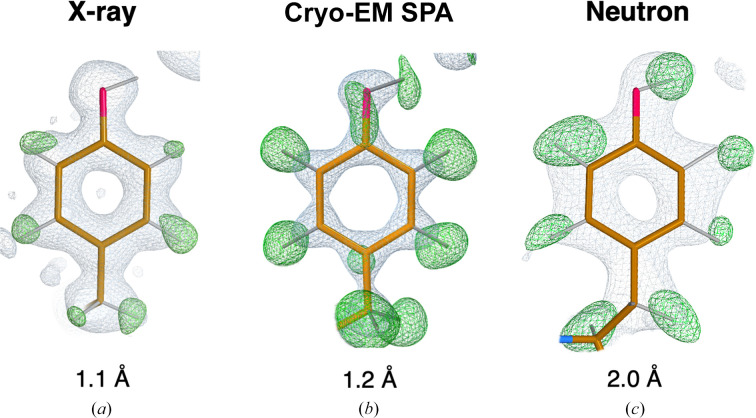
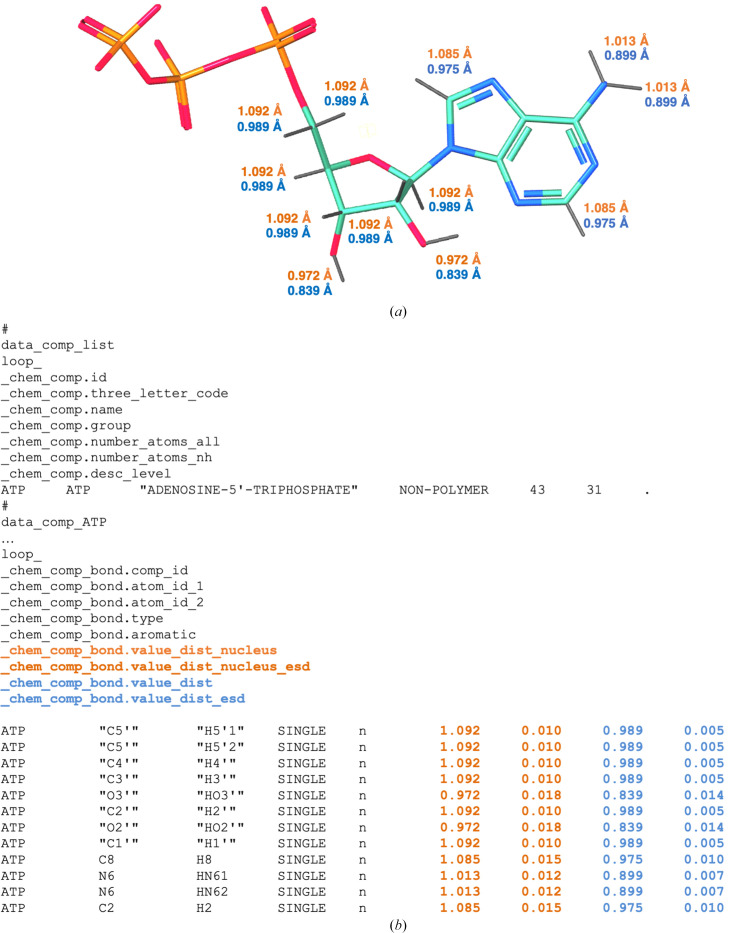
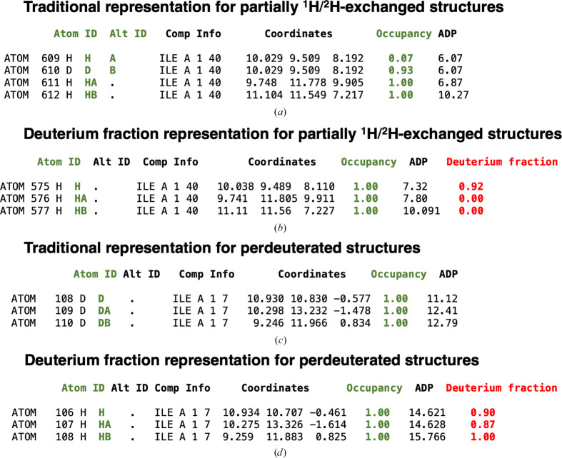
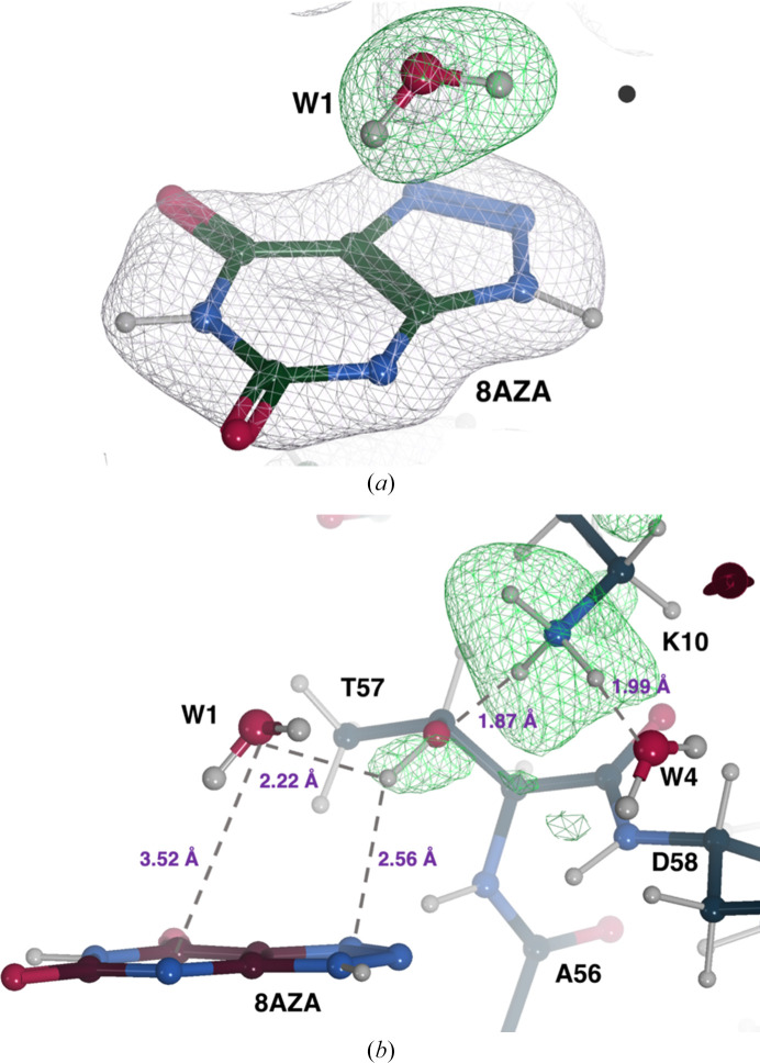
Hydrogen (H) atoms are abundant in macromolecules and often play critical roles in enzyme catalysis, ligand-recognition processes and protein-protein interactions. However, their direct visualization by diffraction techniques is challenging. Macromolecular X-ray crystallography affords the localization of only the most ordered H atoms at (sub-)atomic resolution (around 1.2 Å or higher). However, many H atoms of biochemical significance remain undetectable by this method. In contrast, neutron diffraction methods enable the visualization of most H atoms, typically in the form of deuterium (2H) atoms, at much more common resolution values (better than 2.5 Å). Thus, neutron crystallography, although technically demanding, is often the method of choice when direct information on protonation states is sought. REFMAC5 from the Collaborative Computational Project No. 4 (CCP4) is a program for the refinement of macromolecular models against X-ray crystallographic and cryo-EM data. This contribution describes its extension to include the refinement of structural models obtained from neutron crystallographic data. Stereochemical restraints with accurate bond distances between H atoms and their parent atom nuclei are now part of the CCP4 Monomer Library, the source of prior chemical information used in the refinement. One new feature for neutron data analysis in REFMAC5 is refinement of the protium/deuterium (1H/2H) fraction. This parameter describes the relative 1H/2H contribution to neutron scattering for hydrogen isotopes. The newly developed REFMAC5 algorithms were tested by performing the (re-)refinement of several entries available in the PDB and of one novel structure (FutA) using either (i) neutron data only or (ii) neutron data supplemented by external restraints to a reference X-ray crystallographic structure. Re-refinement with REFMAC5 afforded models characterized by R-factor values that are consistent with, and in some cases better than, the originally deposited values. The use of external reference structure restraints during refinement has been observed to be a valuable strategy, especially for structures at medium-low resolution.

Keywords: CCP4; H atoms; REFMAC5; crystallographic refinement; neutron macromolecular crystallography.


Figures

Figure 1
Figure 1
Map examples. (a) Electron density maps for Tyr12 (PDB entry 3kyu, 1.1 Å resolution) show positive peaks for all aromatic H atoms; however, the H atom on the hydroxyl group is not visible. (b) Cryo-EM maps for Tyr32 (PDB entry 7a4m, 1.22 Å resolution) show positive difference peaks for all H atoms. (c) Neutron scattering length density maps for Tyr146 (PDB entry 1cq2, 2.0 Å resolution) show positive difference peaks for all H atoms (in the form of 2H), including the H atom on the hydroxyl group. Electron and neutron scattering length density maps were calculated using REFMAC5 (Murshudov et al., 2011 ▸), contoured at the +1.0σ (2mF oDF c in grey) and +3.0σ (mF oDF c in green) levels. Cryo-EM weighted and sharpened F o (grey) and omit (F oF c, green) maps were calculated using Servalcat (Yamashita et al., 2021 ▸) and contoured at the +1.5σ and +3.0σ levels, respectively. Molecular-graphics representations were produced with Coot 1.0 (Emsley et al., 2010 ▸).
Figure 2
Figure 2
Example of a dictionary mmCIF file from the updated version of AceDRG. (a) 3D representation of the adenosine triphosphate (ATP) monomer. N, C, O and P atoms are shown in blue, teal, red and orange, respectively. X—H bonds are represented by grey sticks with their nuclear and X-ray diffraction-derived bond lengths (in Å) highlighted in orange and light blue, respectively. (b) Extract from the monomer description of the ATP component dictionary. The category _chem_comp_bond describes the bonded atoms, bond types and the ideal values of bond lengths and uncertainties associated with them. In this example, we show the ideal X—H bond lengths and standard deviations for nucleus positions (_chem_comp_bond.value_dist_nucleus and chem_comp_bond.value_dist_nucleus_esd; orange) and electron positions (_chem_comp_bond.value_dist and _chem_comp_bond.value_dist_esd; light blue).
Figure 3
Figure 3
Comparison between the traditional representation of partially 1H/2H-exchanged structures and perdeuterated structures and the deuterium fraction representation in mmCIF files. (a) Traditional exchangeable 1H/2H sites representation extracted from the mmCIF file of PDB entry 1vcx. The 1H atom bonded to the main-chain N atom of Ile40 is partially exchanged with 2H. 1H and 2H isotopes have separate atom rows in the atom table with alternative locations A and B (green). The sum of their total occupancy (green) is set to 1.0 (the occupancy values of the 1H and 2H atoms are 0.07 and 0.93, respectively). The H atoms bonded to CA and CB of Ile40 are not exchanged during the partial deuteration procedure; their occupancy value is equal to 1.0. (b) Deuterium fraction representation created by REFMAC5. A new column has been created that specifies the fraction of the deuterium substitution (where 100% is fully deuterated) for the exchanged H atoms. 2H atoms are not present in the atom table, only 1H atoms with the corresponding deuterium fraction parameters (red). The 1H atom bonded to the main-chain N atom of Ile40 has a deuterium fraction value of 0.92, while the 1H atoms bonded to CA and CB of Ile40 are not exchanged, hence the deuterium fraction for these H atoms is zero. (c) Traditional perdeuterated sites representation extracted from the mmCIF file of PDB entry 3rz6. Here, all of the H atoms of Ile7 have been substituted with 2H atoms. There are no 1H atoms in the traditional perdeuterated structures. The occupancy of the 2H atoms is set to 1.0 by convention. (d) Deuterium fraction representation for perdeuterated structures. All 2H atoms are converted to 1H atoms, the corresponding deuterium fractions are refined and values close to 1.0 are obtained.
Figure 4
Figure 4
Neutron structure of the UOX:8AZA complex. (a) W1 is present as a neutral H2O molecule. A 2mF oDF c neutron scattering length density map for 8AZA and the O atom of W1 is shown in grey at the +1.0σ level. An omit mF o − DF c neutron map indicates the presence of two deuterons (H1 and H2) as suggested by the elongated positive density (in green at the +3.0σ level) next to the O atom. (b) Representation of a portion of the active site highlighting the protonation of the Lys10–Thr57 dyad and the hydrogen-bonding network. H difference neutron density for Lys10NZ and Thr57OG1 is shown in green at the +3.0σ and +2.5σ levels, respectively. Hydrogen bonds are shown as grey dashed lines and their distances are shown in purple.
Figure 5
Figure 5
FutA (ferric state) determined by neutron diffraction at 2.1 Å resolution. The iron-binding site is formed by four tyrosines (Tyr13, Tyr143, Tyr199 and Tyr200), a solvent molecule (W1) and Arg103 in the second coordination shell. The positive density (green mesh, mF oDF c omit map at the +3.0σ level) indicates that these atoms have undergone 1H/2H exchange and suggests that Arg103 is positively charged whilst W1 is neutral. The side chain of Arg203 is not oriented towards the binding site and does not engage in polar interactions. N, C and O atoms are shown in blue, dark green and red, respectively. Iron is shown in gold and H atoms are in grey.

Similar articles

Cited by

References

    1. Adams, P. D., Mustyakimov, M., Afonine, P. V. & Langan, P. (2009). Acta Cryst. D65, 567–573. - PMC - PubMed
    1. Afonine, P. V., Grosse-Kunstleve, R. W., Echols, N., Headd, J. J., Moriarty, N. W., Mustyakimov, M., Terwilliger, T. C., Urzhumtsev, A., Zwart, P. H. & Adams, P. D. (2012). Acta Cryst. D68, 352–367. - PMC - PubMed
    1. Afonine, P. V., Mustyakimov, M., Grosse-Kunstleve, R. W., Moriarty, N. W., Langan, P. & Adams, P. D. (2010). Acta Cryst. D66, 1153–1163. - PMC - PubMed
    1. Afonine, P. V., Poon, B. K., Read, R. J., Sobolev, O. V., Terwilliger, T. C., Urzhumtsev, A. & Adams, P. D. (2018). Acta Cryst. D74, 531–544. - PMC - PubMed
    1. Agirre, J., Atanasova, M., Bagdonas, H., Ballard, C. B., Baslé, A., Beilsten-Edmands, J., Borges, R. J., Brown, D. G., Burgos-Mármol, J. J., Berrisford, J. M., Bond, P. S., Caballero, I., Catapano, L., Chojnowski, G., Cook, A. G., Cowtan, K. D., Croll, T. I., Debreczeni, J. É., Devenish, N. E., Dodson, E. J., Drevon, T. R., Emsley, P., Evans, G., Evans, P. R., Fando, M., Foadi, J., Fuentes-Montero, L., Garman, E. F., Gerstel, M., Gildea, R. J., Hatti, K., Hekkelman, M. L., Heuser, P., Hoh, S. W., Hough, M. A., Jenkins, H. T., Jiménez, E., Joosten, R. P., Keegan, R. M., Keep, N., Krissinel, E. B., Kolenko, P., Kovalevskiy, O., Lamzin, V. S., Lawson, D. M., Lebedev, A. A., Leslie, A. G. W., Lohkamp, B., Long, F., Malý, M., McCoy, A. J., McNicholas, S. J., Medina, A., Millán, C., Murray, J. W., Murshudov, G. N., Nicholls, R. A., Noble, M. E. M., Oeffner, R., Pannu, N. S., Parkhurst, J. M., Pearce, N., Pereira, J., Perrakis, A., Powell, H. R., Read, R. J., Rigden, D. J., Rochira, W., Sammito, M., Sánchez Rodríguez, F., Sheldrick, G. M., Shelley, K. L., Simkovic, F., Simpkin, A. J., Skubak, P., Sobolev, E., Steiner, R. A., Stevenson, K., Tews, I., Thomas, J. M. H., Thorn, A., Valls, J. T., Uski, V., Usón, I., Vagin, A., Velankar, S., Vollmar, M., Walden, H., Waterman, D., Wilson, K. S., Winn, M. D., Winter, G., Wojdyr, M. & Yamashita, K. (2023). Acta Cryst. D79, 449–461.