. 2023 Dec;39(12):2531-2543.
doi: 10.1007/s10554-023-02978-9. Epub 2023 Nov 3.

CCTA-based CABG SYNTAX Score: a tool to evaluate completeness of coronary segment revascularization after bypass surgery

Affiliations

CCTA-based CABG SYNTAX Score: a tool to evaluate completeness of coronary segment revascularization after bypass surgery

Nozomi Kotoku et al. Int J Cardiovasc Imaging. 2023 Dec.

Abstract

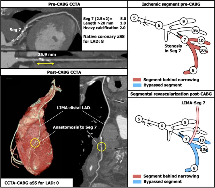
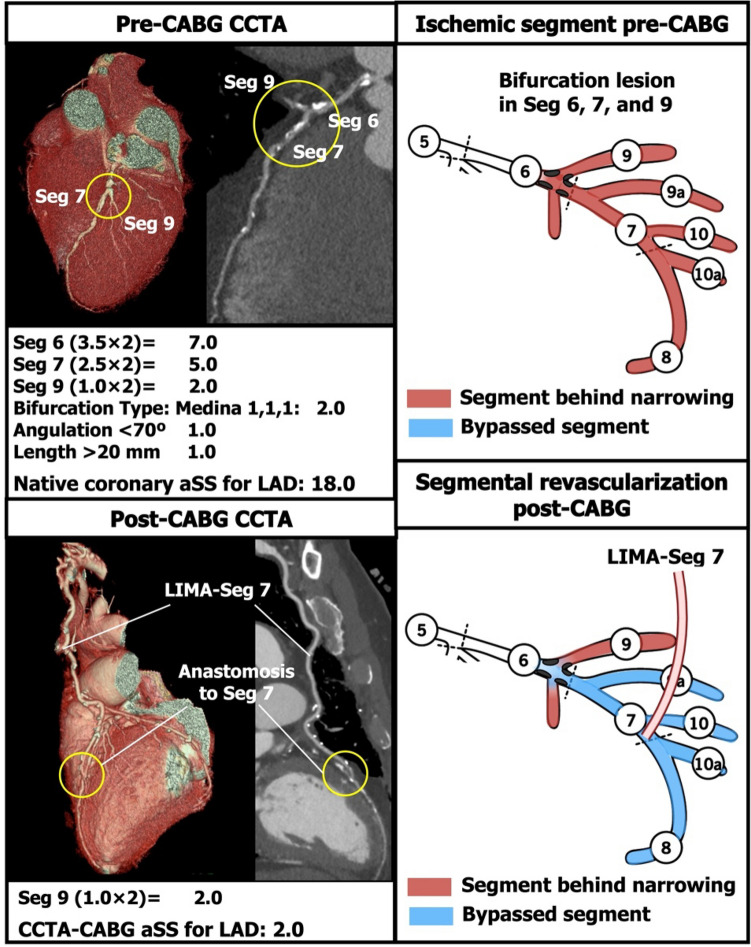
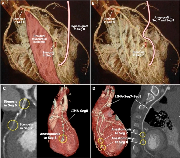
To describe the updated coronary computed tomographic angiography (CCTA)-based coronary artery bypass graft (CABG) anatomic SYNTAX Score (aSS) and assess its utility and reproducibility for assessing the completeness of revascularization after CABG. The CCTA-CABG aSS is a visual assessment using CCTA post-CABG which quantifies the failure in effectively grafting stenotic coronary segments, and therefore assesses the completeness of surgical revascularization. It is calculated by subtracting the aSS of successfully anastomosed coronary segments from the aSS of the native coronary tree. The inter-observer reproducibility of the CCTA-CABG aSS was evaluated in 45 consecutive patients with three-vessel disease with or without left main disease who underwent a CCTA 30 days (± 7 days) after CABG. The CCTA-CABG aSS was evaluated in 45 consecutive patients with 117 bypass grafts and 152 anastomoses. The median native coronary aSS was 35.0 [interquartile range (IQR) 27.0-41.0], whilst the median CCTA-CABG aSS was 13.0 (IQR 9.0-20.5). The inter-observer level of agreement for the native coronary aSS and the CCTA-CABG aSS were both substantial with respective Kappas of 0.67 and 0.61. The CCTA-CABG aSS was feasible in all patients who underwent CABG for complex coronary artery disease with substantial inter-observer reproducibility, and therefore can be used to quantify the completeness of revascularization after CABG.

Keywords: Completeness of revascularization; Coronary artery bypass graft; Coronary artery disease; Coronary computed tomographic angiography; SYNTAX Score.


Conflict of interest statement

Dr Kotoku has received a grant for studying overseas from Fukuda Foundation for Medical Technology. Dr Serruys has received consultancy fees from Philips/Volcano, SMT, Novartis, Xeltis, Merillife. Dr Masuda received a grant from Terumo Corporation outside the submitted work. Dr Narula is on the Scientific Advisory Board of HeartFlow Inc.

Figures

Fig. 1
Fig. 1
Example of points subtracted from the native coronary aSS in a left anterior descending artery (LAD) with a single lesion. If there was a lesion in segment (Seg) 7 and a bypass was placed on the distal LAD, the aSS of Seg 7 would be subtracted. aSS anatomical SYNTAX Score, CABG coronary artery bypass graft, CCTA coronary computed tomographic angiography, LAD left artery descending artery, LIMA left internal mammary artery
Fig. 2
Fig. 2
Example of the points subtracted from the native coronary aSS in a left anterior descending artery (LAD) with sequential lesions. If there were sequential lesions in segment (Seg) 6 and 7, and a bypass graft was anastomosed in the distal LAD, only the aSS of Seg 7 would be subtracted. T.O. total occlusion. Other abbreviations as in Fig. 1
Fig. 3
Fig. 3
Example of subtraction of points from the native coronary aSS in a left circumflex (LCX) with sequential lesions. If sequential lesions in segment (Seg) 11 and Seg 12a were bypassed with a single anastomosis to Seg 12a, the aSS points of Seg 11 remain. Ra radial artery, T.O. total occlusion. Other abbreviations as in Fig. 1
Fig. 4
Fig. 4
Example of subtracted points from the native coronary aSS in a left anterior descending artery (LAD) with a bifurcation lesion. If the bifurcation lesion had a Medina class of 1-1-1 and was bypassed with a single anastomosis to the distal LAD, then the aSS points for the LAD would be deducted, but not those for the diagonal branch. In this case, the remaining points were (1 × 2) = 2 for the stenosis in the diagonal branch. Abbreviations as in Fig. 1
Fig. 5
Fig. 5
A variety of different scenarios showing the points subtracted from the native coronary aSS when an obstructed or occluded native vessel has an occluded, stenosed or patent bypass graft. In cases of an occluded native vessel with a stenosed bypass graft (Panel C), the segment-weighting factor was reduced from ×5 to ×2 to signify the net improvement in flow to the myocardial bed had improved. Abbreviations as in Fig. 1
Fig. 6
Fig. 6
Distribution of the native coronary aSS and CCTA-based CABG aSS. aSS anatomical SYNTAX Score, CABG coronary artery bypass graft, CCTA coronary computed tomographic angiography
Fig. 7
Fig. 7
Difference in residual ischemic burden between a single graft and a jump graft on a left artery descending artery (LAD) with serial lesions. Epicardial arteries (> 400 μm) represent only 5% of the volume of the coronary tree. Pre-arterioles, arterioles, and capillaries represent 95% of the remaining coronary volume, and each coronary segment subtends a well-delineated mass-volume of the myocardium. The post-mortem cast of the coronary circulation depicts the abundance of the microvasculature and helps in understanding the difference in residual myocardial ischemia between a single (A, C) and jump graft (B, D) for an LAD with serial lesions. LIMA left internal mammary artery, Seg segment. The picture on Panel A and B was originally published in Journal of Nuclear Medicine (Camici PG, Rimoldi OE. The Clinical Value of Myocardial Blood Flow Measurement. Journal of Nuclear Medicine 2009;50:1076. © 2009 by the Society of Nuclear Medicine, Inc) [19]
Fig. 8
Fig. 8
Assessment of myocardial blood flow and FFRCT after CABG. A Pre-CABG maximum intensity projection (MIP) and curved multiplanar reconstruction (MPR) images: yellow circles indicate significant stenoses in segments (Seg) 1 and 3. B Simulation of myocardial blood flow to the left ventricle yields a quantitative assessment of the myocardium at risk, and the area subtended by each coronary segment and its side branches. C and D FFRCT values before CABG. E Post-CABG volume rendering (VR) and MPR. F Post-CABG FFRCT based on a simulation of both flows from the aorta and bypass graft anastomosed on Seg 4

References

    1. Farooq V, Girasis C, Magro M, et al. The CABG SYNTAX score-an angiographic tool to grade the complexity of coronary disease following coronary artery bypass graft surgery: from the SYNTAx left main angiographic (SYNTAX-LE MANS) substudy. EuroIntervention. 2013;8:1277–1285. doi: 10.4244/EIJV8I11A196. - DOI - PubMed
    1. Andreini D, Collet C, Leipsic J, et al. Pre-procedural planning of coronary revascularization by cardiac computed tomography: an expert consensus document of the Society of cardiovascular computed tomography. EuroIntervention. 2022;18:e872–e887. doi: 10.4244/EIJ-E-22-00036. - DOI - PMC - PubMed
    1. Serruys WP, Kotoku N, Nørgaard LB, et al. Computed tomographic angiography in coronary artery disease. EuroIntervention. 2023;18:e1307–e1327. doi: 10.4244/EIJ-D-22-00776. - DOI - PMC - PubMed
    1. Serruys PW, Hara H, Garg S, et al. Coronary computed tomographic angiography for complete assessment of coronary artery disease: JACC state-of-the-art review. J Am Coll Cardiol. 2021;78:713–736. doi: 10.1016/j.jacc.2021.06.019. - DOI - PubMed
    1. Papadopoulou SL, Girasis C, Dharampal A, et al. CT-SYNTAX score: a feasibility and reproducibility study. JACC Cardiovasc Imaging. 2013;6:413–415. doi: 10.1016/j.jcmg.2012.09.013. - DOI - PubMed

MeSH terms