. 2023 Nov 28;42(11):113355.
doi: 10.1016/j.celrep.2023.113355. Epub 2023 Nov 1.

Functional screening of amplification outlier oncogenes in organoid models of early tumorigenesis

Affiliations

Functional screening of amplification outlier oncogenes in organoid models of early tumorigenesis

Ameen A Salahudeen et al. Cell Rep. .

Abstract

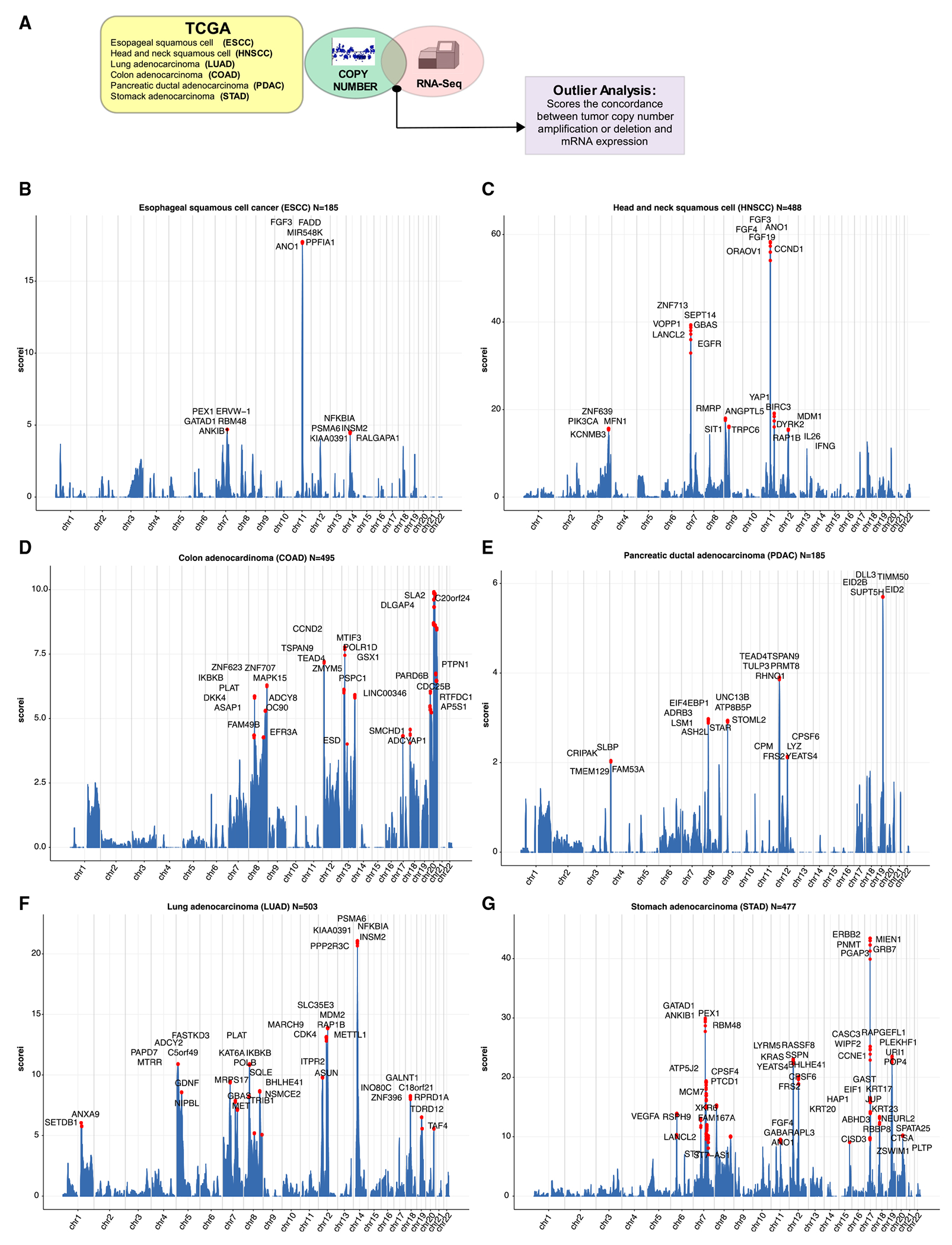
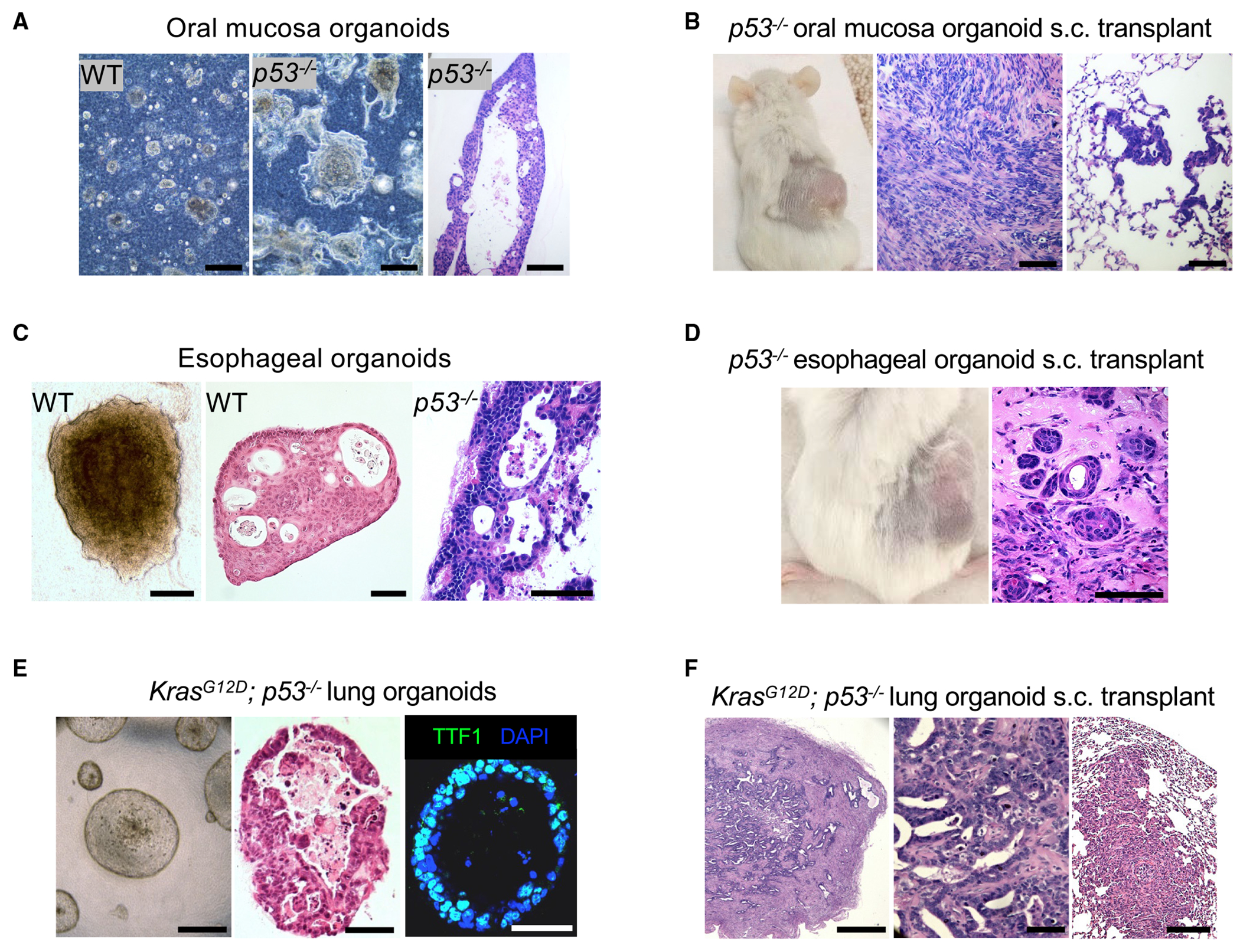
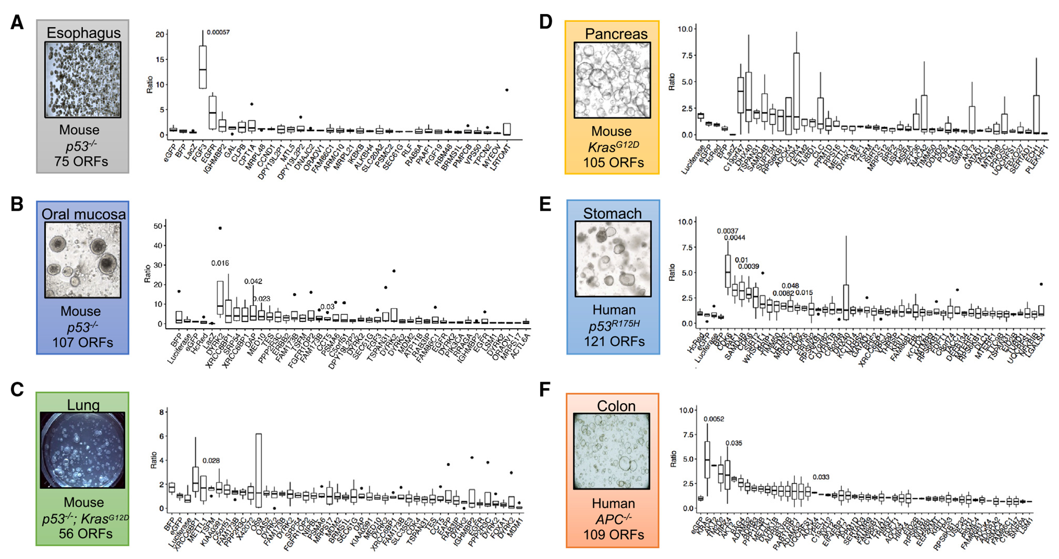
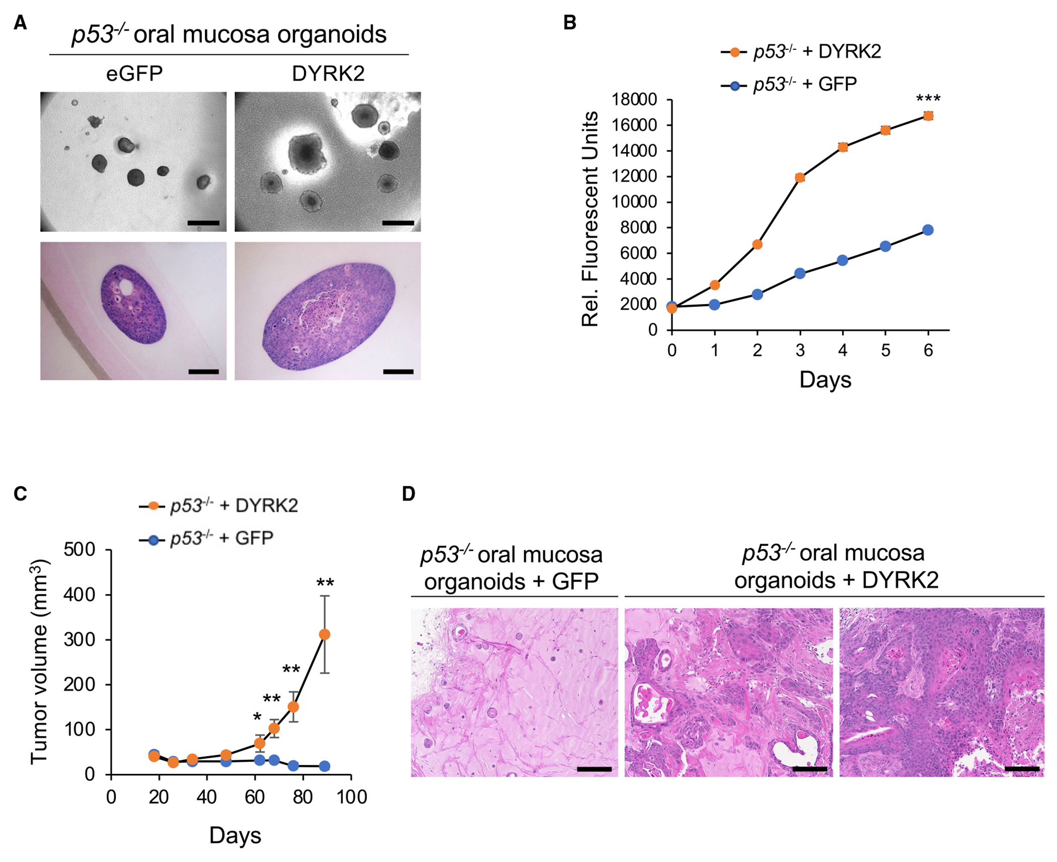
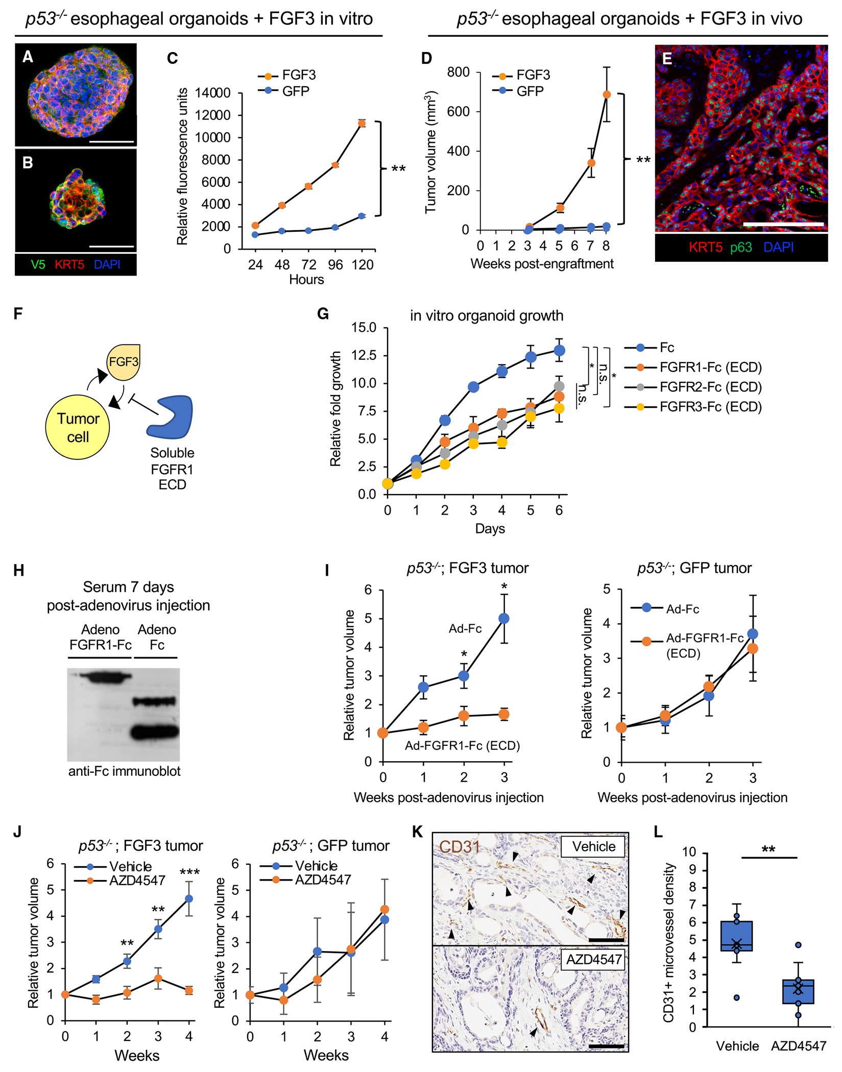
Somatic copy number gains are pervasive across cancer types, yet their roles in oncogenesis are insufficiently evaluated. This inadequacy is partly due to copy gains spanning large chromosomal regions, obscuring causal loci. Here, we employed organoid modeling to evaluate candidate oncogenic loci identified via integrative computational analysis of extreme copy gains overlapping with extreme expression dysregulation in The Cancer Genome Atlas. Subsets of "outlier" candidates were contextually screened as tissue-specific cDNA lentiviral libraries within cognate esophagus, oral cavity, colon, stomach, pancreas, and lung organoids bearing initial oncogenic mutations. Iterative analysis nominated the kinase DYRK2 at 12q15 as an amplified head and neck squamous carcinoma oncogene in p53-/- oral mucosal organoids. Similarly, FGF3, amplified at 11q13 in 41% of esophageal squamous carcinomas, promoted p53-/- esophageal organoid growth reversible by small molecule and soluble receptor antagonism of FGFRs. Our studies establish organoid-based contextual screening of candidate genomic drivers, enabling functional evaluation during early tumorigenesis.

Keywords: CP: Cancer; DYRK2; FGF3; amplification; cancer; cancer driver; copy number alteration; functional genomics; organoid; precision oncology; squamous cancer.


Conflict of interest statement

Declaration of interests A.A.S. has served as a consultant for Boehringer Ingelheim and Pharmacosmos and is a former employee of Tempus Labs. C.J.K. is a scientific advisory board member for Surrozen, Inc., Mozart Therapeutics, and NextVivo. C.C. has served as a scientific advisory board member/consultant for Genentech, Grail, DeepCell, Nanostring, and Viosera. W.C.H. is a consultant for Thermo Fisher, Solasta Ventures, MPM Capital, KSQ Therapeutics, Tyra Biosciences, Frontier Medicine, Jubilant Therapeutics, RAPPTA Therapeutics, Hexagon Bio, Serinus Biosciences, Function Oncology, and Calyx.

Figures

Figure 1.
Figure 1.. Overview of integrative analysis for expression and copy number amplification events as pan-cancer outliers
(A) Schematic of integrative analysis to nominate outlier gene candidates. (B) Genomic landscape of selected TCGA esophageal squamous cell carcinoma (ESCC) outliers where y axis (iscore) represents the number of outliers for each specific gene. (C) Genomic landscape of selected TCGA head and neck squamous cell carcinoma (HNSCC) outliers where y axis (iscore) represents the number of outliers for each specific gene. (D) Genomic landscape of selected TCGA colon adenocarcinoma (COAD) outliers where y axis (iscore) represents the number of outliers for each specific gene. (E) Genomic landscape of selected TCGA pancreatic ductal adenocarcinoma (PDAC) outliers where y axis (iscore) represents the number of outliers for each specific gene. (F) Genomic landscape of selected TCGA NSCLC adenocarcinoma (LUAD) outliers where y axis (iscore) represents the number of outliers for each specific gene. (G) Genomic landscape of selected TCGA stomach adenocarcinoma (STAD) outliers where y axis (iscore) represents the number of outliers for each specific gene.
Figure 2.
Figure 2.. Generation and characterization of murine p53−/− oral mucosa, esophageal, and KrasG12D p53−/− lung adenocarcinoma organoids
(A) Organoid bright-field imaging of wild-type (WT) (left panel) and p53−/− (middle panel) mouse oral mucosa organoids cultured in ALI for 7 days. H&E staining (right panel) of p53−/− mouse oral mucosa organoids cultured in ALI for 34 days. Scale bar: 100 μm. (B) Tumor formation of p53−/− mouse oral mucosa organoids 7 months after implantation (right panel) and H&E of primary tumor (middle panel) and lung metastasis (right panel) (WT; n = 3, p53−/−; n = 3). Scale bar: 100 μm. (C) Bright field (left panel) and H&E staining of wild-type (middle panel) versus p53−/− mouse esophageal organoids (right panel). Scale bar: 250 μm. (D) p53−/− mouse esophageal organoid tumor formation (left panel) and H&E stain (right panel) 6 months post implantation. Scale bar: 250 μm. (E) Phase contrast microscopy (left panel), H&E staining (middle panel), and TTF-1 immunofluorescence (right panel) of KrasG12D; p53−/− mouse lung organoids after 4 weeks of culture in basal F12 medium. Scale bar: 250 μm. (F) H&E of primary tumor (left panel), high magnification (middle panel), and lung metastasis (right panel) 8 weeks post subcutaneous implantation of KrasG12D; p53−/− mouse lung organoids. Scale bars: 500, 100, and 500 μm, respectively.
Figure 3.
Figure 3.. Screening pan-cancer candidate amplification outliers in organoids with corresponding tissue context
(A–F) Barcode ratios of terminal:initial time points from NGS demonstrating relative enrichments from pooled lentiviral ORF screens. Boxplots represent four technical replicates with the exception of (F), which was performed with two technical replicates. Terminal time points are as follows: A = day 53, B = day 52, C = day 32, D = day 50, E = day 55, and F = day 56.
Figure 4.
Figure 4.. DYRK2 and FGF3 overexpression induces proliferation and tumorigenicity of p53−/− mouse oral mucosa and esophageal organoids, respectively
(A) Morphology of p53−/−; eGFP and p53−/−; DYRK2 organoids cultured in ALI for 14 days. Scale bar: bright field; 500 μm (top panel), H&E staining; 100 μm (bottom panel). (B) In vitro proliferation of p53−/−; eGFP and p53−/−; DYRK2 organoids generated from oral mucosal tissue from a different donor mouse in ALI assessed by resazurin reduction. Data represent mean ± SEM. ***p < 0.001, two-tailed Student’s t test. (C) Tumor volume of p53−/−; eGFP (n = 6) and p53−/−; DYRK2 (n = 7) organoids. Data represent mean ± SEM. *p < 0.05, **p < 0.01, two-tailed Student’s t test. (D) H&E staining of p53−/−; eGFP and p53−/−; DYRK2 organoid tumors. Scale bar: 100 μm.
Figure 5.
Figure 5.. Targeting oncogenic FGF3 amplification with soluble FGFR-ECD or FGFR-specific tyrosine kinase inhibitors in p53−/− mouse esophageal squamous organoids
(A) Multicolor immunofluorescence of p53−/− esophageal organoids with lentiviral expression of FGF3 with a C-terminal V5 tag. Scale bar: 100 μm. (B) Multicolor immunofluorescence of p53−/− esophageal organoids with lentiviral expression of GFP with a C-terminal V5 tag. Scale bar: 100 μm. (C) In vitro proliferation of FGF3 versus GFP-expressing p53−/− esophageal organoids by resazurin reduction. Data represent mean ± SEM. **p < 0.01, two-tailed Student’s t test. (D) In vivo tumor formation and growth upon subcutaneous transplantation of FGF3- versus GFP-expressing p53−/− esophageal organoids in immunodeficient NOG mice. Each group has n = 10 biological replicate mice. Data represent mean ± SEM. *p < 0.05, **p < 0.01, two-tailed Student’s t test. (E) Multicolor immunofluorescence of p53−/−;FGF3 esophageal organoid subcutaneous tumor sections. Scale bar: 100 μm. (F) Schematic of inhibition of FGF3-driven oncogenesis by soluble ligand-binding extracellular domains of FGF receptors. (G) Proliferation of FGF3 p53−/− esophageal organoids upon incubation with Fc or FGFR 1, 2, or 3 ECD-Fc fusions at 10 μg/mL assessed by resazurin reduction. Data represent mean ± SEM. *p < 0.05, two-tailed Student’s t test. (H) Circulating serum expression of FGFR1-Fc fusion protein after intravenous injection of immunodeficient NSG mice infected with the corresponding recombinant adenoviruses. Western blot, anti-Fc. (I) Serial measurements of subcutaneous tumor growth subsequent to adenoviral infection of FGFR1 ECD-Fc versus Fc controls in FGF3-expressing (n = 6) (left panel) or GFP-expressing (n = 5) (right panel) p53−/− esophageal organoid subcutaneous tumors. Data represent mean ± SEM. *p < 0.05, two-tailed Student’s t test. (J) Serial measurements of FGF3-expressing (left panel) or GFP-expressing (right panel) p53−/− esophageal organoid subcutaneous tumors subsequent to daily oral administration of vehicle or the FGFR inhibitor AZD4547. FGF3-expressing organoids have n = 9 biological replicate mice in each treatment group. GFP-expressing organoids have n = 8 in the vehicle treatment group and n = 7 in the AZD4547 treatment group. **p < 0.01, ***p < 0.001, two-tailed Student’s t test. (K) Representative CD31 immunohistochemistry staining of subcutaneous tumors from FGF3-expressing p53−/− esophageal organoids in (E). Scale bar: 200 μm. (L) Chalkley quantitation of CD31+ microvessel density in (F), where each group is three technical replicates for each mouse subcutaneous tumor. **p < 0.01, two-tailed Student’s t test.

References

    1. Beroukhim R, Mermel CH, Porter D, Wei G, Raychaudhuri S, Donovan J, Barretina J, Boehm JS, Dobson J, Urashima M, et al. (2010). The landscape of somatic copy-number alteration across human cancers. Nature 463, 899–905. 10.1038/nature08822. - DOI - PMC - PubMed
    1. Zack TI, Schumacher SE, Carter SL, Cherniack AD, Saksena G, Tabak B, Lawrence MS, Zhsng CZ, Wala J, Mermel CH, et al. (2013). Pan-cancer patterns of somatic copy number alteration. Nat. Genet 45, 1134–1140, [doi]. 10.1038/ng.2760. - DOI - PMC - PubMed
    1. Oh DY, and Bang YJ (2020). HER2-targeted therapies - a role beyond breast cancer. Nat. Rev. Clin. Oncol 17, 33–48. 10.1038/s41571-019-0268-3. - DOI - PubMed
    1. Kryukov GV, Wilson FH, Ruth JR, Paulk J, Tsherniak A, Marlow SE, Vazquez F, Weir BA, Fitzgerald ME, Tanaka M, et al. (2016). MTAP deletion confers enhanced dependency on the PRMT5 arginine methyltransferase in cancer cells. Science 351, 1214–1218. 10.1126/science.aad5214. - DOI - PMC - PubMed
    1. Huang A, Garraway LA, Ashworth A, and Weber B (2020). Synthetic lethality as an engine for cancer drug target discovery. Nat. Rev. Drug Discov 19, 23–38. 10.1038/s41573-019-0046-z. - DOI - PubMed

Publication types

Substances