. 2023 Nov 3;14(1):7068.
doi: 10.1038/s41467-023-42683-8.

Structural basis for the toxicity of Legionella pneumophila effector SidH

Affiliations

Structural basis for the toxicity of Legionella pneumophila effector SidH

Rahul Sharma et al. Nat Commun. .

Abstract

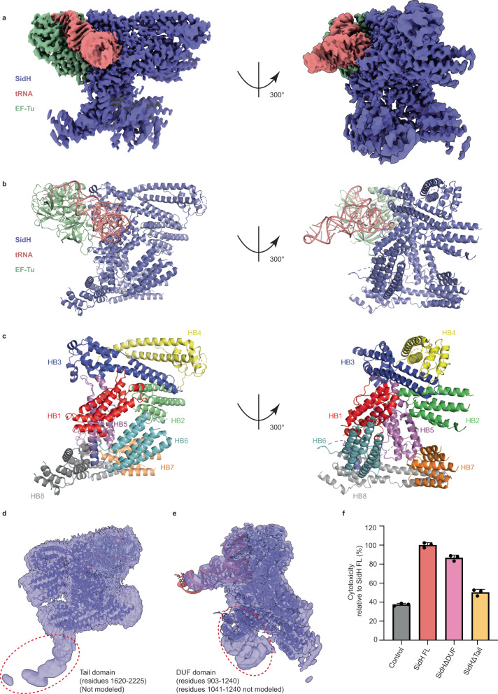
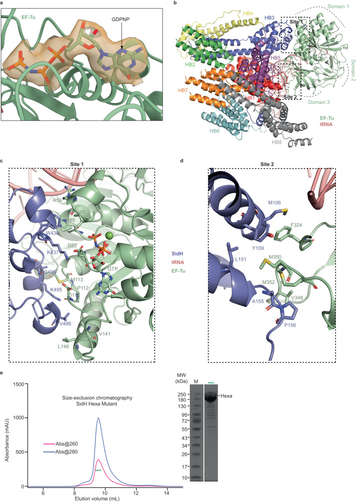
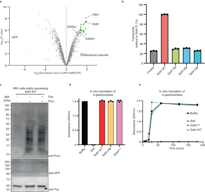
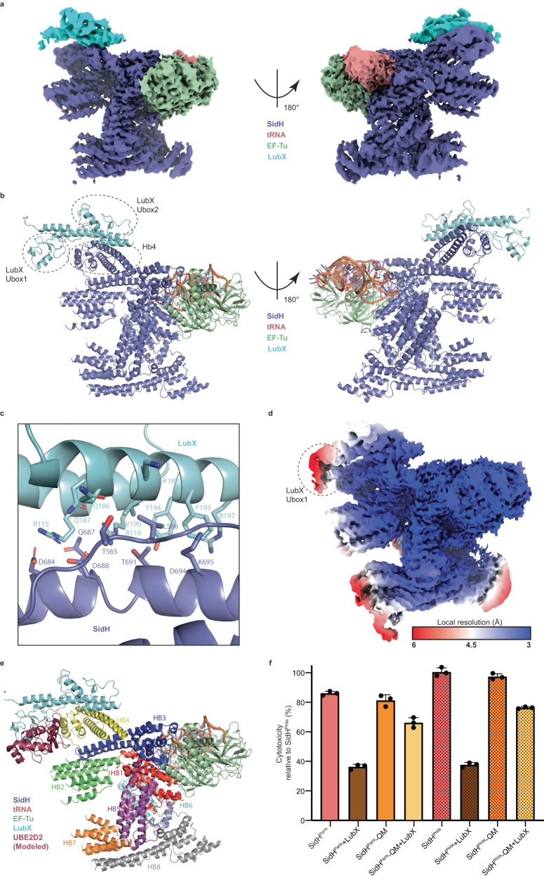
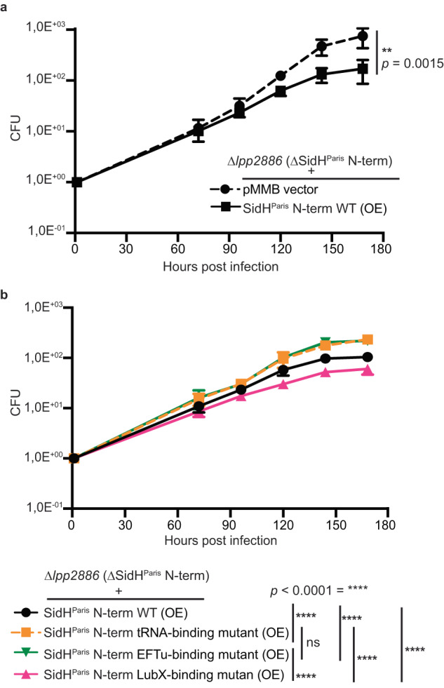
Legionella pneumophila (LP) secretes more than 300 effectors into the host cytosol to facilitate intracellular replication. One of these effectors, SidH, 253 kDa in size with no sequence similarity to proteins of known function is toxic when overexpressed in host cells. SidH is regulated by the LP metaeffector LubX which targets SidH for degradation in a temporal manner during LP infection. The mechanism underlying the toxicity of SidH and its role in LP infection are unknown. Here, we determined the cryo-EM structure of SidH at 2.7 Å revealing a unique alpha helical arrangement with no overall similarity to known protein structures. Surprisingly, purified SidH came bound to a E. coli EF-Tu/t-RNA/GTP ternary complex which could be modeled into the cryo-EM density. Mutation of residues disrupting the SidH-tRNA interface and SidH-EF-Tu interface abolish the toxicity of overexpressed SidH in human cells, a phenotype confirmed in infection of Acanthamoeba castellani. We also present the cryo-EM structure of SidH in complex with a U-box domain containing ubiquitin ligase LubX delineating the mechanism of regulation of SidH. Our data provide the basis for the toxicity of SidH and into its regulation by the metaeffector LubX.


Conflict of interest statement

The authors declare no competing interests.

Figures

Fig. 1
Fig. 1. Overall structure of SidH in complex with tRNAPhe and EF-Tu.
a A cryo-EM map of the complex showing densities for SidH (blue), tRNAPhe (pink), and EF-Tu (green). b A complete model of the complex showing cartoon representation of SidH (blue), tRNAPhe (pink), and EF-Tu (green). The residues not modeled in the structure are shown by dashed lines. c The model of SidH is colored according to the distinct helical bundles (Hb1-Hb8). d Unsharpened EM map shows poor resolution in the region of Tail domain (circled with red dotted line). The SidH residues (1620-2225) could not be modeled in this region. e Unsharpened EM map shows poor resolution in the region of DUF domain (circled with red dotted line), which includes residues from 903 to 1240 of SidH. Although an AlphaFold model for the residues 903-1040 could be fitted, an almost 2/3rd part (1041-1240) of this region could not be modeled. f Cytotoxicity assay shows toxicity levels of SidHΔDUF and SidHΔTail relative to the toxicity of SidH full-length (FL). Data represents the mean ± SD of three independent reactions. Source data are provided as a Source Data file.
Fig. 2
Fig. 2. SidH binding to tRNA is sequence independent.
a The pie chart shows different type of tRNAs came in bound state to purified SidH from E. coli. tRNA isotypes with their percentage of abundance and color scheme (right panel). Source data are provided as a Source Data file. b The electrostatic potential surface projection of SidH shows that T-loop of tRNA (pink) inserts into positively charged groove (blue) of SidH. c The cartoon representation of the complex shows that the binding groove for tRNA is composed of distinct helical bundles colored and labeled as explained in the main text. d The detailed view of tRNA-binding region of SidH shows that the side chains of positively charged residues are pointed towards negatively charged phosphodiester backbone of tRNA to make ionic interactions.
Fig. 3
Fig. 3. Binding interface between SidH and EF-Tu.
a A molecule of GDPNP (stick representation) bound to EF-Tu (cartoon). The electron density for the GDPNP molecule is shown. b Extensive interaction interface between SidH and EF-Tu is shown in black dotted rectangles as site1 and site2. c Detailed view of interactions at site1- key residues (stick representation) which are involved in interaction between domain1 of EF-Tu (green) and Hb3 of SidH (purple) are shown. d Similar view as panel C highlighting the interactions at site2- interaction between domain 2 of EF-Tu (green) and Hb1 of SidH (blue). Stick representation of key residues are shown. e Size-exclusion chromatography (SEC) of Hexa mutant (a tRNA binding deficient mutant of SidH)- the ratio of absorbance (260/280 nm) shows the absence of tRNA. The labelled peak fraction (a horizontal green bar) was loaded on SDS-PAGE gel (right panel), which shows absence of EF-Tu as well, as there is no protein band at around 43 kDa of molecular weight marker. Source data are provided as a Source Data file.
Fig. 4
Fig. 4. Split SidH in Legionella pneumophila Paris strain.
a Schematic representation of split SidH gene- lpp2883 encodes for C-terminal (SidHParis C-term) while lpp2886 encodes for N-terminal (SidHParis C-term) of SidH from strain Paris. lpp2884 and 2885 are the two transposons. b Analytical SEC shows interaction between N-terminal and C-terminal of SidHParis. The highlighted fraction (green horizontal bar) of SidHParis(N-term + C-term) (blue colored curve) sample is shown on SDS-PAGE (right panel). c Cytotoxicity assay in HEK293T cells shows toxicity levels of different constructs of SidHParis relative to the toxicity of SidH FL from Philadelphia strain. Data represents the mean ± SD of three independent reactions. d Analytical SEC shows that SidHParis N-term apo protein has more affinity for pre-formed complex of EF-Tu/tRNA. Fractions of peak1, and 2 from each chromatogram were loaded onto SDS-PAGE (right panel). Source data are provided as a Source Data file for (bd).
Fig. 5
Fig. 5. SidH does not affect host protein synthesis.
a GFP-SidH or GFP alone was expressed in HEK293T cells and isolated proteins were subjected to quantitative mass spectrometry analysis. A volcano plot is shown here for the GFP-SidH interaction proteomics. n = 3 biologically independent experiments. A protein was annotated as a hit with a false discovery rate (fdr) smaller than 5 % and a fold-change of at least 100% and as a candidate with a fdr below 20 % and a fold-change of at least 50 %. b The quantification of cell death shows that TM, HM and OM are not toxic in comparison to the SidH WT. Data represents the mean ± SD of three independent reactions. (SidH WT represents SidH from strain Philadelphia in the Fig. 5b–e) c HEK Cells stably expressing GFP-SidH WT subjected to puromycin incorporation analyses. Doxycyclin was used to induce the expression of GFP-SidH WT. Immunoblotting of lysates was performed using antibodies against puromycin, GFP and Tubulin. d Cell free translation of β-galactosidase in human cell extracts in the presence of purified His-tagged SidI/SidH WT/SidH HM/SidHParis. The reactions were carried out for 30 min. Graph depicts the activity of synthesized β-galactosidase on its substrate ONPG. Data represents mean ± standard error of the mean; n = 3 replicates per treatment condition. e Cell free translation of β-galactosidase in the presence of different proteins shows that SidH (both from Paris and Philadelphia strains) is unable to repress the host protein synthesis at different time points. Source data are provided as a Source Data file for (be).
Fig. 6
Fig. 6. LubX binds to N-terminal of SidH.
a The EM map for LubX in complex with SidH/EF-Tu/tRNA. The density for LubX is shown in Cyan. The rest of the color coding is same as in Fig. 1. b The model for the complex shows that Ubox2 of LubX interacts with Hb4 of SidH. c The detailed view of SidH-LubX interaction interface shows stick representation of key residues involved in the interaction. d Local resolution cryo-EM map shows poor electron density for the Ubox1 of LubX (circled). e Modeled UBE2D2 (E2-enzyme) lies near the Hb2 of SidH. f The cytotoxicity assay with SidH or SidH QM and LubX constructs expressed in HEK293T cells validates the interaction surface between SidH and LubX. Data represents the mean ± SD of three independent reactions. Source data are provided as a Source Data file.
Fig. 7
Fig. 7. SidH, tRNA/EF-Tu binding is relevant in infection.
Intracellular growth kinetics of L. pneumophila in Acanthamoeba castellani. The number of viable bacteria within amoebae was evaluated by the standard plate count assay. Each time point represents the mean of three biological replicates. Infections were performed at 20 °C. a Infection of Acanthamoeba castellani with Δlpp2886 L. pneumophila Paris strain complemented with empty pMMB vector or with the one expressing SidHParis N-term WT. b Infection of Acanthamoeba castellani with Δlpp2886 L. pneumophila Paris strain complemented with the indicated mutants (black- WT; yellow- tRNA-binding mutant; green- EFTu-binding mutant and pink- LubX-binding mutant). Data presented here in Fig. 7 is represented by mean ± SD of three independent replicates. Statistical analysis used two-way ANOVA, with reported P values for significant comparisons. (p < 0.0001 - ****), ns- non-significant. Source data are provided as a Source Data file.

References

    1. Fields BS. The Molecular ecology of Legionellae. Trends Microbiol. 1996;4:286–290. doi: 10.1016/0966-842X(96)10041-X. - DOI - PubMed
    1. Mondino S, et al. Legionnaires’ Disease: State of the Art Knowledge of Pathogenesis Mechanisms of Legionella. Annu. Rev. Pathol. Mech. Dis. 2020;15:439–466. doi: 10.1146/annurev-pathmechdis-012419-032742. - DOI - PubMed
    1. Escoll P, Mondino S, Rolando M, Buchrieser C. Targeting of host organelles by pathogenic bacteria: A Sophisticated subversion strategy. Nat. Rev. Microbiol. 2016;14:5–19. doi: 10.1038/nrmicro.2015.1. - DOI - PubMed
    1. Sahr T, et al. Translocated Legionella Pneumophila Small RNAs Mimic Eukaryotic MicroRNAs Targeting the host immune response. Nat. Commun. 2022;13:762. doi: 10.1038/s41467-022-28454-x. - DOI - PMC - PubMed
    1. O’Connor TJ, Boyd D, Dorer MS, Isberg RR. Aggravating genetic interactions allow a solution to redundancy in a bacterial pathogen. Science. 2012;338:1440–1444. doi: 10.1126/science.1229556. - DOI - PMC - PubMed

Publication types

MeSH terms

Substances