. 2023 Nov 3;13(1):18993.
doi: 10.1038/s41598-023-46395-3.

Expression of G protein-coupled receptor GPR19 in normal and neoplastic human tissues

Affiliations

Expression of G protein-coupled receptor GPR19 in normal and neoplastic human tissues

Lorena Gerlach et al. Sci Rep. .

Abstract

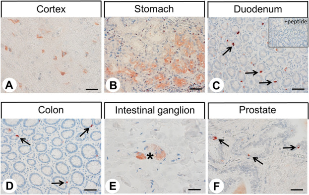
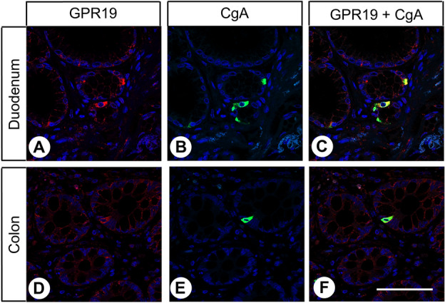
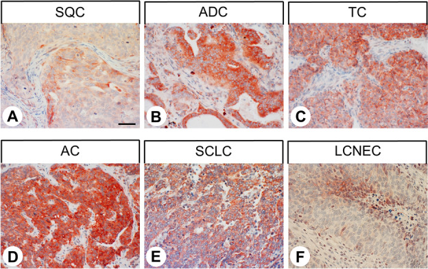
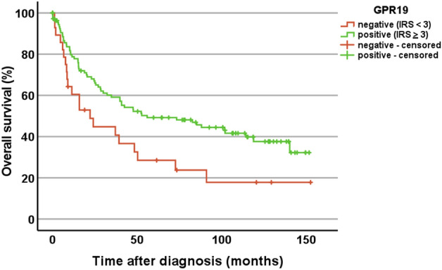
Little is known about the expression of the orphan G protein-coupled receptor GPR19 at the protein level. Therefore, we developed a rabbit antibody, targeting human GPR19. After verification of the antibody specificity using GPR19-expressing cell lines and a GPR19-specific siRNA, the antibody was used for immunohistochemical staining of a variety of formalin-fixed, paraffin-embedded normal and neoplastic human tissue samples. In normal tissues, GPR19 expression was detected in a distinct cell population within the cortex, in single cells of the pancreatic islets, in intestinal ganglia, gastric chief cells, and in endocrine cells of the bronchial tract, the gastrointestinal tract, and the prostate. Among the 30 different tumour entities investigated, strong GPR19 expression was found in adenocarcinomas, typical and atypical carcinoids of the lung, and small cell lung cancer. To a lesser extent, the receptor was also present in large cell neuroendocrine carcinomas of the lung, medullary thyroid carcinomas, parathyroid adenomas, pheochromocytomas, and a subpopulation of pancreatic neuroendocrine neoplasms. In lung tumours, a negative correlation with the expression of the proliferation marker Ki-67 and a positive interrelationship with patient survival was observed. Overall, our results indicate that in adenocarcinomas and neuroendocrine tumours of the lung GPR19 may serve as a suitable diagnostic or therapeutic target.


Conflict of interest statement

Daniel Kaemmerer received support for travelling to meetings by the companies IPSEN and PFIZER. Stefan Schulz is the founder and scientific advisor of 7TM Antibodies GmbH, Jena, Germany, and declares no competing non-financial interests but competing financial interests. All other authors declare that there is no conflict of interest that could be perceived as prejudicing the impartiality of the research reported.

Figures

Figure 1
Figure 1
Verification of the specificity of the anti-GPR19 antibody by immunocytochemistry analyses. (A) OH-1 cells or (D) NCI-h82 cells expressing GPR19 endogenously were fixed and stained with the anti-GPR19 antibody, followed by an Alexa Fluor 488-conjugated anti-rabbit secondary antibody. (B, E) For analysis of the specificity of the antibody, GPR19 expression was silenced in OH-1 cells or NCI-h82 cells using a GPR19-specific siRNA. (C, F) For adsorption controls, the anti-GPR19 antibody was preincubated for 2 h with 10 µg/ml of the peptide used for immunisations of the rabbits. Green, GPR19 immunosignal; blue, 4′,6-diamidino-2-phenylindole (DAPI) staining of DNA. Scale bar, 50 µm (AF).
Figure 2
Figure 2
Verification of the specificity of the anti-GPR19 antibody by Western blot analyses. Left lane: Western blot analysis of membrane preparations of OH-1 cells that endogenously express GPR19 (“control”). Middle lane: Western blot analysis of membrane preparations of OH-1 cells after transfection with siRNA targeting GPR19 (“+ siRNA”). Right lane: For adsorption control, the antibody was preincubated for 2 h with 10 µg/ml of the immunising peptide (“+ peptide”). Ladder indicates migration of protein molecular weight markers (kDa). All results are representative of three independent experiments.
Figure 3
Figure 3
Immunohistochemical detection of GPR19 localisation in normal human tissues. Immunohistochemical staining (red-brown colour) and counterstaining with haematoxylin. Scale bar, 30 µm (A,B,E), 50 µm (C,D,F). Inset in (C) adsorption control, in which the anti-GPR19 antibody was preincubated for 2 h with the peptide used to immunise the rabbits (+ peptide). Arrows in (C,D,F), endocrine cells; asterisk in (E), intestinal ganglion.
Figure 4
Figure 4
Immunohistochemical detection of GPR19 localisation in human tumour entities. Immunohistochemical staining (red-brown colour) and counterstaining with haematoxylin. Scale bar, 50 µm (AF). Insets in (AF) represent adsorption controls, in which the anti-GPR19 antibody was preincubated for 2 h with the peptide used to immunise the rabbits (+ peptide).
Figure 5
Figure 5
Double-labelling immunohistochemical analysis of GPR19 expression and the expression of chromogranin A (CgA) in human duodenum and colon tissue. Labelling of GPR19 was visualised using Cy3-conjugated goat anti-rabbit antibody (red). Labelling of CgA was visualised using Alexa Fluor 488-conjugated donkey anti-mouse antibody (green). Overlapping expression is represented by orange/yellow colour. Blue colour represents 4′,6-diamidino-2-phenylindole (DAPI)-stained DNA. Scale bar: 100 µm.
Figure 6
Figure 6
GPR19 expression pattern in different lung tumour entities. Immunohistochemical staining (red-brown colour) and counterstaining with haematoxylin. Scale bar, 50 µm (AF).
Figure 7
Figure 7
Expression profile of GPR19 in different lung tumour entities. (A) Percentage of GPR19-positive cases within the different lung tumour entities. Tumours were only considered positive with Immunoreactivity Score (IRS) values ≥ 3. (B) GPR19 expression levels (IRS values) in the different lung tumour entities. Median values, upper and lower quartiles, minimum and maximum values, are shown. SQC, squamous cell carcinomas of the lung; ADC, adenocarcinomas of the lung; TC, typical carcinoids of the lung; AC, atypical carcinoids of the lung; SCLC, small cell lung cancers; LC-NEC, large cell neuroendocrine carcinomas of the lung.
Figure 8
Figure 8
Overall survival of patients with GPR19-positive (IRS ≥ 3) or -negative lung tumours. Log-rank test, p = 0.020. Censored: for the Kaplan–Meier curves, the small vertical ticks mark individual patients whose survival times have been “right censored” because they were still alive at the end of the observation period.

References

    1. Odoemelam CS, Percival B, Wallis H, Chang MW, Ahmad Z, Scholey D, Burton E, Williams IH, Kamerlin CL, Wilson PB. G-Protein coupled receptors: Structure and function in drug discovery. RSC Adv. 2020;10:36337. doi: 10.1039/D0RA08003A. - DOI - PMC - PubMed
    1. Yang D, Zhou Q, Labroska V, Qin S, Darbalaei S, Wu Y, Yuliantie E, Xie L, Tao H, Cheng J, Liu Q, Zhao S, Shui W, Jiang Y, Wang MW. G protein-coupled receptors: Structure- and function-based drug discovery. Signal Transduct. Target. Ther. 2021;6:7. doi: 10.1038/s41392-020-00435-w. - DOI - PMC - PubMed
    1. Tang XL, Wang Y, Li DL, Luo J, Liu MY. Orphan G protein-coupled receptors (GPCRs): Biological functions and potential drug targets. Acta Pharmacol. Sin. 2012;33:363–371. doi: 10.1038/aps.2011.210. - DOI - PMC - PubMed
    1. Stockert JA, Devi LA. Advancements in therapeutically targeting orphan GPCRs. Front. Pharmacol. 2015;6:100. doi: 10.3389/fphar.2015.00100. - DOI - PMC - PubMed
    1. O’Dowd BF, et al. A novel gene codes for a putative G protein-coupled receptor with an abundant expression in brain. FEBS Lett. 1996;394:325–329. doi: 10.1016/0014-5793(96)00901-5. - DOI - PubMed

Publication types

Substances