. 2023 Nov 3;14(1):7038.
doi: 10.1038/s41467-023-42927-7.

Structure of a membrane-bound menaquinol:organohalide oxidoreductase

Affiliations

Structure of a membrane-bound menaquinol:organohalide oxidoreductase

Lorenzo Cimmino et al. Nat Commun. .

Abstract

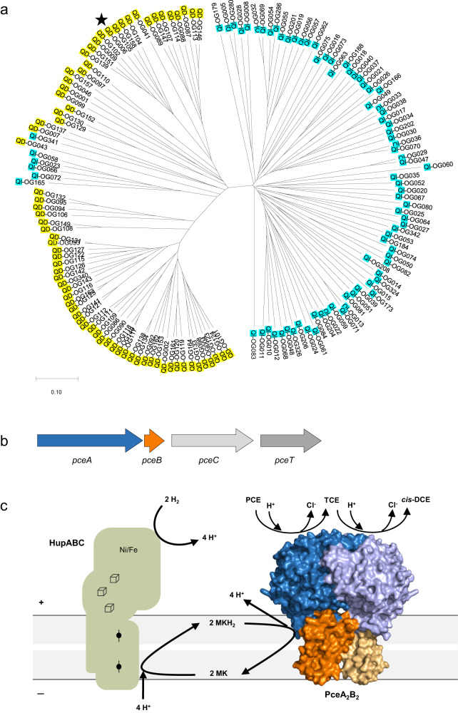
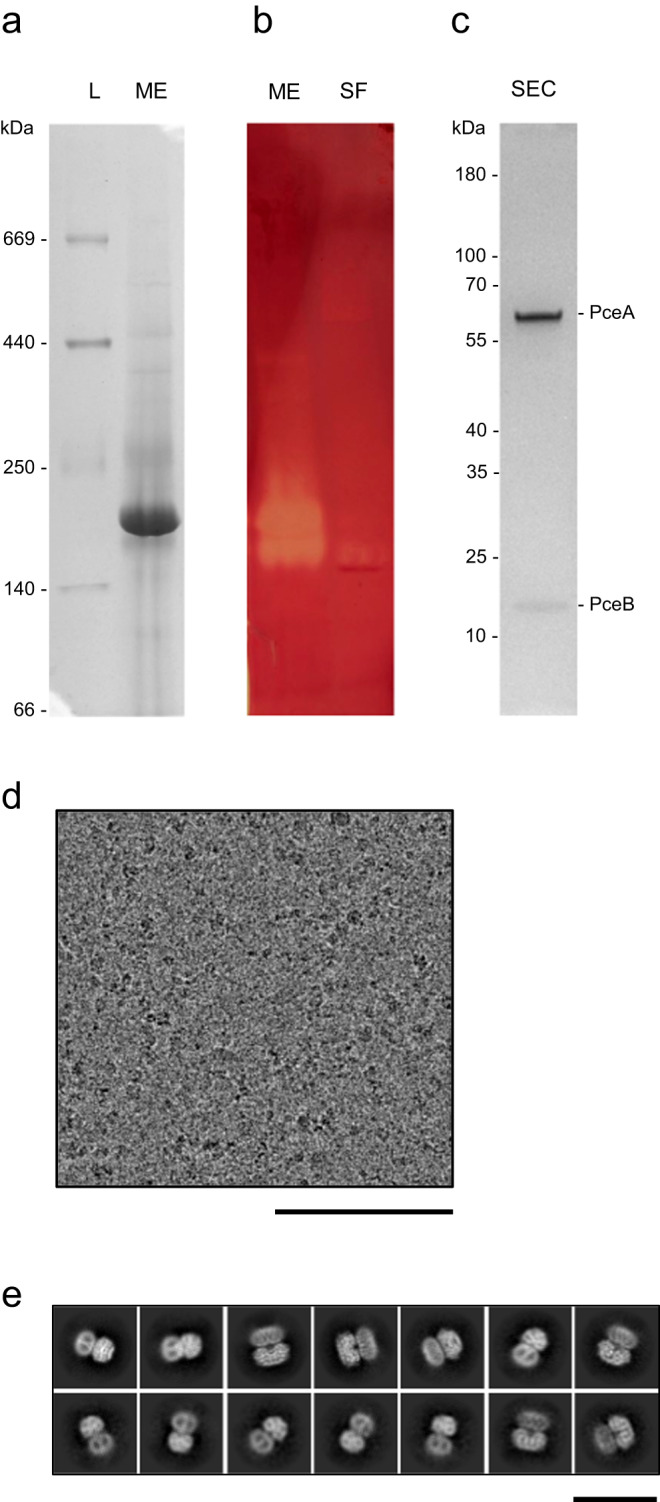
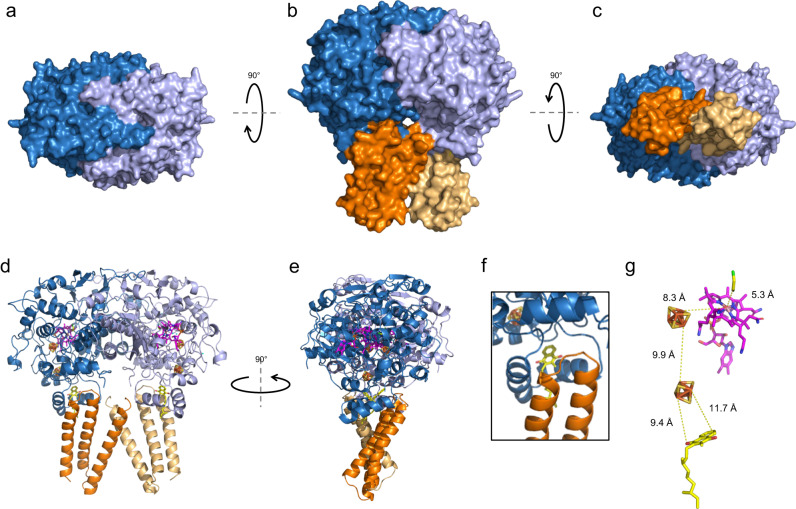
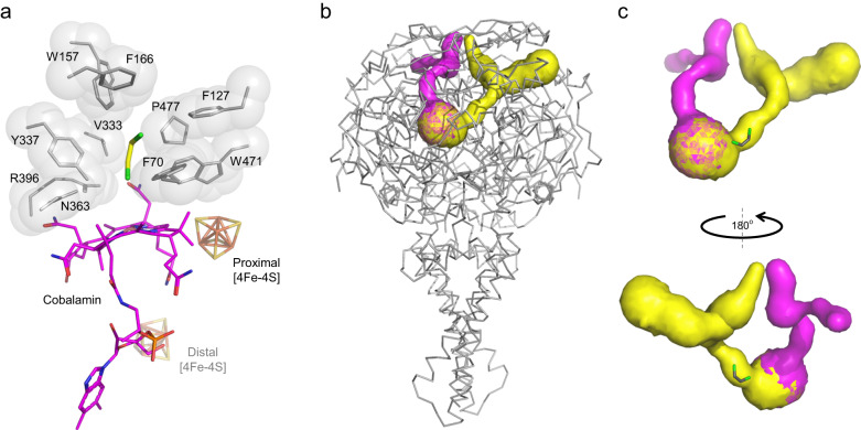
Organohalide-respiring bacteria are key organisms for the bioremediation of soils and aquifers contaminated with halogenated organic compounds. The major players in this process are respiratory reductive dehalogenases, corrinoid enzymes that use organohalides as substrates and contribute to energy conservation. Here, we present the structure of a menaquinol:organohalide oxidoreductase obtained by cryo-EM. The membrane-bound protein was isolated from Desulfitobacterium hafniense strain TCE1 as a PceA2B2 complex catalysing the dechlorination of tetrachloroethene. Two catalytic PceA subunits are anchored to the membrane by two small integral membrane PceB subunits. The structure reveals two menaquinone molecules bound at the interface of the two different subunits, which are the starting point of a chain of redox cofactors for electron transfer to the active site. In this work, the structure elucidates how energy is conserved during organohalide respiration in menaquinone-dependent organohalide-respiring bacteria.


Conflict of interest statement

The authors declare no competing interests.

Figures

Fig. 1
Fig. 1. Reductive dehalogenases.
a Phylogenetic tree of RdhA proteins. The star indicates PceA from Desulfitobacterium hafniense strain TCE1. b The pce gene cluster from D. hafniense strain TCE1. c Model for the electron transfer chain in organohalide respiration by quinone-dependent bacteria growing with H2 and PCE as electron donor and acceptor, respectively. The model displays the PceA2B2 reductive dehalogenase complex obtained in the present study. Legend: OG, orthologous group (as defined earlier); QD: RdhA sequences from quinone-dependent bacteria; QI: RdhA sequences from quinone-independent bacteria; PCE, tetrachloroethene; TCE, trichloroethene; cis-DCE, cis-dichloroethene; Hup: uptake hydrogenase.
Fig. 2
Fig. 2. Reductive dehalogenase complex sample characterisation.
a, b One representative Clear-Native PAGE gel of the membrane extract (ME) and soluble fraction (SF): a stained by Coomassie and b stained by the in-gel enzymatic activity. Lanes represented in a and b were issued from the same gel and cut for staining. This experiment has been performed three additional times with similar results. c SDS-PAGE of the major protein peak after size-exclusion chromatography (SEC) of the ME sample. This experiment has been performed once. d One representative cryo-EM micrograph (out of 13783 images), scale bar: 100 nm. e Selected class averages of particles of the complex, scale bar: 20 nm. Source data are provided as a Source Data file.
Fig. 3
Fig. 3. Cryo-EM structure of the PceA2B2 complex from D. hafniense strain TCE1.
Representation of the surface of the complex from a the top, b the front and c the bottom view. Cartoon representations of d the front and e the side view of the protein complex. f Detail of the menaquinone-binding pocket in front view. g Cofactor arrangement in one PceAB heterodimer indicating the estimated distances between the cofactors. Colour code: PceA subunits are depicted in blue and light purple, PceB subunits in orange and beige; the menaquinone is in yellow, both [4Fe-4S] clusters in orange/yellow, the cobalamin in magenta and the dechlorination product (cis-1,2-dichloroethene) in yellow/green.
Fig. 4
Fig. 4. Active site and channels in the PceA2B2 complex.
a The active site of PceA, and b, c the possible channels (in yellow and pink) for substrate and product diffusion in and out of the active site.
Fig. 5
Fig. 5. The menaquinone-binding pocket of the PceAB heterodimer.
a Cartoon representation of the four-helix bundle involved in binding the menaquinone. b PceA and c PceB amino acids likely involved in binding the quinone head and tail (here resolved with only two isoprene subunits), respectively. d Cartoon representation of the menaquinone-binding pocket highlighting conserved protonatable residues in PceA (in blue) and in PceB (in orange) that could be involved in proton transfer.

References

    1. Atashgahi S, Häggblom MM, Smidt H. Organohalide respiration in pristine environments: implications for the natural halogen cycle. Environ. Microbiol. 2018;20:934–948. doi: 10.1111/1462-2920.14016. - DOI - PubMed
    1. Jugder B-E, et al. Organohalide respiring bacteria and reductive dehalogenases: key tools in organohalide bioremediation. Front. Microbiol. 2016;7:249. doi: 10.3389/fmicb.2016.00249. - DOI - PMC - PubMed
    1. Wang S, et al. Electron transport chains in organohalide-respiring bacteria and bioremediation implications. Biotechnol. Adv. 2018;36:1194–1206. doi: 10.1016/j.biotechadv.2018.03.018. - DOI - PubMed
    1. Holliger C, Wohlfarth G, Diekert G. Reductive dechlorination in the energy metabolism of anaerobic bacteria. FEMS Microbiol. Rev. 1998;22:383–398. doi: 10.1111/j.1574-6976.1998.tb00377.x. - DOI
    1. Kunze C, Diekert G, Schubert T. Subtle changes in the active site architecture untangled overlapping substrate ranges and mechanistic differences of two reductive dehalogenases. FEBS J. 2017;284:3520–3535. doi: 10.1111/febs.14258. - DOI - PubMed

Publication types