Review
. 2024 Apr;21(4):219-237.
doi: 10.1038/s41569-023-00946-3. Epub 2023 Nov 3.

The role of the NLRP3 inflammasome and pyroptosis in cardiovascular diseases

Affiliations
Review

The role of the NLRP3 inflammasome and pyroptosis in cardiovascular diseases

Stefano Toldo et al. Nat Rev Cardiol. 2024 Apr.

Abstract

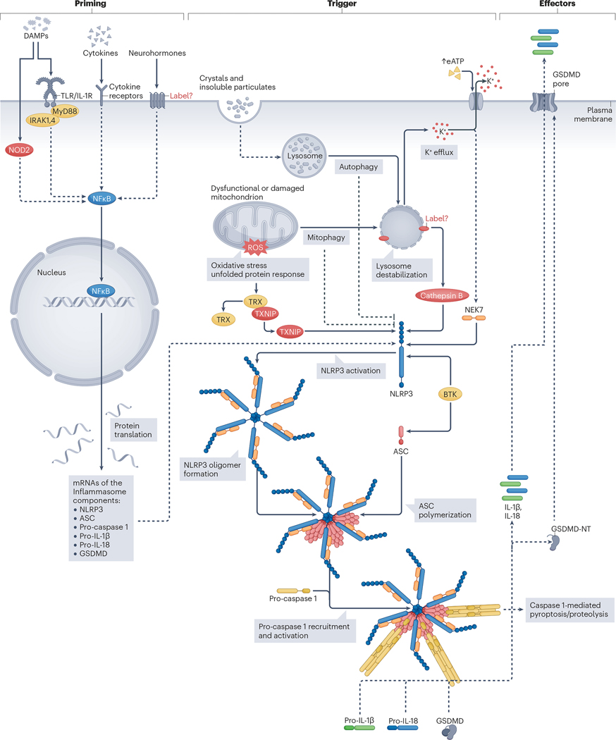
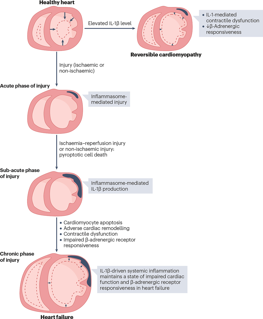
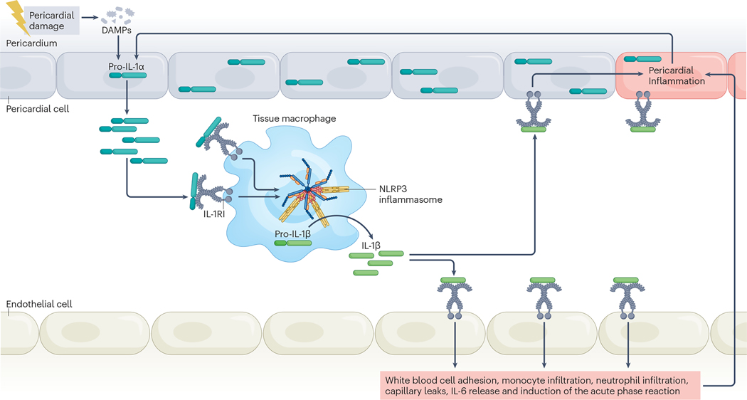
An intense, stereotyped inflammatory response occurs in response to ischaemic and non-ischaemic injury to the myocardium. The NACHT, LRR and PYD domains-containing protein 3 (NLRP3) inflammasome is a finely regulated macromolecular protein complex that senses the injury and triggers and amplifies the inflammatory response by activation of caspase 1; cleavage of pro-inflammatory cytokines, such as pro-IL-1β and pro-IL-18, to their mature forms; and induction of inflammatory cell death (pyroptosis). Inhibitors of the NLRP3 inflammasome and blockers of IL-1β and IL-18 activity have been shown to reduce injury to the myocardium and pericardium, favour resolution of the inflammation and preserve cardiac function. In this Review, we discuss the components of the NLRP3 inflammasome and how it is formed and activated in various ischaemic and non-ischaemic cardiac pathologies (acute myocardial infarction, cardiac dysfunction and remodelling, atherothrombosis, myocarditis and pericarditis, cardiotoxicity and cardiac sarcoidosis). We also summarize current preclinical and clinical evidence from studies of agents that target the NLRP3 inflammasome and related cytokines.


Conflict of interest statement

Competing interests

A.A. has served as a consultant to Cardiol, Implicit Bioscience, Janssen, Kiniksa, Novo Nordisk, Olatec, R-Pharm, Sanofi and Serpin Pharma. S.T. has received research grants from Cardiol, Kiniksa and Olatec.

Figures

Fig. 1 |
Fig. 1 |. Activation of the NLRP3 inflammasome pathways in cardiomyocytes.
The NACHT, LRR and PYD domains-containing protein 3 (NLRP3) inflammasome forms in myocardial cells following a two-step process. Firstly, ‘priming’ that leads to transcription and translation of the NLRP3 inflammasome components and substrates (IL-1β and IL-18). Nuclear factor-kB (NF-κB) activation drives the priming process, initiated by the membrane Toll-like receptors (TLRs) or IL-1 receptor (IL-1R) family. Downstream signalling is mediated by myeloid differentiation primary response protein MyD88 (MyD88) and interleukin-1 receptor-associated kinases (IRAKs). Nucleotide-binding oligomerization domain-containing protein 2 (NOD2) promotes a similar priming signal. Secondly, activation or ‘triggering’ of NLRP3. NLRP3 activation is mediated by extracellular and intracellular pathways. Increasing the concentration of extracellular ATP (eATP) activates the P2X purinoceptor 7 and leads to K+ efflux, a step that triggers the activation of NLRP3. The serine/threonine-protein kinase NEK7 senses K+ efflux and binds to NLRP3, allowing its activation. Lysosomal destabilization by crystals and indigestible material represent another mechanism that leads to NLRP3 activation, through leakage of the lysosomal enzyme cathepsin B and by induction of K+. Mitochondrial damage and dysfunction produce reactive oxygen species (ROS), leading to dissociation of thioredoxin-interacting protein (TXNIP) from thioredoxin (TRX), and TXNIP binding to NLRP3. Ineffective clearance of dysfunctional mitochondria through mitophagy contributes to lysosomal destabilization. By contrast, effective mitophagy and autophagy limit the activation of NLRP3. Tyrosine-protein kinase BTK (also known as Bruton kinase) also binds to NLRP3,as well as to apoptosis speck-like protein containing a caspase recruiting domain (ASC), contributing to inflammasome activation. The active NLRP3 bound to NEK7 oligomerizes and forms a disc-like structure that is platform for the polymerization of ASC into a central filament, facilitating assembly of caspase 1 into filaments that form a star-like structure. Active caspase 1 cleaves the inactive pro-IL-1β and pro-IL-18 into the active forms, IL-1 and IL-18. Caspase 1 also cleaves gasdermin D (GSDMD) producing N-terminal fragments (GSDMD-NT) that oligomerize and form pores in the cell membrane, leading to the extracellular release of active IL-1β and IL-18 and pyroptosis. DAMPs, damage-associated molecular patterns.
Fig. 2 |
Fig. 2 |. Temporal expression of the NLRP3 inflammasome components and window of opportunity for effective inhibition in ischaemia–reperfusion injury.
Progressive expression of the NACHT, LRR and PYD domains-containing protein 3 (NLRP3) inflammasome components (black line) and of infarct area growth (orange line) over time are shown. The infarct area (orange) in the ventricular wall is shown below the graph. For the initial hour after ischaemia–reperfusion injury, the expression of NLRP3 components and the inflammasome activity in the myocardium remains low. The expression of the inflammasome components increases between 1 and 3 hours after injury, leading to effective priming and facilitation of inflammasome formation during this time and in subsequent hours. The size of the infarct continues to grow after reperfusion. The time that precedes the activation of the inflammasome represents a therapeutic window for intervention with NLRP3 inhibitors before the inflammasome forms.
Fig. 3 |
Fig. 3 |. Prevention of NLRP3 inflammasome formation reduces damage in animal models of ischaemia–reperfusion injury.
Coronary plaque growth and instability are promoted by NACHT, LRR and PYD domains-containing protein 3 (NLRP3) inflammasome formation. An atherothrombotic event causes ischaemic ventricular damage, leading to the death of cardiac muscle. During reperfusion, inflammasome-mediated damage promotes further damage and infarct growth. Preventing the formation of the inflammasome — by deleting genes of the inflammasome components (NLRP3, apoptosis speck-like protein containing a caspase recruiting domain (ASC) or caspase 1), by reducing expression of NLRP3 using small interfering RNA (siRNA) or by administering selective NLRP3 inhibitors — reduces infarct size.
Fig. 4 |
Fig. 4 |. Role of IL-1β in acute injury and progression to heart failure.
The left and right ventricular cavities in diastole and in systole (dotted lines) are shown. IL-1β decreases myocardial contractility and the response to β-adrenergic receptor agonists in cardiomyocytes, even in the absence of acute myocardial injury. In the acute phase that follows ischaemic or non-ischaemic myocardial injury, NACHT, LRR and PYD domains-containing protein 3 (NLRP3) inflammasome formation promotes myocardial cell death due to pyroptosis. In the subacute phase, IL-1β induces cardiomyocyte apoptosis, favouring adverse cardiac remodelling and heart failure. Enhanced IL-1β activity in the subacute and chronic phases contribute to impaired myocardial contractility and β-adrenergic responsiveness in heart failure.
Fig. 5 |
Fig. 5 |. Mechanism of action of NLRP3 inflammasome inhibitors tested in experimental models of ischaemic and non-ischaemic injury.
Several NACHT, LRR and PYD domains-containing protein 3 (NLRP3) inflammasome inhibitors have been tested in animal models of acute myocardial infarction. Colchicine can act downstream of NLRP3 by inhibiting the polymerization of apoptosis speck-like protein containing a caspase recruiting domain (ASC). OLT1177, BAY 11–7082, INF4E, MCC950 and CY-09 inhibit the ATPase activity of NLRP3. ZYIL1, selnoflast, DFV890 and JC-121 (also known as 16673–34-0) prevent oligomerization of NLRP3 and the interaction with ASC. Ibrutinib inhibits activation of NLRP3 mediated by tyrosine-protein kinase BTK (also known as Bruton kinase). RRx-001 binds NLRP3 and inhibits its interaction with serine/threonine-protein kinase NEK7. IL-1β activity is blocked by the IL-1 inhibitors anakinra (a recombinant IL-1 receptor antagonist), canakinumab (an anti-IL-1β antibody), rilonacept (an IL-1 trap) and goflikicept (a different IL-1 trap). IL-18 activity is blocked by tadekinig α (a recombinant form of IL-18 binding protein; IL-18BP) and by GSK1070806 (an anti-IL-18 antibody). eATP, extracellular ATP.
Fig. 6 |
Fig. 6 |. Role of IL-1α and IL-1β in the pathophysiology of recurrent pericarditis.
Injury to the pericardial cells induces the release of intracellular contents. (Pro)-IL-1α released outside the cell is already active, binds the IL-1 receptor type I (IL-1RI) on macrophages and activates the signal, functioning as an alarmin. Macrophages respond to the alarmins by forming and triggering the NACHT, LRR and PYD domains-containing protein 3 (NLRP3) inflammasome that processes and releases active IL-1β in its active form. This process amplifies the inflammatory response and induces further injury, which in turn leads to the release of more pro-IL-1α. IL-1β also induces the release of IL-6, which mediates the acute phase reaction associated with inflammation. DAMPs, damage-associated molecular patterns.
Fig. 7 |
Fig. 7 |. Overview of phase II–III trials of IL-1 blockers in acute myocardial infarction and heart failure.
a, The CANTOS trial. Canakinumab 150 mg every 3 months significantly reduced the incidence of cardiovascular death, non-fatal myocardial infarction and stroke in patients with prior myocardial infarction (MI) and residual inflammatory risk. b, A pooled analysis of the VCU-ART trials. Anakinra 100 mg daily (or twice daily) for 14 days significantly reduced the incidence of heart failure or death in patients with ST-segment elevation MI (STEMI). c, The MRC-ILA Heart trial. Anakinra 100 mg daily for 14 days was associated with an unexpected increase in late cardiac and cerebral ischaemic events in patients with non-ST segment elevation MI (NSTEMI). d, The REDHART trial. Anakinra 100 mg daily for 12 weeks, but not for 2 weeks only, showed a favourable rate of hospital readmission for heart failure in patients with recently decompensated systolic heart failure as compared with placebo and an observational registry.

References

    1. Tsao CW et al. Heart Disease and Stroke Statistics-2022 Update: A Report From the American Heart Association. Circulation 145, e153–e639 (2022). - PubMed
    1. Lenz A, Franklin GA & Cheadle WG Systemic inflammation after trauma. Injury 38, 1336–1345 (2007). - PubMed
    1. Abbate A. et al. Interleukin-1 and the Inflammasome as Therapeutic Targets in Cardiovascular Disease. Circ. Res 126, 1260–1280 (2020). - PMC - PubMed
    1. Toldo S. & Abbate A. The NLRP3 inflammasome in acute myocardial infarction. Nat. Rev. Cardiol 15, 203–214 (2018). - PubMed
    1. Westman PC et al. Inflammation as a Driver of Adverse Left Ventricular Remodeling After Acute Myocardial Infarction. J. Am. Coll. Cardiol 67, 2050–2060 (2016). - PubMed

Publication types

LinkOut - more resources