. 2024 Jan 19;65(1):128-141.
doi: 10.1093/pcp/pcad132.

Glutathione Transferase Photoaffinity Labeling Displays GST Induction by Safeners and Pathogen Infection

Affiliations

Glutathione Transferase Photoaffinity Labeling Displays GST Induction by Safeners and Pathogen Infection

Maria Font Farre et al. Plant Cell Physiol. .

Abstract

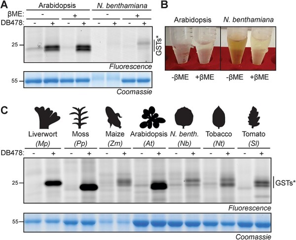
Glutathione transferases (GSTs) represent a large and diverse enzyme family involved in the detoxification of small molecules by glutathione conjugation in crops, weeds and model plants. In this study, we introduce an easy and quick assay for photoaffinity labeling of GSTs to study GSTs globally in various plant species. The small-molecule probe contains glutathione, a photoreactive group and a minitag for coupling to reporter tags via click chemistry. Under UV irradiation, this probe quickly and robustly labels GSTs in crude protein extracts of different plant species. Purification and mass spectrometry (MS) analysis of labeled proteins from Arabidopsis identified 10 enriched GSTs from the Phi(F) and Tau(U) classes. Photoaffinity labeling of GSTs demonstrated GST induction in wheat seedlings upon treatment with safeners and in Arabidopsis leaves upon infection with avirulent bacteria. Treatment of Arabidopsis with salicylic acid (SA) analog benzothiadiazole (BTH) induces GST labeling independent of NPR1, the master regulator of SA. Six Phi- and Tau-class GSTs that are induced upon BTH treatment were identified, and their labeling was confirmed upon transient overexpression. These data demonstrate that GST photoaffinity labeling is a useful approach to studying GST induction in crude extracts of different plant species upon different types of stress.

Keywords: Agrochemical; Chemical proteomics; GST; Glutathione transferase; Immunity; Photoaffinity labeling; Safener.


Figures

Fig. 1
Fig. 1
Photoaffinity labeling of GSTs. (A) The structure of the GST photoaffinity probe DB478, which was originally named GSTABP-G (Stoddard et al. 2017). (B) The procedure of photoaffinity labeling of GSTs. (C) Structures of the used reporters. (D) Photoaffinity labeling of Arabidopsis leaf extracts requires probe, UV light and click chemistry. An Arabidopsis leaf extract was incubated with and without a 5-μM probe and irradiated with UV light for 45 min and coupled to a fluorophore via click chemistry. Fluorescent proteins were visualized by in-gel fluorescence scanning. Coomassie staining is shown as loading control. Arrowheads: Specific labeling of GSTs; *aspecific labeling, probably from the large subunit of rubisco (RbcL).
Fig. 2
Fig. 2
GST labeling depends on conditions. (A) Photoaffinity labeling saturates at 5-μM probe concentration. Arabidopsis leaf extracts were incubated with various probe concentrations and exposed to UV for 30 min. (B) Photoaffinity labeling requires >30 min UV treatment. Arabidopsis leaf extracts were incubated with a 5-μM probe and exposed to UV at various times. (C) Photoaffinity labeling requires pH > 6. (D) GST inhibitors differentially suppress photoaffinity labeling. Arabidopsis leaf extracts were incubated with a 5-μM probe at various pH and exposed to UV for 30 min. Samples were further analyzed as described in Fig. 1D. Plotted are signal intensities from three independent replicates normalized to the signal with the highest intensity.
Fig. 3
Fig. 3
Proteomics confirms GST enrichment upon labeling. (A) GSTs are enriched in probe-labeled samples. Arabidopsis leaf extracts were labeled with and without a 5-μM probe, crosslinked and coupled to biotin via click chemistry and enriched on streptavidin beads. Beads were treated with trypsin, and released peptides were analyzed by MS. Plotted are the MS protein intensities plotted against the fold change for all proteins that were detected in all three replicates of the probe-labeled samples. Highlighted are all detected GST proteins (circles), abundant non-enriched proteins (RbcL, RbcS) and endogenously biotinylated proteins BCCP, MCCP and ACC1. FDR was calculated by the MaxLFQ algorithm of MaxQuant. (B) High protein coverage of GSTs. Unique and shared peptides identified by proteomics (A) were mapped onto the GST protein sequences. The predicted MW in kDa for each GST is on the left. (C) Identified GSTs highlighted in phylogeny of Arabidopsis GSTs. Tree is adapted from Wagner et al. (2002). Unrooted bootstrapped tree (n = 5000) is based on a multiple sequence alignment by ClustalX of the full-length protein sequences of all Arabidopsis GSTs. (D) In-solution digest (ISD) of the same Arabidopsis leaf extracts used for labeling. Shown are the proteins detected in all three replicates, ranked on their average LFQ intensity, with the detected GSTs indicated. GSTs that are enriched upon labeling are printed in bold.
Fig. 4
Fig. 4
Putative GST labeling of other plant species. (A) Photoaffinity labeling of putative GSTs in N. benthamiana requires extraction with a reducing agent. Leaf extracts of Arabidopsis and N. benthamiana were generated with and without 3 mM βME, incubated with and without a 5-μM probe and exposed to UV light and visualized by fluorescence scanning and Coomassie staining. (B) βME avoids sample browning during labeling. (C) Putative GST labeling in leaf extracts of various plant species in the presence of βME. Leaf extracts were generated with 3 mM βME, incubated with and without a 5-μM probe and exposed to UV light and visualized by fluorescence scanning and Coomassie staining. *putative GST-derived signals.
Fig. 5
Fig. 5
Safener induces GSTs in wheat. (A) Structures of safener and herbicide used in this experiment. (B) Experimental procedure of agrochemical treatment. (C) Safener treatment induces GST activities. Leaf extracts of seedlings incubated with/out safener and/or herbicide were labeled with DB478 and fluorescently labeled proteins were detected from protein gels by fluorescence scanning and quantified. Coomassie staining is shown as a loading control. (D) Quantified fluorescence of the 22–24 kDa signals from three biological replicates, analyzed by ANOVA. Groups a and b are statistically different from each other (P < 0.05). Error bars represent the standard deviation of three biological replicates.
Fig. 6
Fig. 6
Pathogens and BTH induce GSTs independently of NPR1. (A) Avirulent P. syringae causes increased GST labeling in Arabidopsis. Arabidopsis leaves were infiltrated with PtoDC3000 wild-type (WT) or carrying plasmids encoding avrRpt2 or avrRpm1. Total extracts generated on day 2 were labeled with DB478, coupled to a fluorophore and analyzed by fluorescent scanning of protein gels. (B) BTH-treated plants. Adult Arabidopsis plants were watered with and without BTH for 3 d and proteins were extracted and analyzed by Western blot using anti-PR2 antibodies and Ponceau staining. (C) BTH-induced GST labeling is not dependent on NPR1. The samples of Col-0 wild-type and npr1-1 mutant plants treated with water/BTH for 3 d were labeled with 5 µM DB478, coupled to a fluorophore with click chemistry, followed by in-gel fluorescence scanning and Coomassie staining.
Fig. 7
Fig. 7
Transient overexpression of BTH-induced GSTs. (A) BTH-induced GSTs. Leaf extracts from water- and BTH-treated Arabidopsis plants were labeled with DB478 in triplicate. Labeled proteins were coupled to biotin-azide via click chemistry and enriched on streptavidin beads. On-bead digested proteins were analyzed by MS and plotted in volcano plots where log2 of fold change (log2FC) was plotted against their significance (-log10FDR) for three biological replicates. FDR was calculated by the MaxLFQ algorithm of MaxQuant. Proteins were significantly reduced (blue) induced (red) or unchanged (gray) upon BTH treatment. GSTs and PR1 are highlighted. (B) Labeling of GST–GFP fusion proteins upon agroinfiltration. Arabidopsis GSTs were fused to a C-terminal GFP tag and expressed in N. benthamiana by agroinfiltration. Extracts of agroinfiltrated leaves containing βME taken at 4 d upon agroinfiltration were labeled with and without 5 µM DB478, coupled to a fluorophore via click chemistry and visualized by in-gel fluorescence scanning. Fusions were detected by Western blot with anti-GFP antibody. Highlighted are labeled GFP-tagged GSTs (*) and endogenous GSTs (arrowheads). (C) Labeling of GST-His fusion proteins upon agroinfiltration. Arabidopsis GSTs were fused to a C-terminal His tag and expressed in N. benthamiana by agroinfiltration. Extracts of agroinfiltrated leaves containing βME taken at 4 d upon agroinfiltration were labeled with and without 5 µM DB478, coupled to a fluorophore via click chemistry and visualized by in-gel fluorescence scanning. Fusions were detected by Western blot with anti-GFP antibody. Highlighted are labeled GFP-tagged GSTs (*) and endogenous GSTs (arrowheads).

Similar articles

References

    1. Allocati N., Masulli M., Di Ilio C. and Federici L. (2018) Glutathione transferases: substrates, inhibitors and pro-drugs in cancer and neurodegenerative diseases. Oncogenesis 7: 1–15. - PMC - PubMed
    1. Bally J., Jung H., Mortimer C., Naim F., Philips J.G., Hellens R., et al. (2018) The rise and rise of Nicotiana benthamiana: a plant for all reasons. Annu. Rev. Phytopathol. 56: 405–426. - PubMed
    1. Basantani M. and Srivastava A. (2007) Plant glutathione transferases—a decade falls short. Can. J. Bot. 85: 443–456.
    1. Behringer C., Bartsch K. and Schaller A. (2011) Safeners recruit multiple signalling pathways for the orchestrated induction of the cellular xenobiotic detoxification machinery in Arabidopsis. Plant Cell Environ. 34: 1970–1985. - PubMed
    1. Blanco F., Garretón V., Frey N., Dominguez C., Pérez-Acle T., Van der Straeten D., et al. (2005) Identification of NPR1-dependent and independent genes early induced by salicylic acid treatment in Arabidopsis. Plant Mol. Biol. 59: 927–944. - PubMed

MeSH terms