. 2023 Dec 7;110(12):2003-2014.
doi: 10.1016/j.ajhg.2023.10.005. Epub 2023 Nov 3.

Efficient in vivo prime editing corrects the most frequent phenylketonuria variant, associated with high unmet medical need

Affiliations

Efficient in vivo prime editing corrects the most frequent phenylketonuria variant, associated with high unmet medical need

Dominique L Brooks et al. Am J Hum Genet. .

Abstract

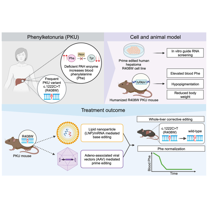
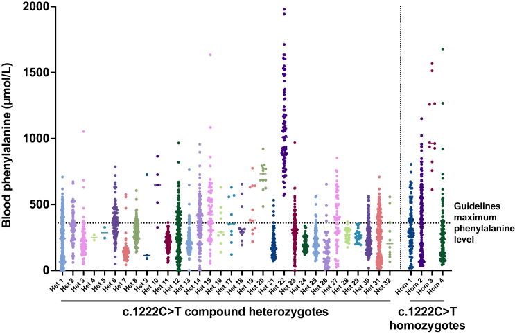
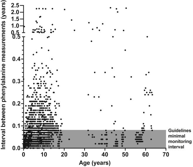
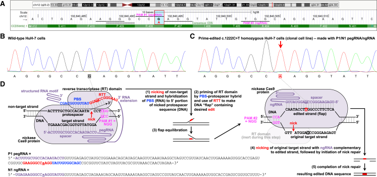
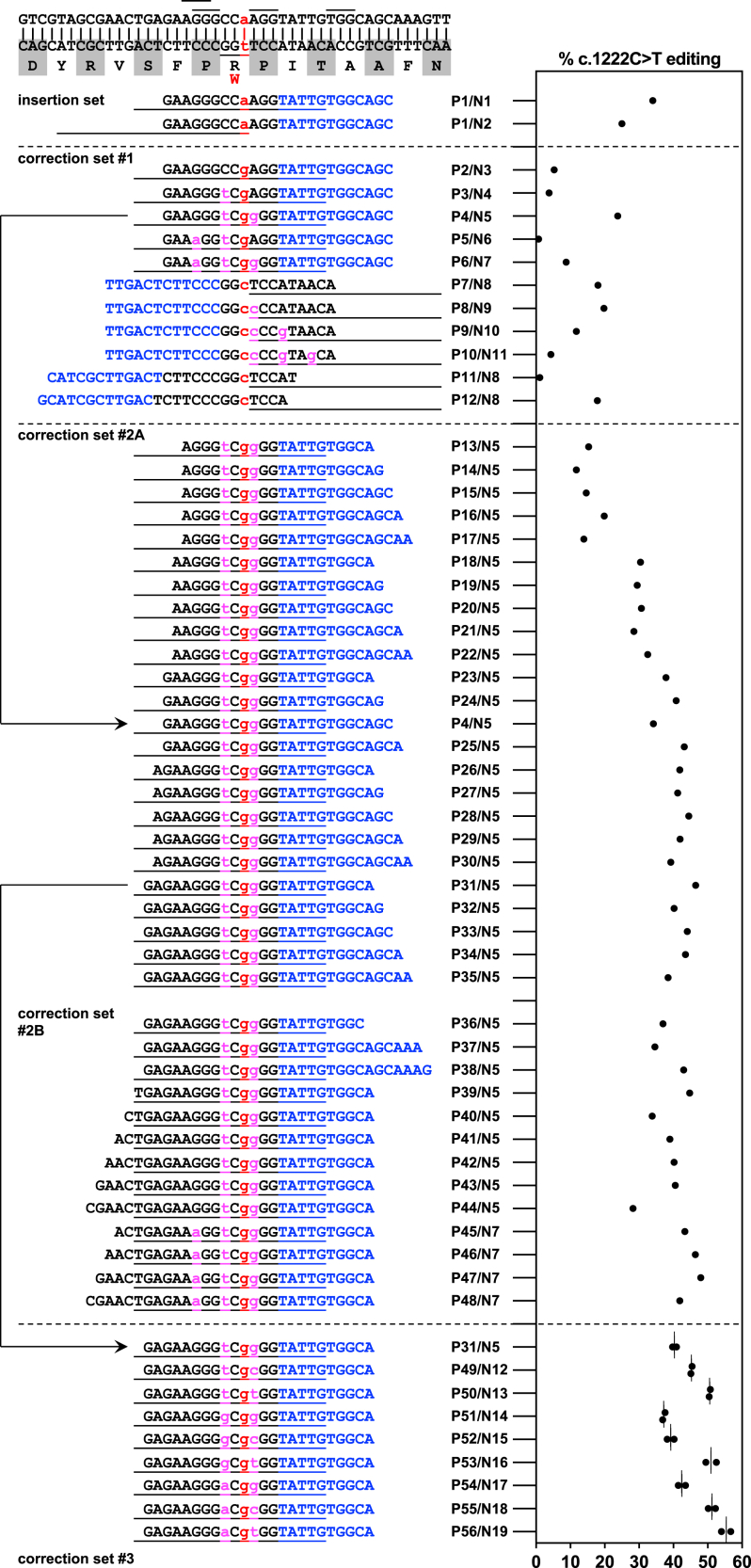
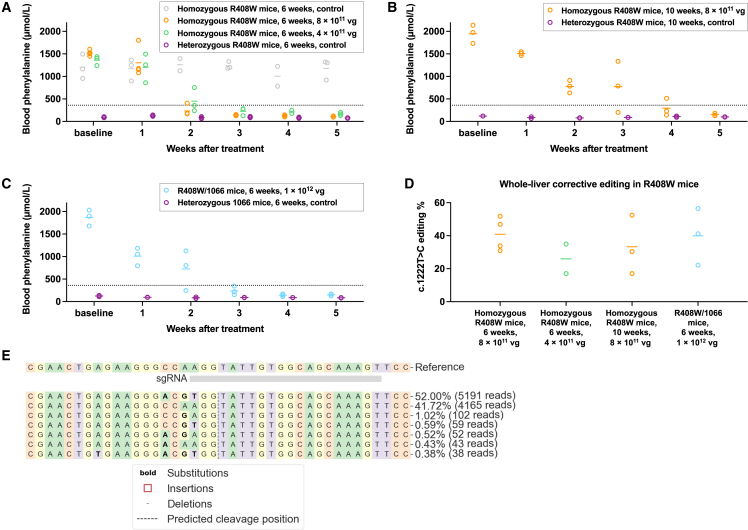
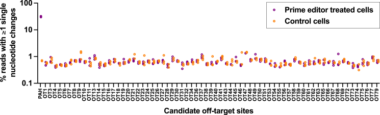
The c.1222C>T (p.Arg408Trp) variant in the phenylalanine hydroxylase gene (PAH) is the most frequent cause of phenylketonuria (PKU), the most common inborn error of metabolism. This autosomal-recessive disorder is characterized by accumulation of blood phenylalanine (Phe) to neurotoxic levels. Using real-world data, we observed that despite dietary and medical interventions, most PKU individuals harboring at least one c.1222C>T variant experience chronic, severe Phe elevations and do not comply with Phe monitoring guidelines. Motivated by these findings, we generated an edited c.1222C>T hepatocyte cell line and humanized c.1222C>T mouse models, with which we demonstrated efficient in vitro and in vivo correction of the variant with prime editing. Delivery via adeno-associated viral (AAV) vectors reproducibly achieved complete normalization of blood Phe levels in PKU mice, with up to 52% whole-liver corrective PAH editing. These studies validate a strategy involving prime editing as a potential treatment for a large proportion of individuals with PKU.

Keywords: CRISPR; gene editing; genome editing; inborn error of metabolism; metabolic disease; phenylketonuria; prime editing; rare disease.


Conflict of interest statement

Declaration of interests K.M. is an advisor to and holds equity in Verve Therapeutics and Variant Bio and is an advisor to LEXEO Therapeutics. R.C.A.-N. is an advisor to Latus Bio. All other authors have no financial conflicts of interests to disclose.

Figures

None
Graphical abstract
Figure 1
Figure 1
Lifetime recorded measurements of blood phenylalanine concentrations in PKU individuals with PAH c.1222C>T variants All available Phe concentrations from 32 PKU individuals harboring the c.1222C>T variant in trans with a second pathogenic allele are shown on the left, where all available levels from four individuals homozygous for c.1222C>T are shown on the right. Most individuals had several recorded concentrations above 360 μmol/L, the maximum recommended concentration per PKU guidelines, as indicated by the dotted line.
Figure 2
Figure 2
Intervals between measurements of blood phenylalanine concentrations in PKU individuals with PAH c.1222C>T variants The durations of intervals between Phe-monitoring checks are plotted by age. Recommended guidelines for how often Phe should be checked varies with age and is indicated in gray. Most individuals did not check Phe concentrations frequently enough, as evidenced by the fact that most monitoring intervals fell outside the recommended range.
Figure 3
Figure 3
Prime editing to introduce PAH c.1222C>T variant in human hepatocytes in vitro (A) Schematic depiction of the genomic site of the PAH c.1222C>T variant, adapted from the UCSC Genome Browser (GRCh38/hg38). The vertical blue bar outlined by the red box indicates the G altered to A (in red) by the variant on the antisense strand. The four horizontal lines near the site of the variant indicate four potential NGG PAM sequences for use with pegRNAs or ngRNAs; the horizontal line to the far right of the variant indicates a potential NGG PAM for use with an ngRNA only. The labeled horizontal lines (PAM #1, PAM #2) correspond to the PAMs used in the schematic shown in (D). (B) Sanger sequencing chromatogram of the genomic site of the PAH c.1222C>T variant in wild-type HuH-7 cells. The boxed nucleotide is at the site of the variant. (C) Chromatogram of the site in PAH c.1222C>T homozygous HuH-7 cell line. The boxed nucleotide is at the site of the edited variant, in the position indicated by the red arrow. (D) Schematic depiction of prime editing with the P1/N1 pegRNA/ngRNA combination to insert the c.1222C>T variant into the PAH locus in the human genome. Boxes or underlines indicate nucleotides at the site of the variant.
Figure 4
Figure 4
Prime editing to correct the PAH c.1222C>T variant in human hepatocytes in vitro Efficiencies of PAH c.1222C>T editing by various pegRNA/ngRNA combinations (Px/Nx, refer to Tables S1 and S2 for sequences); the first two combinations in the table/graph were for insertion of the c.1222C>T variant in wild-type HuH-7 cells (used to generate the PAH c.1222C>T homozygous HuH-7 cell line in Figure 3), and the remainder for correction of the variant in homozygous HuH-7 cells. For each letter sequence, blue indicates the PBS of the pegRNA, and black indicates the RTT of the pegRNA. Red lowercase letters indicate edits at the site of the variant; magenta lowercase letters indicate additional synonymous edits. The short lines at the top of the table indicate NGG PAM sequences used for pegRNAs and/or ngRNAs. Each long line immediately below a pegRNA PBS/RTT sequence in the table indicates the span of the protospacer sequence of the ngRNA used with that pegRNA. n = 1 biological replicate for each pegRNA/ngRNA combination except for the last set, for which n = 2 biological replicates. Lines in the graph = mean values.
Figure 5
Figure 5
Prime editing to correct PAH c.1222C>T variant in humanized mice (A) Changes in blood phenylalanine levels in homozygous PKU mice after treatment with 8 × 1011 vg AAV dose (n = 4 animals) or with 4 × 1011 vg AAV dose (n = 3 animals); concentrations at various timepoints up to 5 weeks after treatment were compared to concentrations in untreated homozygous PKU control (n = 3 animals) and untreated heterozygous non-PKU control (n = 3 animals) age-matched (6 weeks of age) colony mates (one blood sample per timepoint). (B) Changes in blood phenylalanine concentrations in homozygous PKU mice after treatment with 8 × 1011 vg AAV dose (n = 3 animals). Concentrations at various timepoints up to 5 weeks after treatment to were compared to concentrations in untreated heterozygous non-PKU control (n = 2 animals) age-matched (10 weeks of age) colony mates (one blood sample per timepoint). (C) Changes in blood phenylalanine concentrations in compound-heterozygous PKU mice after treatment with 1 × 1012 vg AAV dose (n = 3 animals). Concentrations at various timepoints up to 5 weeks after treatment were compared to concentrations in untreated heterozygous non-PKU control (n = 2 animals) age-matched (6 weeks of age) colony mates (one blood sample per timepoint). (D) Corrective PAH c.1222C>T editing (determined from genomic DNA) in the whole liver (mean value from eight liver samples) in each of the treated groups of mice. For compound-heterozygous mice, each displayed number is the estimated percentage of edited c.1222C>T alleles (editable alleles). (E) Standard CRISPResso output for a liver sample from the treated homozygous PKU mouse with the highest level of editing. Lines in graphs = mean values.
Figure 6
Figure 6
Assessment of off-target editing On-target or off-target editing at top ONE-seq-nominated or in-silico-nominated candidate sites calculated as the proportion of aligned sequencing reads with alteration of ≥1 base pair within the target sequence (ranging from the position proximal to the pegRNA nick site to the distal position spanned by the RTT sequence, capturing both single-nucleotide substitutions and indels) in PAH c.1222C>T homozygous HuH-7 cells that underwent plasmid transfection with the lead pegRNA/ngRNA (P56/N19) combination (n = 3 treated and 3 untreated biological replicates; y axis is log10 scale). Sites with unsuccessful sequencing are omitted. Refer to Table S4 for candidate site sequences and numerical values.

Comment in

References

    1. Vockley J., Andersson H.C., Antshel K.M., Braverman N.E., Burton B.K., Frazier D.M., Mitchell J., Smith W.E., Thompson B.H., Berry S.A., American College of Medical Genetics and Genomics Therapeutics Committee Phenylalanine hydroxylase deficiency: diagnosis and management guideline. Genet. Med. 2014;16:188–200. doi: 10.1038/gim.2013.157. - DOI - PubMed
    1. Jurecki E.R., Cederbaum S., Kopesky J., Perry K., Rohr F., Sanchez-Valle A., Viau K.S., Sheinin M.Y., Cohen-Pfeffer J.L. Adherence to clinic recommendations among patients with phenylketonuria in the United States. Mol. Genet. Metabol. 2017;120:190–197. doi: 10.1016/j.ymgme.2017.01.001. - DOI - PubMed
    1. Hillert A., Anikster Y., Belanger-Quintana A., Burlina A., Burton B.K., Carducci C., Chiesa A.E., Christodoulou J., Đorđević M., Desviat L.R., et al. The genetic landscape and epidemiology of phenylketonuria. Am. J. Hum. Genet. 2020;107:234–250. doi: 10.1016/j.ajhg.2020.06.006. - DOI - PMC - PubMed
    1. Leuders S., Wolfgart E., Ott T., du Moulin M., van Teeffelen-Heithoff A., Vogelpohl L., Och U., Marquardt T., Weglage J., Feldmann R., Rutsch F. Influence of PAH genotype on sapropterin response in PKU: results of a single-center cohort study. JIMD Rep. 2014;13:101–109. doi: 10.1007/8904_2013_263. - DOI - PMC - PubMed
    1. Burton B.K., Longo N., Vockley J., Grange D.K., Harding C.O., Decker C., Li M., Lau K., Rosen O., Larimore K., et al. Pegvaliase for the treatment of phenylketonuria: results of the phase 2 dose-finding studies with long-term follow-up. Mol. Genet. Metabol. 2020;130:239–246. doi: 10.1016/j.ymgme.2020.06.006. - DOI - PubMed