Multicenter Study
. 2023 Nov 4;8(1):426.
doi: 10.1038/s41392-023-01685-0.

SUMOylation-triggered ALIX activation modulates extracellular vesicles circTLCD4-RWDD3 to promote lymphatic metastasis of non-small cell lung cancer

Affiliations
Multicenter Study

SUMOylation-triggered ALIX activation modulates extracellular vesicles circTLCD4-RWDD3 to promote lymphatic metastasis of non-small cell lung cancer

Xiayao Diao et al. Signal Transduct Target Ther. .

Abstract

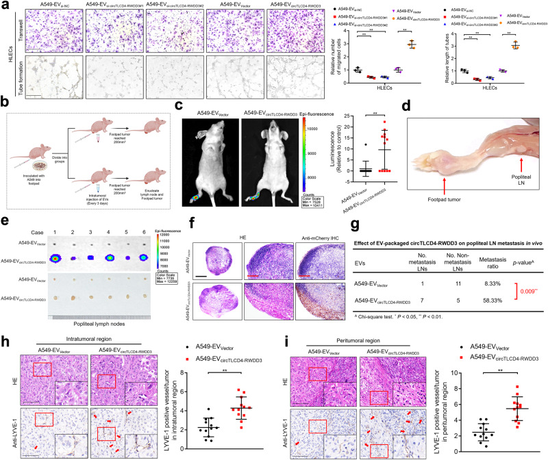
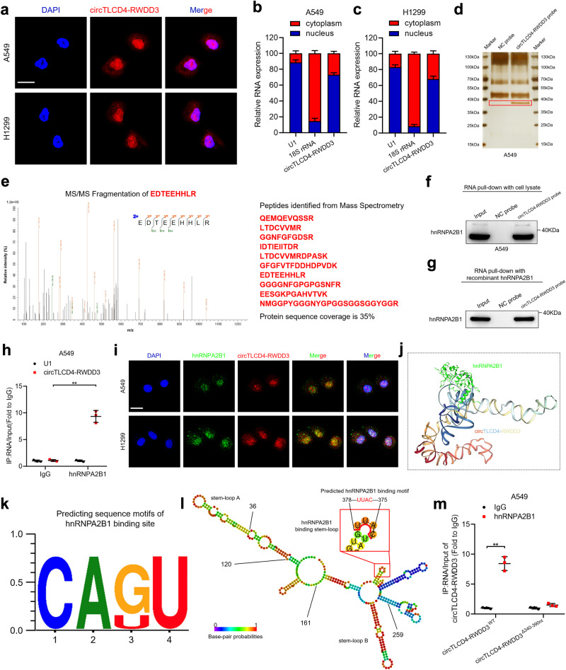
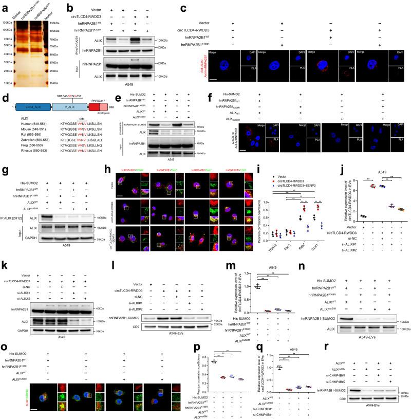
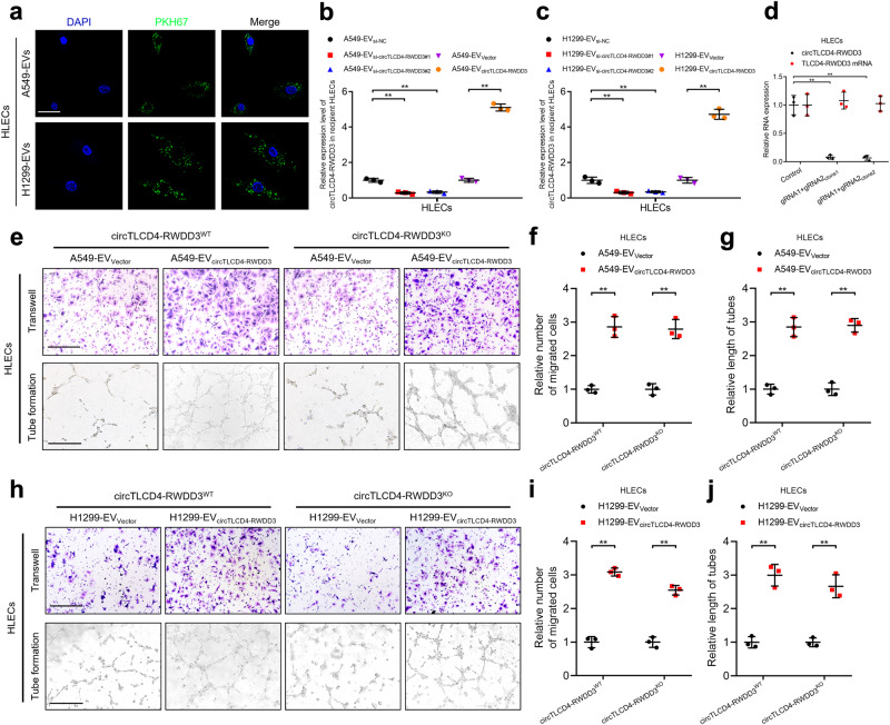
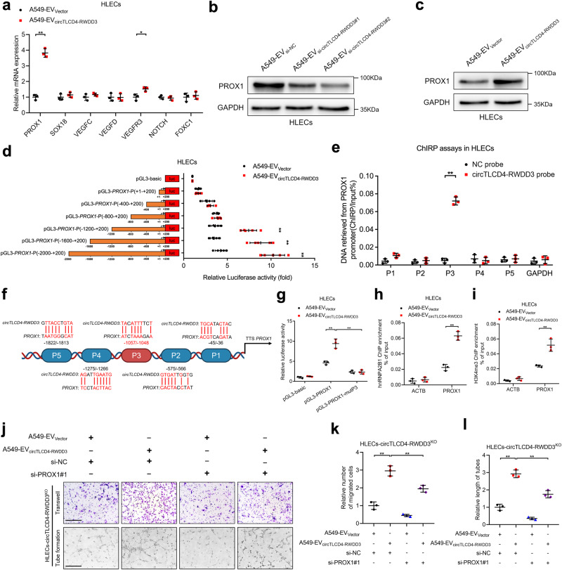
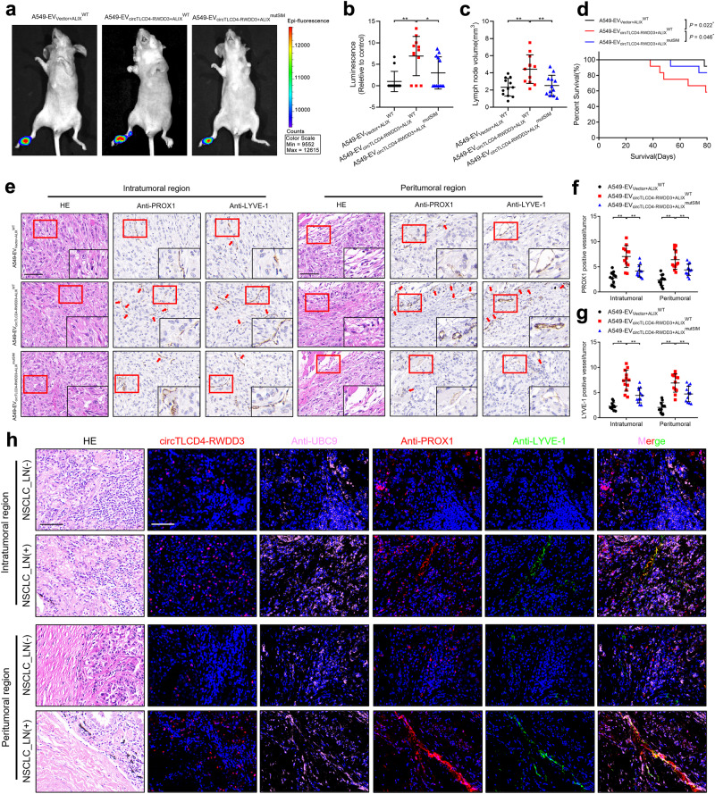
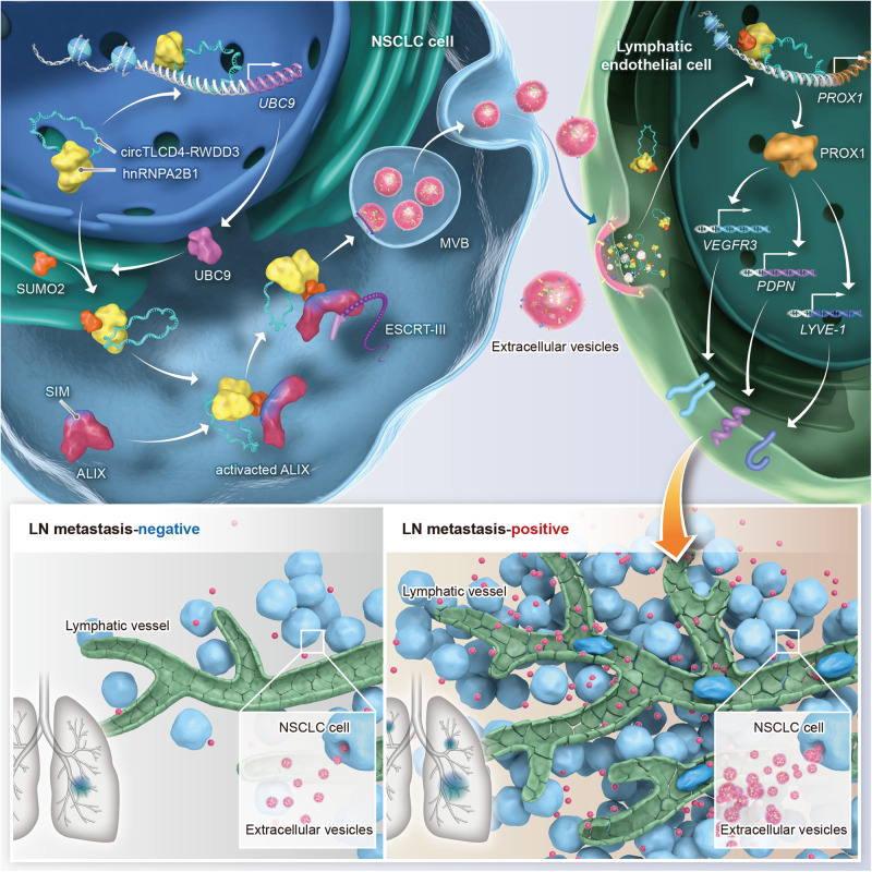
Lymph node (LN) metastasis is one of the predominant metastatic routes of non-small cell lung cancer (NSCLC) and is considered as a leading cause for the unsatisfactory prognosis of patients. Although lymphangiogenesis is well-recognized as a crucial process in mediating LN metastasis, the regulatory mechanism involving lymphangiogenesis and LN metastasis in NSCLC remains unclear. In this study, we employed high-throughput sequencing to identify a novel circular RNA (circRNA), circTLCD4-RWDD3, which was significantly upregulated in extracellular vesicles (EVs) from LN metastatic NSCLC and was positively associated with deteriorated OS and DFS of patients with NSCLC from multicenter clinical cohort. Downregulating the expression of EV-packaged circTLCD4-RWDD3 inhibited lymphangiogenesis and LN metastasis of NSCLC both in vitro and in vivo. Mechanically, circTLCD4-RWDD3 physically interacted with hnRNPA2B1 and mediated the SUMO2 modification at K108 residue of hnRNPA2B1 by upregulating UBC9. Subsequently, circTLCD4-RWDD3-induced SUMOylated hnRNPA2B1 was recognized by the SUMO interaction motif (SIM) of ALIX and activated ALIX to recruit ESCRT-III, thereby facilitating the sorting of circTLCD4-RWDD3 into NSCLC cell-derived EVs. Moreover, EV-packaged circTLCD4-RWDD3 was internalized by lymphatic endothelial cells to activate the transcription of PROX1, resulting in the lymphangiogenesis and LN metastasis of NSCLC. Importantly, blocking EV-mediated transmission of circTLCD4-RWDD3 via mutating SIM in ALIX or K108 residue of hnRNPA2B1 inhibited the lymphangiogenesis and LN metastasis of NSCLC in vivo. Our findings reveal a precise mechanism underlying SUMOylated hnRNPA2B1-induced EV packaging of circTLCD4-RWDD3 in facilitating LN metastasis of NSCLC, suggesting that EV-packaged circTLCD4-RWDD3 could be a potential therapeutic target against LN metastatic NSCLC.


Conflict of interest statement

The authors declare no competing interests.

Figures

Fig. 1
Fig. 1
EV-packaged circTLCD4-RWDD3 positively correlates with LN metastasis of NSCLC. a Flowchart of steps to identify circRNAs upregulated in NSCLC relative to NATs and upregulated in LN-positive NSCLC relative to LN-negative cancer. b, c Heatmap of circRNAs differentially expressed in NSCLC tissues and NATs and in NSCLC tissues with or without LN metastasis. d qRT-PCR analysis of circTLCD4-RWDD3 expression in NSCLC cell lines and human bronchial epithelial cell line. e–h qRT-PCR analysis of circTLCD4-RWDD3 expression in LN-positive and LN-negative NSCLC tissues from PUMCH, SYSMH, SYSUCC, and combined cohort, respectively. i Representative FISH images and percentages of circTLCD4-RWDD3 expression in LN-positive or LN-negative NSCLC tissues and NATs. White arrows indicated the extracellular expression of circTLCD4-RWDD3. Scale bars, 50 µm. j Representative images and percentages of circTLCD4-RWDD3 expression and LYVE-1-indicated lymphatic vessels in NSCLC tissues. Scale bars, 50 µm. k, l Kaplan–Meier curves for OS (k) and DFS (l) of NSCLC patients with low versus high circTLCD4-RWDD3 expression. The cutoff value is the median. m, n TEM (m) and NTA (n) identified the characteristics of A549 cell-derived EVs. Scale bar, 200 nm. o Western blotting analysis of EV markers in cell lysates or A549 cell-derived EVs. p qRT-PCR analysis of circTLCD4-RWDD3 expression in EVs derived from NSCLC tissues and paired NATs. q qRT-PCR analysis of circTLCD4-RWDD3 expression in EVs derived from LN-positive and LN-negative NSCLC tissues. r qRT-PCR analysis of circTLCD4-RWDD3 expression in NSCLC cell lines and human bronchial epithelial cell line and in their corresponding EVs. The statistical difference was assessed through nonparametric Mann–Whitney U test in (e–h, p, and q); and Chi-square test in (i, j); and one-way ANOVA followed by Dunnett tests in (d, r); and unpaired Student’s t-test in (r). Error bars show the SD from three independent experiments. *P < 0.05; **P < 0.01
Fig. 2
Fig. 2
EV-packaged circTLCD4-RWDD3 promotes lymphangiogenesis and LN metastasis in NSCLC in vitro and in vivo. a Representative images and quantification of the tube formation and Transwell migration of HLECs treated with circTLCD4-RWDD3-downregulating or -upregulating A549 cell-derived EVs. Scale bars, 100 µm. b Schematic diagram of the construction of a nude mice popliteal LN metastasis model. Schematic was created with BioRender (www.biorender.com). c Representative images and quantification of bioluminescence of the popliteal LN metastasis in nude mice (n = 12 per group). d Representative image of the footpad primary tumor and popliteal metastatic LN in a nude mouse. e Representative bioluminescence image of excised popliteal LNs from the nude mice (n = 12 per group). f Representative anti-mCherry IHC images of nude mice popliteal LNs (n = 12 per group). Scale bars, 500 µm (black); scale bars, 100 µm (red). g The percentage of metastatic popliteal LN in the nude mice (n = 12 per group). h, i Representative IHC images and percentages of LYVE-1-indicated lymphatic vessel density in intratumoral (h) and peritumoral region (i) of footpad primary tumor tissues. Scale bars, 50 µm. The statistical difference was assessed with one-way ANOVA followed by Dunnett tests in (a); and unpaired Student’s t-test in (a, c, h, and i); and Chi-square test in (g). Error bars show the SD from three independent experiments. *P < 0.05; **P < 0.01
Fig. 3
Fig. 3
circTLCD4-RWDD3 directly interacts with hnRNPA2B1 in NSCLC cells. a–c Detection of intracellular localization of circTLCD4-RWDD3 in NSCLC cells using FISH assays (a) and subcellular fraction assays (b, c). Scale bars, 5 µm. d Silver staining image of RNA pull-down assay with circTLCD4-RWDD3 and control probes in A549 cells. e Mass spectrometry analysis of circTLCD4-RWDD3-binding proteins after RNA pull-down assay. f, g Western blotting analysis after RNA pull-down assay to investigate the interaction between circTLCD4-RWDD3 and hnRNPA2B1. h RIP assays with anti-hnRNPA2B1 revealing the enrichment of circTLCD4-RWDD3 by hnRNPA2B1 in A549 cells. i Detection of intracellular co-localization of circTLCD4-RWDD3 and hnRNPA2B1 in NSCLC cells. Scale bars, 5 µm. j 3D schematic diagram predicting the interaction between circTLCD4-RWDD3 and hnRNPA2B1. k hnRNPA2B1-binding motif predicted by RBPmap. l The stem-loop structure of hnRNPA2B1-binding motifs in circTLCD4-RWDD3. m RIP assays after mutating of the 340–390 nt region of circTLCD4-RWDD3 in A549 cells. The statistical difference was assessed by unpaired Student’s t-test in (h, m). Error bars show the SD from three independent experiments. *P < 0.05; **P < 0.01
Fig. 4
Fig. 4
circTLCD4-RWDD3 induces SUMO2 modification at K108 residue of hnRNPA2B1 by upregulating UBC9. a Western blotting analysis of hnRNPA2B1 expression in A549 cells treated with various inhibitors of PTM. PYR-41 for ubiquitylation, MK-8719 for O-GlcNAcylation, 2-D08 for SUMOylation, MLN4924 for NEDDylation, Calyculin A for phosphorylation, Tunicamycin for N-linked glycosylation, SGC707 for arginine methylation, CI-amidine for deimination and 2-BP for palmitoylation. b Co-IP assay to investigate the SUMOylation type of hnRNPA2B1 in A549 cells. c Co-IP assay to assess SUMO2 modification of hnRNPA2B1 after 2-D08 treatment or overexpressing SENP3. d Schematic illustration of the SUMOylation sites on hnRNPA2B1 predicted by GPS-SUMO. e Sanger sequencing to confirm the hnRNPA2B1K108R and hnRNPA2B1K125R mutations. f Western blotting analysis to confirm the SUMO2 modification site on hnRNPA2B1. g Western blotting analysis of SUMO2 modification on hnRNPA2B1 in A549 cells with or without circTLCD4-RWDD3 overexpression. h qRT-PCR analysis for SUMOylation-related enzyme expression in A549 cells with or without circTLCD4-RWDD3 overexpression. I, j Western blotting analysis to confirm UBC9 expression after circTLCD4-RWDD3 downregulation (i) or overexpression (j) in A549 cells. k Western blotting analysis to investigate SUMO2 modification on hnRNPA2B1 in circTLCD4-RWDD3-overexpressing A549 cells with or without knocking down UBC9. l Transcriptional activity of UBC9 in circTLCD4-RWDD3-overexpressing A549 cells transfected with truncated UBC9 promoter luciferase plasmids. m ChIRP assays to investigate the circTLCD4-RWDD3-associated chromatin fragments of the UBC9 promoter in A549 cells. n Schematic illustration of the DNA-RNA triplex structure between circTLCD4-RWDD3 and the UBC9 promoter. Schematic was created with BioRender (www.biorender.com). o Luciferase activity detected in circTLCD4-RWDD3-overexpressing A549 cells with or without mutating the circTLCD4-RWDD3-binding site on UBC9 promoter. p, q ChIP-qPCR of hnRNPA2B1 (p) and H3K4me3 (q) enrichment on UBC9 promoter after upregulating circTLCD4-RWDD3 in A549 cells. (r) Western blotting analysis of UBC9 expression in circTLCD4-RWDD3-overexpressing A549 cells with or without knocking down hnRNPA2B1. (s) ChIP-qPCR analysis of H3K4me3 enrichment on UBC9 promoter in circTLCD4-RWDD3-overexpressing A549 cells with or without knocking down hnRNPA2B1. The statistical difference was assessed by unpaired Student’s t-test in (h, l, m, p, and q); and one-way ANOVA followed by Dunnett tests in (o, s). Error bars show the SD from three independent experiments. *P < 0.05; **P < 0.01
Fig. 5
Fig. 5
SUMOylated hnRNPA2B1 activates ALIX to trigger circTLCD4-RWDD3 loading into EVs in ESCRT-III-dependent manner. a Silver staining after co-IP assay to detect SUMOylated hnRNPA2B1-interacting proteins. b Western blotting analysis to assess the interaction between hnRNPA2B1 and ALIX in circTLCD4-RWDD3-overexpressing A549 cells with or without mutating K108 residue of hnRNPA2B1. c Representative confocal images of PLA between hnRNPA2B1 and ALIX (red signal) in circTLCD4-RWDD3-overexpressing A549 cells with or without mutating K108 residue of hnRNPA2B1. Scale bars, 5 μm. d Schematic illustrating conserved SIM in ALIX sequence. e Western blotting analysis after co-IP assays to investigate the interaction between hnRNPA2B1 and ALIX after mutating K108 residue of hnRNPA2B1 or SIM in ALIX. f Confocal images of PLA between hnRNPA2B1 and ALIX (red signal) in A549 cells with hnRNPA2B1K108R or mutation of the SIM in ALIX. Scale bars, 5 μm. g Western blotting analysis after co-IP assays to assess the activation of ALIX in A549 cells with hnRNPA2B1K108R or mutation of the SIM in ALIX. h Representative confocal images of the co-localization between hnRNPA2B1 and the indicated subcellular markers in circTLCD4-RWDD3-overexpressing A549 cells with or without SENP3 treatment. Scale bars, 5 μm. i Pearson correlation coefficients were calculated from the A549 cells expressing hnRNPA2B1 and the indicated subcellular markers. j qRT-PCR analysis for circTLCD4-RWDD3 expression in EVs derived from circTLCD4-RWDD3-overexpressing A549 cells with or without knocking down ALIX. k, l Western blotting analysis for hnRNPA2B1 expression in circTLCD4-RWDD3-overexpressing A549 cells with or without knocking down ALIX (k) or in EVs derived from the indicated A549 cells (l). m qRT-PCR analysis for circTLCD4-RWDD3 expression in EVs derived from A549 cells with hnRNPA2B1K108R or mutation of the SIM in ALIX. n Western blotting analysis of hnRNPA2B1 expression in EVs derived from A549 cells with hnRNPA2B1K108R or mutation of the SIM in ALIX. o Detection of intracellular co-localization of ALIX and CHMP4B in A549 cells with hnRNPA2B1K108R or mutation of the SIM in ALIX. Scale bars, 5 μm. p Pearson correlation coefficients were calculated from the indicated A549 cells expressing ALIX and CHMP4B. q qRT-PCR analysis for circTLCD4-RWDD3 expression in EVs derived from A549 cells with mutation of the SIM in ALIX or knocking down CHMP4B. r Western blotting analysis of hnRNPA2B1 expression in EVs derived from A549 cells with mutation of the SIM in ALIX or knocking down CHMP4B. The statistical difference was assessed by the unpaired Student’s t-test in (j); and one-way ANOVA followed by Dunnett tests in (i, m, p, and q). Error bars show the SD from three independent experiments. *P < 0.05; **P < 0.01
Fig. 6
Fig. 6
EV-packaged circTLCD4-RWDD3 is internalized by HLECs to facilitate lymphangiogenesis. a Representative fluorescence images of HLECs after incubating with PKH67-labeled EVs. Scale bars, 5 μm. b, c qRT-PCR analysis of circTLCD4-RWDD3 expression in HLECs treated with EVs. d qRT-PCR analysis of circTLCD4-RWDD3 and TLCD4-RWDD3 mRNA expression in circTLCD4-RWDD3KO HLECs. e–g Representative images (e) and quantification of the Transwell migration (f) and tube formation (g) of circTLCD4-RWDD3WT and circTLCD4-RWDD3KO HLECs treated with A549-EVVector or A549-EVcircTLCD4-RWDD3. Scale bars, 100 µm. h–j Representative images (h) and quantification of the Transwell migration (i) and tube formation (j) of circTLCD4-RWDD3WT and circTLCD4-RWDD3KO HLECs treated with H1299-EVVector or H1299-EVcircTLCD4-RWDD3. Scale bars, 100 µm. The statistical difference was assessed with one-way ANOVA followed by Dunnett tests in b–d; and unpaired Student’s t-test in (b, c, f, g, i, and j). Error bars show the SD from three independent experiments. *P < 0.05; **P < 0.01
Fig. 7
Fig. 7
EV-packaged circTLCD4-RWDD3 promotes PROX1 expression in HLECs. a qRT-PCR analysis of lymphangiogenesis-related gene expression in A549-EVVector- or A549-EVcircTLCD4-RWDD3-treated HLECs. b Western blotting analysis to verify PROX1 expression in HLECs treated with A549-EVsi-NC, A549-EVsi-circTLCD4-RWDD3#1, or A549-EVsi-circTLCD4-RWDD3#2. c Western blotting analysis to verify PROX1 expression in HLECs treated with A549-EVVector or A549-EVcircTLCD4-RWDD3. d Transcriptional activity of PROX1 in A549-EVcircTLCD4-RWDD3-treated HLECs transfected with truncated PROX1 promoter luciferase plasmids. e ChIRP assays to investigate the circTLCD4-RWDD3-associated chromatin fragments of the PROX1 promoter in HLECs. f Schematic representation of the DNA-RNA triplex structure between circTLCD4-RWDD3 and the PROX1 promoter. Schematic was created with BioRender (www.biorender.com). g Luciferase activity detected in A549-EVVector- or A549-EVcircTLCD4-RWDD3-treated HLECs with or without mutating the circTLCD4-RWDD3-binding site on PROX1 promoter. h, i ChIP-qPCR of the enrichment of hnRNPA2B1 (h) and H3K4me3 (i) on PROX1 promoter in A549-EVVector- or A549-EVcircTLCD4-RWDD3-treated HLECs. j–l Representative images (j) and quantification of Transwell migration (k) and tube formation (l) of A549-EVVector- or A549-EVcircTLCD4-RWDD3-treated circTLCD4-RWDD3KO HLECs with or without knocking down PROX1. Scale bars, 100 µm. The statistical difference was assessed by the unpaired Student’s t-test in (a, d, e, h, and i); and one-way ANOVA followed by Dunnett tests in (g, k, and l). Error bars show the SD from three independent experiments. *P < 0.05; **P < 0.01
Fig. 8
Fig. 8
Blocking the transmission of EV-packaged circTLCD4-RWDD3 suppresses LN metastasis of NSCLC. a, b Representative bioluminescence images (a) and quantification (b) of popliteal metastatic LNs from nude mice treated with EVs secreted by control or circTLCD4-RWDD3-overexpressing A549 cells with or without mutating SIM in ALIX (n = 12 per group). (c) Quantification of popliteal LN volume of nude mice treated with EVs (n = 12 per group). d Kaplan–Meier curves show the survival of nude mice treated with EVs secreted by control or circTLCD4-RWDD3-overexpressing A549 cells with or without mutating SIM in ALIX. e–g Representative IHC images and percentages of PROX1- or LYVE-1-indicated lymphatic vessel density in footpad primary tumor tissues. Scale bars, 50 µm. h Representative fluorescence images to confirm the correlation for circTLCD4-RWDD3 expression, UBC9 and PROX1 expression, and LYVE-1-indicated MLD in the serial section of NSCLC tissues from the multicenter cohorts (n = 312). Scale bars, 50 μm. The statistical difference was assessed through one-way ANOVA followed by Dunnett tests in (b, c, f, and g). Error bars show the SD from three independent experiments. *P < 0.05; **P < 0.01
Fig. 9
Fig. 9
Schematic illustrating the potential mechanism by which EV-packaged circTLCD4-RWDD3 promotes LN metastasis of NSCLC

References

    1. Sung H, et al. Global cancer statistics 2020: GLOBOCAN estimates of incidence and mortality worldwide for 36 cancers in 185 countries. CA Cancer J. Clin. 2021;71:209–249. doi: 10.3322/caac.21660. - DOI - PubMed
    1. Chen Z, Fillmore CM, Hammerman PS, Kim CF, Wong KK. Non-small-cell lung cancers: a heterogeneous set of diseases. Nat. Rev. Cancer. 2014;14:535–546. doi: 10.1038/nrc3775. - DOI - PMC - PubMed
    1. Asamura H, et al. The international association for the study of lung cancer lung cancer staging project: proposals for the revision of the N descriptors in the forthcoming 8th edition of the TNM classification for lung cancer. J. Thorac. Oncol. 2015;10:1675–1684. doi: 10.1097/JTO.0000000000000678. - DOI - PubMed
    1. Li Z, et al. Predictive value of folate receptor-positive circulating tumor cells for the preoperative diagnosis of lymph node metastasis in patients with lung adenocarcinoma. Small Methods. 2021;5:e2100152. doi: 10.1002/smtd.202100152. - DOI - PubMed
    1. Karaman S, Detmar M. Mechanisms of lymphatic metastasis. J. Clin. Invest. 2014;124:922–928. doi: 10.1172/JCI71606. - DOI - PMC - PubMed

Publication types

MeSH terms