. 2023 Nov 4;14(1):7086.
doi: 10.1038/s41467-023-42831-0.

Alternative lengthening of telomeres (ALT) cells viability is dependent on C-rich telomeric RNAs

Affiliations

Alternative lengthening of telomeres (ALT) cells viability is dependent on C-rich telomeric RNAs

Ilaria Rosso et al. Nat Commun. .

Abstract

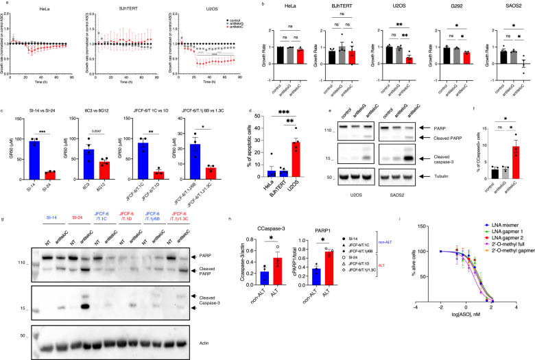
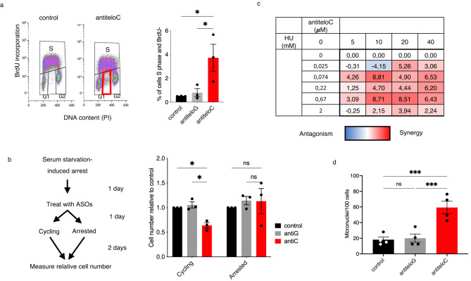
Alternative lengthening of telomeres (ALT) is a telomere maintenance mechanism activated in ~10-15% of cancers, characterized by telomeric damage. Telomeric damage-induced long non-coding RNAs (dilncRNAs) are transcribed at dysfunctional telomeres and contribute to telomeric DNA damage response (DDR) activation and repair. Here we observed that telomeric dilncRNAs are preferentially elevated in ALT cells. Inhibition of C-rich (teloC) dilncRNAs with antisense oligonucleotides leads to DNA replication stress responses, increased genomic instability, and apoptosis induction selectively in ALT cells. Cell death is dependent on DNA replication and is increased by DNA replication stress. Mechanistically, teloC dilncRNA inhibition reduces RAD51 and 53BP1 recruitment to telomeres, boosts the engagement of BIR machinery, and increases C-circles and telomeric sister chromatid exchanges, without increasing telomeric non-S phase synthesis. These results indicate that teloC dilncRNA is necessary for a coordinated recruitment of DDR factors to ALT telomeres and it is essential for ALT cancer cells survival.


Conflict of interest statement

F.R. and F.d.A.d.F. are inventors on the patent applications PCT/EP2013/059753 and PCT/EP2016/068162. C.J.W. and J.A. are inventors on the patent application PCT/ EP2016/068162. The remaining authors declare no competing interests.

Figures

Fig. 1
Fig. 1. ALT cells display elevated levels of telomeric dilncRNAs.
a Telomeric dilncRNAs were quantified by strand-specific RT-qPCR and normalized on RPLP0 gene; average values for each cell line were quantified in three biologically independent experiments; data are presented as mean values +/− SEM among n = 2 non-ALT and n = 3 ALT cell lines; two-tailed unpaired t-test, df = 3, t = 1.174 teloG, t = 3.718 teloC; *p = 0.0339. b Northern blot analysis of total RNA. Radiolabeled C-rich and G-rich telomeric oligonucleotides were used to probe for teloG and teloC dilncRNAs, respectively. EtBr-stained 18S RNA is shown as loading control; n = 2 biologically independent experiments, one representative image shown. c Telomeric dilncRNAs were quantified and reported as in (a); mean values for each cell line were quantified in n = 2 or 3 biologically independent experiments; data are presented as mean values +/− SEM among n = 5 non-ALT and ALT cell lines; two-tailed unpaired t-test, df = 8, t = 3.295 teloG, t = 4.127 teloC; *p = 0.0109, **p = 0.0033. d HeLa Long Telomere (LT) cells were transfected with siSCR (control) or siASF1a/b siRNAs twice per week, and telomeric dilncRNAs were quantified as in (a) 14 days after first siRNA transfection; data are presented as mean values +/− SEM, n = 3 biologically independent experiments; two-tailed unpaired t-test, df = 4, t = 4.208 teloG, t = 3.432 teloC; for teloG *p = 0.0136, for teloC *p = 0.0265. Source data are provided as a Source data file.
Fig. 2
Fig. 2. TeloC dilncRNAs are essential to maintain ALT cells viability.
a Cells were transfected with 10 nM ASO, and growth rate was calculated by Incucyte; data are presented as mean values +/− SEM, n = 3 biologically independent experiments; two-way ANOVA, for HeLa control vs antiteloC 24 h *p = 0.0334, 28 h **p = 0.0063, 32 h *p = 0.0372; for HeLa antiteloG vs antiteloC 24 h *p = 0.0133, 28 h ***p = 0.0009, 32 h **p = 0.0092; for U2OS control vs antiteloC from 24 h to 72 h ****p < 0.0001; for U2OS antiteloG vs antiteloC from 24 h to 32 h ****p < 0.000, 36 h ***p = 0.003, 40 h ***p = 0.0002, 44 h ***p = 0.0007, 48 h ***p = 0.0004, from 52 h to 64 h ***p = 0.0002, 68 h ***p = 0.0003, 72 h ***p = 0.0005. b Cells were transfected with 20 nM ASO and growth rate was calculated three days later by resazurin; data are presented as mean values +/− SEM, n = 3 biologically independent experiments for HeLa (df = 8, F = 2.865) and G292 (df = 8, F = 8.454), n = 4 biologically independent experiments for BJhTERT (df = 11, F = 0.5318), U2OS (df = 11, F = 14.50) and SAOS2 (df = 11, F = 7.493); one-way ANOVA, ns = non-significant, for U2OS control vs antiteloC **p = 0.0024 antiteloG vs antiteloC **p = 0.0041; for G292 control vs antiteloC *p = 0.0224 antiteloG vs antiteloC *p = 0.0356; for SAOS2 control vs antiteloC **p = 0.0206 antiteloG vs antiteloC *p = 0.0212;. c Cells were treated with a range of concentrations of ASO antiteloC without transfection reagent and GR50 was calculated three days later with RealTime-Glo. When not determined, GR50 was underestimated to 100 μM (the highest concentration used in this experiment); data are presented as mean values +/− SEM, n = 3 biologically independent experiments for SI-14 and SI-24 (df = 4, t = 14.37), for JFCF-6/T1C and 1D (df = 4, t = 6.267), and for JFCF-6/T.1J6B and 1.3 C (df = 4, t = 3.349) and n = 4 biologically independent experiments for 6C3 and 8G12 (df = 6, t = 2.382); two-tailed unpaired t-test, for SI-14 vs SI-24 ***p = 0.0001; for JFCF-6/T1C and 1D **p = 0.0033; for JFCF-6/T.1J6B and 1.3 C *p = 0.0286 d Cells treated as in (a), and apoptotic cells counted by Incucyte three days after transfection; values reported as mean values +/- SEM, n = 5 biologically independent experiments; one-way ANOVA, df = 14, F = 34.71, HeLa vs U2OS ***p = 0.0004, BJhTERT vs U2OS **p = 0.0048. e Cells were transfected with 20 nM ASO and two days later analyzed by western blot; n = 2, one representative image shown. f U2OS cells were transfected with 20 nM ASO and fixed two days later for flow cytometry analysis; data are presented as mean values +/− SEM, n = 3 biologically independent experiments; one-way ANOVA, df = 8, F = 11.19; ns = non-significant, control vs antiteloC *p = 0.0135, antiteloG vs antiteloC *p = 0.0169. g Cells were treated with 20 μM of ASO antiteloC without transfection reagent and three days later analyzed by western blot. h The ratios between cleaved caspase 3 over actin and cleaved PARP1 over total PARP1 from the experiment shown in (g) were measured; data are presented as mean values +/− SEM, n = 3 non-ALT and n = 3 ALT cell lines; two-tailed paired t-test, df = 2, t(Ccaspase-3) = 4.458, t(PARP1) = 5.811,CCaspase3 *p = 0.0468, PARP1 *p = 0.0284. i U2OS cells were transfected with the indicated antiteloC ASO at a range of concentrations and live cells were counted by Incucyte three days after transfection; data are presented as mean values +/− SEM, n = 3 biologically independent experiments. Source data are provided as a Source data file.
Fig. 3
Fig. 3. teloC dilncRNAs are required to maintain viability during replication stress.
a U2OS cells were transfected with 20 nM ASO, and two days later pulsed with BrdU for 20 min and analyzed by flow cytometry; data are presented as mean values +/− SEM, n = 3 biologically independent experiments; one-way ANOVA, df = 8, F = 6.762, control vs antiteloC *p = 0.0279, antiteloG vs antiteloC *p = 0.0410; left, representative flow cytometry distribution; right, quantification. b Serum-starved SAOS2 cells were transfected with 20 nM ASO as indicated and relative cell number was quantified with resazurin; data are presented as mean values +/− SEM, n = 3 biologically independent experiments; one-way ANOVA, df = 8, F(cycling) = 21.04, F(arrested) = 0.26, ns = non-significant, control vs antic *p = 0.0444, antiG vs antic *p = 0.0235; left, experimental design; right, quantification. c U2OS cells were treated with a range of concentrations of HU and antiteloC ASO in absence of transfection reagent, and cell number was measured three days later by resazurin. Synergy was calculated according to the excess over Bliss additivism model (ref. ). Lower Bliss values correspond to antagonism and are shown in blue, higher values correspond to synergy and are shown in red; data are presented as mean values, n = 3 biologically independent experiments. d U2OS cells were transfected with 20 nM ASO and, two days later, fixed, stained with DAPI, and micronuclei manually counted; data are presented as mean values +/− SEM, n = 4 biologically independent experiments; one-way ANOVA, df = 15, F = 18.10, ns = non-significant, control vs antiteloC ***p = 0.0005, antiteloG vs antiteloC ***p = 0.0008. Source data are provided as a Source data file.
Fig. 4
Fig. 4. teloC dilncRNA inhibition upregulates unproductive break induced replication.
a U2OS cells were transfected twice with the indicated siRNAs prior to transfection of siRNAs and 20 nM ASO, and cell viability was monitored three days later with resazurin; data are presented as mean values +/− SEM, n = 3 biologically independent experiments; two-way ANOVA, ns = non-significant, siSCR ***p = 0.0008, siBLM **p = 0.0084, siSLX4 *p = 0.0245. b Cells were transfected with 20 nM ASO and relative cell viability was measured three days later with resazurin; data are presented as mean values +/− SEM, n = 3 biologically independent experiments; two-way ANOVA, ns = non-significant, parental **p = 0.0017, BLM KO and RMi1 KO ****p < 0.0001, PML KO **p = 0.0051. ce U2OS cells were transfected with 20 nM ASO as indicated and collected two days later for analysis. c Metaphases were subjected to COFISH and stained with PNA probes; data are presented as mean values +/− SEM, n = 3 biologically independent experiments; two-sided, paired t test, **p = 0.0499. d C-circle levels were analyzed by CCA, signals were normalized on ALU; values reported as the difference between +Φ29 and −Φ29, data are presented as mean values +/− SEM, n = 3 biologically independent experiments; one-way ANOVA, df = 8, F = 20.01, ns=non-significant, control vs antiteloC **p = 0.0044, antiteloG vs antiteloC **p = 0.0032. e U2OS were pulsed for 2 h with 10 μM EdU before fixation. Telomeric non-S DNA synthesis was quantified as colocalization of TRF2 and EdU foci in cells with less than 20 EdU foci; data are presented as mean values +/− SEM, n = 3 biologically independent experiments, one-way ANOVA, ns = non-significant. Source data are provided as a Source data file.
Fig. 5
Fig. 5. teloC dilncRNA inhibition alters the engagement of DDR factors at ALT telomeres.
ag U2OS cells were transfected with 20 nM ASO as indicated and collected two days later for analysis. a Average TRF2 foci size per cell was measured with an automated pipeline in ImageJ; data are presented as mean values +/− SEM, n = 3 biologically independent experiments; one-way ANOVA, *p = 0.049, **p = 0.0041. b Cells were immunostained for RAD52 and TRF2 and colocalizations were scored; data are presented as percentages +/− 95% confidence interval; n =  more than 710 cells examined over 3 biologically independent experiments; two-sided Fisher’s exact test, ****p < 0.0001. c Cells were immunostained for POLD3 and TRF2 and colocalizations were scored; data are presented as percentages +/− 95% confidence interval; n =  more than 680 cells examined over 3 biologically independent experiments; two-sided Fisher’s exact test, ****p < 0.0001. d Cells were immunostained for RAD51 and TRF2 and colocalizations were scored; data are presented as percentages +/− 95% confidence interval; n =  more than 500 cells examined over 3 biologically independent experiments; two-sided Fisher’s exact test, **p = 0.0021, *p = 0.0404. e Cells were immunostained for 53BP1 and TRF2 and colocalizations were scored; data are presented as percentages +/− 95% confidence interval; n= more than 710 cells examined over 3 biologically independent experiments; two-sided Fisher’s exact test, *p = 0.0093. f Cells were immunostained for γH2AX and TRF2 and colocalizations were scored; data are presented as percentages +/− 95% confidence interval; n =  more than 680 cells examined over 3 independent biological replicates; two-sided Fisher’s exact test, ***p = 0.0002, ****p < 0.0001. g Cells were immunostained for RPA and TRF2 and colocalizations were scored; data are presented as percentages +/− 95% confidence interval; n =  more than 325 cells examined over 3 biologically independent experiments; two-sided Fisher’s exact test, **p = 0.0035. Source data are provided as a Source data file.
Fig. 6
Fig. 6. Model of proposed teloC dilncRNA function at ALT telomeres.
a Proposed mechanism in unchallenged ALT cells. b Proposed mechanism in ASO antiteloC treated ALT cells. Created with BioRender.com.

References

    1. de Lange T. Shelterin-mediated telomere protection. Annu Rev. Genet. 2018;52:223–247. doi: 10.1146/annurev-genet-032918-021921. - DOI - PubMed
    1. D’Adda Di Fagagna F, et al. A DNA damage checkpoint response in telomere-initiated senescence. Nature. 2003;426:194–198. doi: 10.1038/nature02118. - DOI - PubMed
    1. Herbig U, Jobling WA, Chen BPC, Chen DJ, Sedivy JM. Telomere shortening triggers senescence of human cells through a pathway involving ATM, p53, and p21CIP1, but not p16INK4a. Mol. Cell. 2004;14:501–513. doi: 10.1016/S1097-2765(04)00256-4. - DOI - PubMed
    1. Karlseder J, Broccoli D, Yumin D, Hardy S, De Lange T. p53- and ATM-dependent apoptosis induced by telomeres lacking TRF2. Science. 1999;283:1321–1325. doi: 10.1126/science.283.5406.1321. - DOI - PubMed
    1. Greider CW, Blackburn EH. Identification of a specific telomere terminal transferase activity in tetrahymena extracts. Cell. 1985;43:405–413. doi: 10.1016/0092-8674(85)90170-9. - DOI - PubMed

Publication types