Artificial Intelligence in Breast Cancer Diagnosis and Personalized Medicine
- PMID: 37926067
- PMCID: PMC10625863
- DOI: 10.4048/jbc.2023.26.e45
Artificial Intelligence in Breast Cancer Diagnosis and Personalized Medicine
Abstract
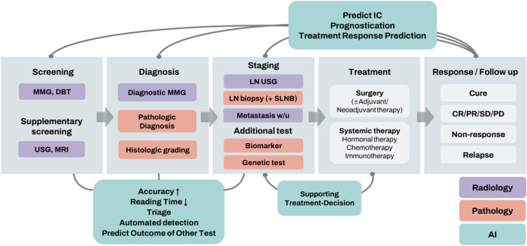
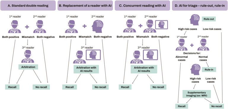
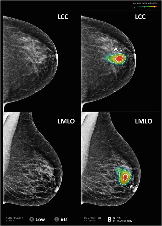
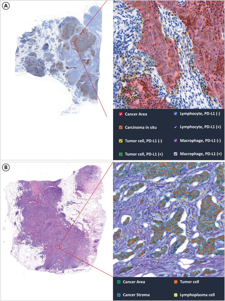
Breast cancer is a significant cause of cancer-related mortality in women worldwide. Early and precise diagnosis is crucial, and clinical outcomes can be markedly enhanced. The rise of artificial intelligence (AI) has ushered in a new era, notably in image analysis, paving the way for major advancements in breast cancer diagnosis and individualized treatment regimens. In the diagnostic workflow for patients with breast cancer, the role of AI encompasses screening, diagnosis, staging, biomarker evaluation, prognostication, and therapeutic response prediction. Although its potential is immense, its complete integration into clinical practice is challenging. Particularly, these challenges include the imperatives for extensive clinical validation, model generalizability, navigating the "black-box" conundrum, and pragmatic considerations of embedding AI into everyday clinical environments. In this review, we comprehensively explored the diverse applications of AI in breast cancer care, underlining its transformative promise and existing impediments. In radiology, we specifically address AI in mammography, tomosynthesis, risk prediction models, and supplementary imaging methods, including magnetic resonance imaging and ultrasound. In pathology, our focus is on AI applications for pathologic diagnosis, evaluation of biomarkers, and predictions related to genetic alterations, treatment response, and prognosis in the context of breast cancer diagnosis and treatment. Our discussion underscores the transformative potential of AI in breast cancer management and emphasizes the importance of focused research to realize the full spectrum of benefits of AI in patient care.
Keywords: Artificial Intelligence; Breast Neoplasms; Diagnostic Imaging; Pathology; Precision Medicine.
© 2023 Korean Breast Cancer Society.
Conflict of interest statement
Jong Seok Ahn, Sangwon Shin, Su-A Yang, Eun Kyung Park, Ki Hwan Kim, Soo Ick Cho, and Chan-Young Ock are employed by Lunit Inc. and/or have stock/stock options in Lunit Inc. No other disclosures were reported.
Figures
References
-
- Giaquinto AN, Sung H, Miller KD, Kramer JL, Newman LA, Minihan A, et al. Breast cancer statistics, 2022. CA Cancer J Clin. 2022;72:524–541. - PubMed
-
- Sung H, Ferlay J, Siegel RL, Laversanne M, Soerjomataram I, Jemal A, et al. Global cancer statistics 2020: GLOBOCAN estimates of incidence and mortality worldwide for 36 cancers in 185 countries. CA Cancer J Clin. 2021;71:209–249. - PubMed
-
- The Royal College of Radiologists. RCR Clinical Radiology Workforce Census 2022. London: The Royal College of Radiologists; 2022.
Publication types
LinkOut - more resources
Full Text Sources
Other Literature Sources