

Successful Ovarian Vein Embolization of a Multiparous Woman with Pelvic Congestion Syndrome
- PMID: 37927827
- PMCID: PMC10624537
- DOI: 10.1055/s-0041-1731272
Successful Ovarian Vein Embolization of a Multiparous Woman with Pelvic Congestion Syndrome
Abstract
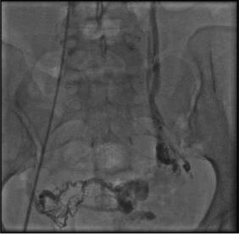
Pelvic congestion syndrome (PCS) is a clinical syndrome supported by specific findings, such as ovarian vein's dilatation, that cause pelvic vein congestion. Although many theories are explaining the pathophysiologies of this condition, the underlying cause remains unknown. The clinical manifestations of PCS are various including chronic pelvic pain (CPP), voiding disturbances, or ureteral obstruction. Imaging modality, such as ultrasonography, computed tomography (CT scan), magnetic resonance imaging (MRI), and venography, are needed to confirm and exclude the differential diagnosis. Currently, American venous forum guidelines recommended endovascular therapy which is percutaneous embolization as the first option therapy of PCS. Here, we reported a 35-year-old woman with PCS who underwent successful percutaneous embolization therapy.
Keywords: chronic pelvic pain; multiparous woman; pelvic congestion syndrome; percutaneous embolization; venography.
International College of Angiology. This article is published by Thieme.
Conflict of interest statement
Conflict of Interest None declared.
Figures


References
-
- Cura M, Cura A. What is the significance of ovarian vein reflux detected by computed tomography in patients with pelvic pain? Clin Imaging. 2009;33(04):306–310. - PubMed
-
- Gutvirtz G, Imterat M, Weintraub Y A. Pelvic congestion syndrome: a current review. Pelviperineology. 2018;37:14–16.
-
- S.J. Park, J.W. Lim, Y.T. Ko, D.H. Lee, Y. Yoon, J.H. Oh, et al. Diagnosis of pelvic congestion syndrome using transabdominal and transvaginal sonography AJR Am J Roentgenol, 182 (3) (2004 Mar), pp.683–688. - PubMed
-
- Phillips D, Deipolyi A R, Hesketh R L, Midia M, Oklu R. Pelvic congestion syndrome: etiology of pain, diagnosis, and clinical management. J Vasc Interv Radiol. 2014;25(05):725–733. - PubMed
