. 2020 May:2020:23-31.
doi: 10.2312/molva.20201098.

Mesoscope: A Web-based Tool for Mesoscale Data Integration and Curation

Affiliations

Mesoscope: A Web-based Tool for Mesoscale Data Integration and Curation

L Autin et al. MolVa (2020). 2020 May.

Abstract

Interest is growing for 3D models of the biological mesoscale, the intermediate scale between the nanometer scale of molecular structure and micrometer scale of cellular biology. However, it is currently difficult to gather, curate and integrate all the data required to define such models. To address this challenge we developed Mesoscope (mesoscope.scripps.edu/beta), a web-based data integration and curation tool. Mesoscope allows users to begin with a listing of molecules (such as data from proteomics), and to use resources at UniProt and the PDB to identify, prepare and validate appropriate structures and representations for each molecule, ultimately producing a portable output file used by CellPACK and other modeling tools for generation of 3D models of the biological mesoscale. The availability of this tool has proven essential in several exploratory applications, given the high complexity of mesoscale models and the heterogeneity of the available data sources.

Keywords: Applied computing → Bioinformatics; Computing methodologies → Graphics systems and interfaces.


Figures

Figure 1:
Figure 1:
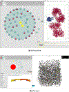
Mesoscope Web Interface
Figure 2:
Figure 2:. Recipe View
Recipe View with three options for labeling and coloring nodes: by name, PDB accession code, and with annotation information. This recipe includes components for exosomes, with hand-drawn sprites by intern Julia Jimenez. The red triangle indicates a component that requires curation, and icons may be dragged interactively between the inner, membrane, and exterior compartments.
Figure 3:
Figure 3:. Recipe Table
Detailed specifications for each molecule in the recipe is included in an editable spreadsheet, to allow curation of the data that will be exported for model building. Updates are immediately propagated to other Mesoscope components.
Figure 4:
Figure 4:. NGL Molecular Viewer
NGL is used to display and manipulate atomic structures of components. Most standard NGL visual representations are supported. A membrane-bound protein is shown here, showing the location of the membrane-spanning portion, which may be tuned using interactive sliders. NGL viewport is synchronized with sequence information from the Sequence features and the ProtVista widget. The mouse click and over position on the sequence displays the corresponding 3D residues (purple and yellow ball and stick) helping refining membrane orientation.
Figure 5:
Figure 5:. Mesoscope Features Currently Under Development
a) Interaction - A prototype capability to define specific interactions between molecules is implemented by connecting icons in the Recipe View, and providing a structure of the complex of the two ingredients. b) Preview - A prototype tool to preview interactive instances of the model showing the number of each ingredient in the current state of the recipe.
Figure 6:
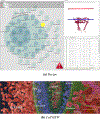
Figure 6:. HIV in Blood Plasma
a) The recipe includes two concentric bounding elements, the inner capsid and the outer envelope membrane, and soluble/fibrous components inside the capsid, in the viral lumen, and outside the viral envelope. The membrane location of the viral matrix protein (highlighted in yellow in the recipe view) is being curated in the NGL window. (b) CellVIEW visualization of a cross-section through the HIV final model, using context-specific labeling methods to identify the major features of the current view.
Figure 7:
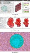
Figure 7:. Insulin Secretory Vesicle
a) The recipe includes membrane-bound and soluble interior ingredients for the vesicle, and a simplified recipe for generic cytoplasm. The recipe also includes specifications for a small crystal of insulin, being previewed in the NGL viewer, which is often found in mature forms of the vesicle. b) Through K-means clustering of atomic position, Mesoscope generates n-beads representing the current structure viewed in NGL. Radii of the beads may be calculated from the cluster boundary (left and center, 3 and 25 beads auto radius) or defined by the user (right, 25 beads with radius of 25 Angstroms). c) cellPACK-gpu visualization of a cross-section through the model, with the vesicle in turquoise and the surrounding cytoplasm in pink. The large circular feature inside the vesicle is the insulin crystalline inclusion.
Figure 8:
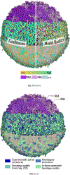
Figure 8:. Mycoplasma Cell
Colors are used to explore confidence levels and provenance of ingredients in a model of an entire mycoplasma cell. These values are curated in the Mesoscope spreadsheet and exported to CellView for display. (a) Color coding of confidence levels for the protein ingredients of the recipe. (b) Color coding of protein ingredients by the source of the molecular structure.
Figure 9:
Figure 9:. Recipe Exported to CellPAINT
A recipe composed of hand-drawn ingredients was designed and curated in Mesoscope, then exported to CellPAINT, a prototype paint program designed for interactive creation of 2.5D mesoscale images.
Figure 10:
Figure 10:. Recipe Exported to CellPAINT-VR
A recipe for the erythrocyte cytoskeleton was designed and curated in Mesoscope. Two domains from spectrin are being curated in the NGL viewer at center, to specify how they will be connected to form a larger fibrous molecule. The recipe was exported to CellPAINT-VR, to allow construction of the network-shaped cytoskeleton using virtual reality viewing and interactive manipulation.

References

    1. AMARO RE, MULHOLLAND AJ: Multiscale Methods in Drug Design Bridge Chemical and Biological Complexity in the Search for Cures. Nat Rev Chem 2, 4 (Apr 2018). [PubMed Central:PMC6445369] [DOI:10.1038/s41570-018-0148] [PubMed:30949587]. 2 - DOI - PMC - PubMed
    1. DANA JM, GUTMANAS A, TYAGI N, QI G, O’DONOVAN C, MARTIN M, VELANKAR S: SIFTS: updated Structure Integration with Function, Taxonomy and Sequences resource allows 40-fold increase in coverage of structure-based annotations for proteins. Nucleic Acids Research 47, D1 (11 2018), D482–D489. URL: 10.1093/nar/gky1114, arXiv:https://academic.oup.com/nar/article-pdf/47/D1/D482/27437256/gky11..., doi:10.1093/nar/gky1114.4 - DOI - DOI - PMC - PubMed
    1. FEIG M, SUGITA Y: Whole-Cell Models and Simulations in Molecular Detail. Annu. Rev. Cell Dev. Biol 35 (10 2019), 191–211. [PubMed Central:PMC6783340] [DOI:10.1146/annurev-cellbio-100617-062542] [PubMed:27957539]. 2 - DOI - PMC - PubMed
    1. FULTON AB: How crowded is the cytoplasm? Cell 30, 2 (Sep 1982), 345–347. [DOI:10.1016/0092-8674(82)90231–8] [PubMed:6754085]. 2 - DOI - PubMed
    1. GARDNER A, AUTIN L, BARBARO B, OLSON AJ, GOODSELL DS: CellPAINT: Interactive Illustration of Dynamic Mesoscale Cellular Environments. IEEE Comput Graph Appl 38, 6 (2018), 51–66. [PubMed Central:PMC6456043] [DOI:10.1109/MCG.2018.2877076] [PubMed:30668455]. 2, 5 - DOI - PMC - PubMed

LinkOut - more resources