. 2023 Oct 20;8(43):40665-40676.
doi: 10.1021/acsomega.3c05703. eCollection 2023 Oct 31.

Antimycobacterial Precatorin A Flavonoid Displays Antibiofilm Activity against Mycobacterium bovis BCG

Affiliations

Antimycobacterial Precatorin A Flavonoid Displays Antibiofilm Activity against Mycobacterium bovis BCG

Esmeralda Ivonne Niño-Padilla et al. ACS Omega. .

Abstract

The aim of this study was to evaluate the potential antibiofilm activity of Rhynchosia precatoria (R. precatoria) compounds over Mycobacterium bovis BCG (M. bovis BCG) as a model for Mycobacterium tuberculosis (Mtb). We evaluated the antibiofilm activity as the ability to both inhibit biofilm formation and disrupt preformed biofilms (bactericidal) of R. precatoria compounds, which have been previously described as being antimycobacterials against Mtb. M. bovis BCG developed air-liquid interface biofilms with surface attachment ability and drug tolerance. Of the R. precatoria extracts and compounds that were tested, precatorin A (PreA) displayed the best biofilm inhibitory activity, as evaluated by biofilm biomass quantification, viable cell count, and confocal and atomic force microscopy procedures. Furthermore, its combination with isoniazid at subinhibitory concentrations inhibited M. bovis BCG biofilm formation. Nonetheless, neither PreA nor the extract showed bactericidal effects. PreA is the R. precatoria compound responsible for biofilm inhibitory activity against M. bovis BCG.


Conflict of interest statement

The authors declare no competing financial interest.

Figures

Figure 1
Figure 1
Experimental timeline for antibiofilm activity evaluation on M. bovis BCG. Antimycobacterial activity of commercial drugs and R. precatoria extract and its compounds was determined via the fREMA assay as shown in (A). Biofilm inhibitory and bactericidal activities of the antimycobacterials were evaluated by determining biofilm biomass and cell viability at the times indicated in (B) and (C); cell morphology was analyzed in biofilm inhibitory assays (C). For evaluation of biofilm inhibitory activity, bacteria were cultured at the MIC of the antimycobacterials, unless otherwise indicated. For bactericidal activity, commercial drugs (RIF, INH, and EMB) were used at MIC, MICx10, and MICx100; RPE, PreA, and Caj at MIC. MBC, minimal bactericidal concentration; fREMA, fluorometric resazurin microplate assay; CV, crystal violet staining; CFU mL–1, colony forming units per milliliter; CLSM, confocal laser scanning microscopy; AFM, atomic force microscopy; and d, days.
Figure 2
Figure 2
M. bovis BCG air–liquid interface biofilm formation. BCG cultured in Middlebrook 7H9 broth supplemented with 10% OADC for 25 days resulted in adherent and highly heterogeneous pellicle formation. Biofilm extraction and staining allowed for the visualization of heavy cording and thick biofilm areas (optic microscopy, 10×) (A). Planktonic cell aggregation facilitated microcolony formation and subsequent air–liquid interface biofilm formation (optic microscopy, 20×) (B). Both planktonic and biofilm strata can be identified by using CLSM (20×) (C).
Figure 3
Figure 3
Biofilm inhibitory activity of antimycobacterials against M. bovis BCG. Bacilli were cultured in the presence of R. precatoria extract (RPE), PreA, and Caj at 31.25 μg mL–1; rifampicin (RIF) at 0.0625 μg mL–1, isoniazid (INH) at 0.20 μg mL–1, and ethambutol (EMB) at 3.13 μg mL–1. Kinetics of biofilm formation was evaluated with crystal violet staining for up to 35 days (A). For inhibitory activity evaluation, biofilm biomass and cell viability were determined at Day 25 (B, C). Representative results of three independent experiments; media and error bars (SD) are shown. Differences between treatments were assessed by using one-way analysis of variance (ANOVA) with Tukey’s post hoc multiple comparison test (p < 0.05).
Figure 4
Figure 4
Distribution of live and dead cells in air–liquid interface biofilms. Twenty-one-day-old M. bovis BCG biofilms generated in the presence of RPE, PreA, and Caj were double-stained with 5 μM CFDA-SE and 2.5 μM PI and analyzed by using CLSM. Micrographs were taken with a 20×/0.8 M27 objective, and images were size scaled to 232 × 232 μm. Representative results of three independent experiments are shown. ESID, electronically switchable illumination and detection; CFDA-SE, carboxyfluorescein succinimidyl ester; and PI, propidium iodide.
Figure 5
Figure 5
Roughness (SQ) variation of M. bovis BCG air–liquid interface biofilms developed in the presence of RPE, PreA, and Caj. Twenty-five-day-old biofilms were collected, washed, and fixed for AFM analysis in noncontact mode. Root mean square height (SQ) values are shown in nm. AFM, atomic force microscopy.
Figure 6
Figure 6
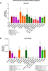
Bactericidal activity over the M. bovis BCG air–liquid interface preformed biofilms. Twenty-day-old preformed biofilms were treated with RPE, PreA, or Caj at MIC or RIF, INH, and EMB at inhibitory (MIC) and superinhibitory concentrations (MIC×10 and MIC×100), either alone or in combination. Analysis of biomass (A) and cell viability (B) was performed after 10 days of treatment. Representative results of three independent experiments; media and error bars (SD) are shown. Differences between treatments were assessed by using one-way analysis of variance (ANOVA) with Tukey’s post hoc multiple comparison test (p < 0.05).
Figure 7
Figure 7
Biofilm inhibitory activity of PreA in combination with antimycobacterial drugs. M. bovis BCG was cultured in the presence of PreA and RIF or INH at inhibitory (MIC) or subinhibitory (MIC/2) concentrations for 20 d. Morphological observations were made (A), as well as biofilm biomass quantification (B) and cell viability (C). The combination of RIF, INH, and EMB at the MIC was used as the maximum inhibition control. Representative results of three independent experiments; media and error bars (SD) are shown. Differences between treatments were analyzed by using one-way analysis of variance (ANOVA) with Tukey’s post hoc multiple comparison test (p < 0.05).

Similar articles

Cited by

References

    1. Torrelles J. B.; Schlesinger L. S. Integrating Lung Physiology, Immunology, and Tuberculosis. Trends Microbiol. 2017, 25 (8), 688–697. 10.1016/j.tim.2017.03.007. - DOI - PMC - PubMed
    1. World Health Organization . Global Tuberculosis Report 2021; World Health Organization: Geneva, Switzerland, 2021.
    1. Yam Y. K.; Alvarez N.; Go M. L.; Dick T. Extreme Drug Tolerance of Mycobacterium abscessus “Persisters. Front. Microbiol. 2020, 11, 359.10.3389/fmicb.2020.00359. - DOI - PMC - PubMed
    1. Greendyke R.; Byrd T. F. Differential Antibiotic Susceptibility of Mycobacterium abscessus Variants in Biofilms and Macrophages Compared to That of Planktonic Bacteria. Antimicrob. Agents Chemother. 2008, 52 (6), 2019–2026. 10.1128/AAC.00986-07. - DOI - PMC - PubMed
    1. McNabe M.; Tennant R.; Danelishvili L.; Young L.; Bermudez L. E. Mycobacterium avium ssp. hominissuis biofilm is composed of distinct phenotypes and influenced by the presence of antimicrobials. Clin. Microbiol. Infec. 2011, 17 (5), 697–703. 10.1111/j.1469-0691.2010.03307.x. - DOI - PMC - PubMed