. 2024 Aug 6;33(16):1379-1390.
doi: 10.1093/hmg/ddad176.

Demonstration of the pathogenicity of a common non-exomic mutation in ABCA4 using iPSC-derived retinal organoids and retrospective clinical data

Affiliations

Demonstration of the pathogenicity of a common non-exomic mutation in ABCA4 using iPSC-derived retinal organoids and retrospective clinical data

Erin R Burnight et al. Hum Mol Genet. .

Abstract

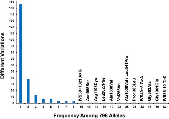
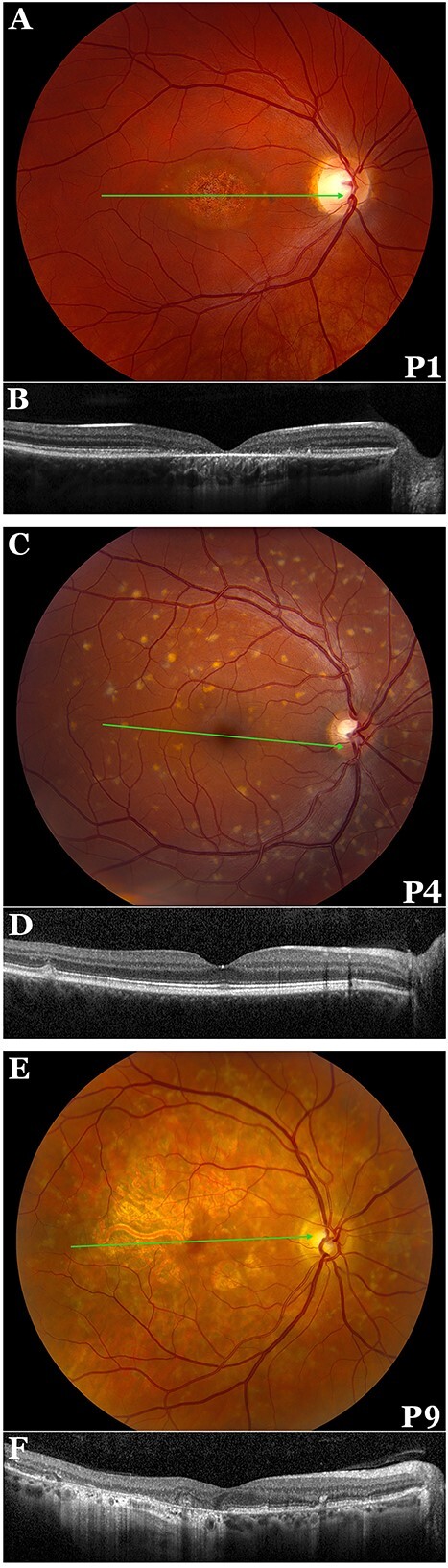
Mutations in ABCA4 are the most common cause of Mendelian retinal disease. Clinical evaluation of this gene is challenging because of its extreme allelic diversity, the large fraction of non-exomic mutations, and the wide range of associated disease. We used patient-derived retinal organoids as well as DNA samples and clinical data from a large cohort of patients with ABCA4-associated retinal disease to investigate the pathogenicity of a variant in ABCA4 (IVS30 + 1321 A>G) that occurs heterozygously in 2% of Europeans. We found that this variant causes mis-splicing of the gene in photoreceptor cells such that the resulting protein contains 36 incorrect amino acids followed by a premature stop. We also investigated the phenotype of 10 patients with compound genotypes that included this mutation. Their median age of first vision loss was 39 years, which is in the mildest quintile of a large cohort of patients with ABCA4 disease. We conclude that the IVS30 + 1321 A>G variant can cause disease when paired with a sufficiently deleterious opposing allele in a sufficiently permissive genetic background.

Keywords: ABCA4; RNA splicing; Stargardt disease; genetic testing; retinal organoids.


Figures

Figure 1
Figure 1
Allelic diversity of ABCA4. This figure shows the number of different disease-causing ABCA4 variants (y-axis) that occur at each frequency (x-axis) in a cohort of 390 probands with clinical features of ABCA4 disease. The unshared alleles of sixteen pseudo-dominant relatives are also included for a total of 796 alleles. Variants seen ten or more times in the cohort are specifically labeled. Of the 242 different plausible disease-causing variants seen in this cohort, 156 (64.5%) were observed only once.
Figure 2
Figure 2
Presence of the IVS30 + 1321 A>G variant increases the rate of pseudo exon 30.1 inclusion in patient iPSC-derived retinal organoids. (A–D) Phase (A and C) and immunohistochemical (B and D) analysis of iPSC-derived retinal organoids (≥10 per line) from a control individual (A–B) and patient P2 (C–D) at 90-days of differentiation. (E) rt-PCR analysis targeted against the photoreceptor cell markers OTX2, NRL, NR2E3, RCVRN and OPN1SW at 90-days of differentiation. (F–I) Phase (A and H) and immunohistochemical (G and I) analysis of iPSC-derived retinal organoids ((≥10 per line) from a control individual (F–G) and the proband (H–I) at 140-days of differentiation. (J and K) Quantitative PCR analysis of control (J) and proband (K) retinal organoids (≥10 per line) using TaqMan probes targeted against the photoreceptor cell markers CRX, RCVRN, and ARR3, at differentiation day 7, 40, 75 and 140, showing normal photoreceptor development. (L) Quantitative PCR analysis of control and proband retinal organoids using TaqMan probes targeted against ABCA4, at differentiation day 7, 75 and 140. (M) Quantitative PCR analysis of control and patient retinal organoids (≥10 per line) using TaqMan probes targeted against ABCA4 pseudo exon 30.1 at differentiation day 140. Data is expressed as a ratio of exon 30.1 to total ABCA4 (probes targeting exons 4–5 and 49–50). Despite normal development and near identical levels of ABCA4 expression, iPSC-derived retinal organoids obtained from the proband express greater than two-fold more pseudo exon 30.1 than those obtained from an unaffected control.
Figure 3
Figure 3
Presence of the IVS30 + 1321 A>G variant increases the rate of pseudo exon 30.1 inclusion in iPSC-derived retinal organoids generated from a second individual. (A–C) Phase micrographs depicting iPSC-derived retinal organoids (≥10 per line) from an additional patient (P1) with IVS30 + 1321 at 140-days of differentiation. By 140 days of differentiation retinal organoids contain photoreceptor cells with outer segment projections. (D–F) Immunocytochemical analysis of P1 iPSC-derived retinal organoids (≥10) at 140-days of differentiation (D: ARR3, OTX2, DAPI. E: OTX2, RCVRN, DAPI. F: RHO, ARR3, DAPI ). (G) Quantitative PCR analysis of control and proband 2 iPSC-derived retinal organoids (≥10 per line) at 140 days of differentiation using TaqMan probes targeted against RCVRN, RHO, ARR3 and ABCA4. Data are expressed as fold change relative to day 0. (H) Quantitative PCR analysis of control and proband 2 retinal organoids (≥10 per line) using TaqMan probes targeted against ABCA4 pseudo exon 30.1 at differentiation day 140. Data is expressed as a ratio of exon 30.1 to total ABCA4. Despite normal development and near identical levels of ABCA4 expression, iPSC-derived retinal organoids obtained from proband 2 express greater than two-fold more pseudo exon 30.1 than those obtained from an unaffected control.
Figure 4
Figure 4
Patient-derived RPE differentiation. (A–C) Phase contrast images of patient-derived RPE cells (D80) from control (A) and two patients (P1—B and P2—C) carrying the IVS30 + 1321 A>G variant. Scale bars = 1000 uM. (D and E) Immunocytochemical analysis of iPSC-derived RPE cells from control (D) and patients (P1—E and P2—F) at D80. ZO-1, DAPI. (G–J) rt-PCR analysis targeted against the RPE cell markers MITF (G), CRALBP (H), BEST1 (I). Patient-derived RPE demonstrate ~3.5 and ~2 fold increased exon 30.1 inclusion (J), respectively. Error bars represent S.E.M. N = 3.
Figure 5
Figure 5
Color fundus photography and OCT obtained from patients with IVS30 + 1321. (A) Color fundus photograph of the right eye of P1, a 27-year-old woman with 20/100 acuity. There is an oval zone of atrophy with a shiny base centered on the foveola and ringed by a few small yellow flecks. (B) OCT of the right eye of P1 reveals central outer retinal loss and an RPE hypertransmission defect corresponding to the atrophy seen on the fundus image. (C) Color fundus photograph of the right eye of P4, a 30-year-old man with 20/15 acuity. There are numerous bright yellow flecks throughout the posterior pole that spare the central 1.5 mm. (D) OCT of the right eye of P4 shows intact retinal layers except for two hyperreflective deposits located between the ellipsoid band and the RPE. (E) Color fundus photograph of the right eye of P9, a 62-year-old man with 20/25-1 acuity. There is extensive macular RPE atrophy circumscribing the fovea with peninsular sparing of the foveal center. More peripherally there are numerous yellow flecks arranged in a reticular pattern, many of which are associated with hyperpigmentation and early atrophy. There is relative sparing of the peripapillary retina. (F) OCT of the right eye of P9 reveals temporal RPE and outer retinal atrophy with a zone of RPE hyper-transmission that reaches the fovea. The nasal retinal laminations are largely preserved with several small foci of incomplete outer retinal atrophy and pigment migration. The green arrow in the fundus photos (A, C, and E) represents the position of the OCT scan.

References

    1. Weng J, Mata NL, Azarian SM. et al. Insights into the function of Rim protein in photoreceptors and etiology of Stargardt's disease from the phenotype in abcr knockout mice. Cell 1999;98:13–23. - PubMed
    1. Quazi F, Lenevich S, Molday RS. ABCA4 is an N-retinylidene-phosphatidylethanolamine and phosphatidylethanolamine importer. Nat Commun 2012;3:925. - PMC - PubMed
    1. Lenis TL, Hu J, Ng SY. et al. Expression of ABCA4 in the retinal pigment epithelium and its implications for Stargardt macular degeneration. Proc Natl Acad Sci U S A 2018;115:E11120–7. - PMC - PubMed
    1. Stone EM, Andorf JL, Whitmore SS. et al. Clinically focused molecular investigation of 1000 consecutive families with inherited retinal disease. Ophthalmology 2017;124:1314–31. - PMC - PubMed
    1. Webster AR, Heon E, Lotery AJ. et al. An analysis of allelic variation in the ABCA4 gene. Invest Ophthalmol Vis Sci 2001;42:1179–89. - PubMed

Publication types

Substances