. 2023 Nov 6;118(1):47.
doi: 10.1007/s00395-023-01017-x.

Activation of the integrated stress response rewires cardiac metabolism in Barth syndrome

Affiliations

Activation of the integrated stress response rewires cardiac metabolism in Barth syndrome

Ilona Kutschka et al. Basic Res Cardiol. .

Abstract

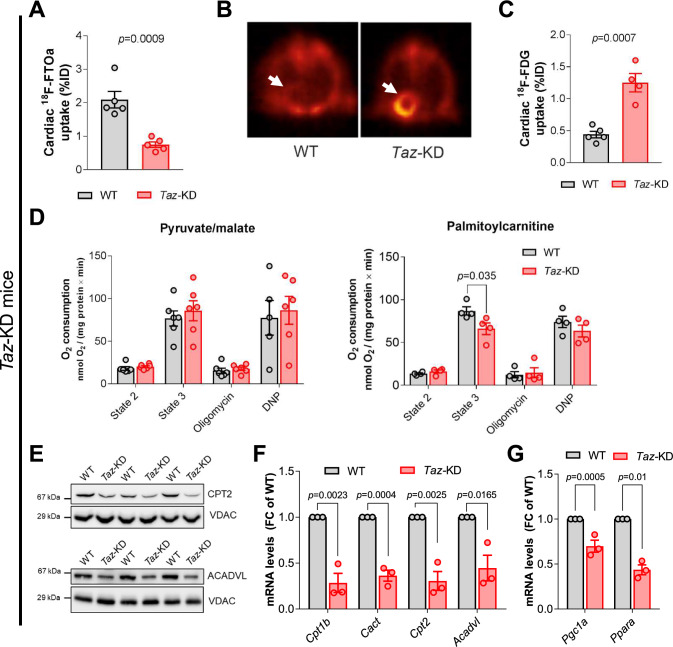
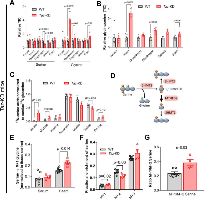
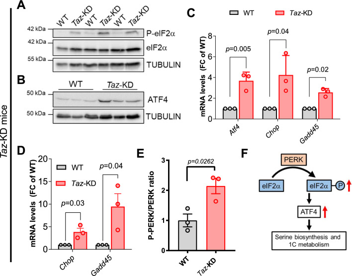
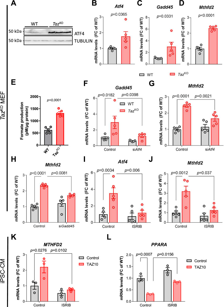
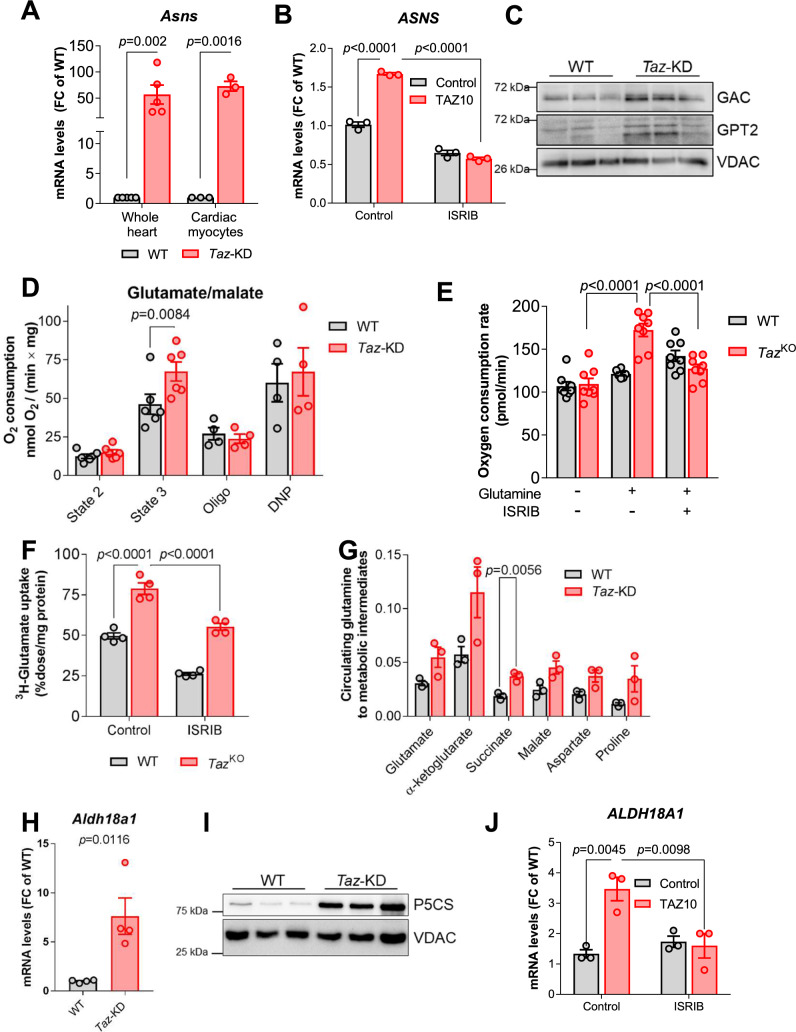
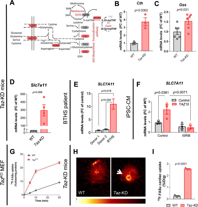
Barth Syndrome (BTHS) is an inherited cardiomyopathy caused by defects in the mitochondrial transacylase TAFAZZIN (Taz), required for the synthesis of the phospholipid cardiolipin. BTHS is characterized by heart failure, increased propensity for arrhythmias and a blunted inotropic reserve. Defects in Ca2+-induced Krebs cycle activation contribute to these functional defects, but despite oxidation of pyridine nucleotides, no oxidative stress developed in the heart. Here, we investigated how retrograde signaling pathways orchestrate metabolic rewiring to compensate for mitochondrial defects. In mice with an inducible knockdown (KD) of TAFAZZIN, and in induced pluripotent stem cell-derived cardiac myocytes, mitochondrial uptake and oxidation of fatty acids was strongly decreased, while glucose uptake was increased. Unbiased transcriptomic analyses revealed that the activation of the eIF2α/ATF4 axis of the integrated stress response upregulates one-carbon metabolism, which diverts glycolytic intermediates towards the biosynthesis of serine and fuels the biosynthesis of glutathione. In addition, strong upregulation of the glutamate/cystine antiporter xCT increases cardiac cystine import required for glutathione synthesis. Increased glutamate uptake facilitates anaplerotic replenishment of the Krebs cycle, sustaining energy production and antioxidative pathways. These data indicate that ATF4-driven rewiring of metabolism compensates for defects in mitochondrial uptake of fatty acids to sustain energy production and antioxidation.

Keywords: Amino acid; Barth syndrome; Fatty acid oxidation; Metabolism; Mitochondria; Oxidative stress.


Conflict of interest statement

CM received speaker and consultancy honoraria from Boehringer Ingelheim, AstraZeneca and Novo Nordisk. TT is founder and shareholder of Cardior Pharmaceuticals (outside of this study). TT filed and licensed patents about the use of noncoding RNAs (outside of the paper).

Figures

Fig. 1
Fig. 1
Metabolic rewiring in BTHS. Summary of changes in gene expression pattern in cardiac samples from Taz-KD mice compared to WT. Red indicates significant upregulation in transcriptomic data described in Fig. 3, genes with a published AARE element in their promoter are indicated with a star. Green indicates significant downregulation, amino acids are indicated in blue. (Ca2+ calcium, OAA ocaloacetate, GCS γ-glutamylcysteine, P5C pyroline-5-carboxylate, G6P glucose-6-phosphate, 3GP 3-phosphoglycerate, 3PHP 3-phosphohydroxypyruvate, 3PS 3-phosphoserine)
Fig. 2
Fig. 2
Uptake and utilization of fatty acids is impaired in tafazzin-deficient mouse hearts. A Cardiac 18F-FTOa uptake normalized to blood levels in WT and Taz-KD mice (12 weeks old). n = 5 per genotype. B Representative PET-CT images of cardiac 18F-FDG uptake in WT and Taz-KD hearts of 12-week-old mice. C Quantification of cardiac 18F-FDG uptake normalized to lung uptake in WT and Taz-KD mice. n = 5 per genotype. D Oxygen consumption rate (OCR) of isolated cardiac mitochondria from 20-week-old WT and Taz-KD mice supplied with pyruvate and malate (left panel, n = 6 per genotype) or palmitoylcarnitine (right panel, n = 4 per genotype) in absence (state 2) and presence (state 3) of ADP (1 mM) before adding oligomycin and 2,4-dinitrophenol (DNP). E Western blot analysis of indicated proteins in isolated mitochondria. F Cardiac mRNA levels of indicated genes normalized to the mtDNA-encoded ribosomal RNA mS12. n = 3 per genotype. G Cardiac mRNA levels of Pgc1a and Ppara normalized to Gapdh. n = 3 per genotype. Data are mean ± SEM; n-numbers are numbers of hearts or animals. Statistical significance was determined with unpaired Student t-test for panels A, C, F, G, and by two-way ANOVA followed by Bonferroni post-test for panel D
Fig. 3
Fig. 3
Key enzymes of serine and 1C metabolism are upregulated in BTHS. A Volcano plot of log2 fold change versus -log p values of all expressed genes in heart muscle of 20-week-old Taz-KD mice in comparison with WT. X axis: log2 transformed fold changes. Y-axis: minus log10 transformed p value. n = 5 per genotype. B Scheme of metabolic pathways with significantly upregulated genes in Taz-KD vs. WT hearts highlighted in red. C mRNA levels in mouse hearts normalized to mS12 or Gapdh, respectively. n = 3 per genotype. D mRNA levels in cardiac myocytes isolated from 12 week old mice by Langendorff perfusion normalized to mS12. n = 3 per genotype. Western blot analysis of indicated proteins in mouse cardiac lysates (E, F) or isolated mouse cardiac mitochondria (G, H). n = 3 per genotype. I mRNA levels normalized to L28 in iPSC-derived cardiac myocytes. n = 3 technical replicates, one representative experiment shown. J Western blot analysis in iPSC-derived cardiac myocytes. One representative experiment shown. Samples loaded in two concentrations. K mRNA levels normalized to L28 in myocardial samples from one BTHS patient and three controls without heart failure. L Western blot analysis in myocardial samples from one healthy control, one BTHS patient, one patient with ischemic heart disease (IHD), and one patient with dilated cardiomyopathy (DCM). Data are mean ± SEM; n-numbers are numbers of hearts or animals for panels A to H and technical replicates in panel I and K; statistical significance was determined with unpaired Student t-test
Fig. 4
Fig. 4
Serine and 1C metabolism are activated in Taz-KD hearts. A Mass spectrometry analysis of serine and glycine levels in different tissues of 10-week-old Taz-KD mice normalized to WT mice. n = 3–7 for WT and n = 3–4 for Taz-KD. B Glycine/serine ratios calculated from measurements in A (TIC, total ion chromatogram). C Cardiac amino acid labeling from [U-13C,15N]glutamine infusion. Metabolite enrichment was normalized to the enrichment of cardiac [15N]glutamine. n = 3 per genotype. D Scheme of [2,3,3-2H]serine metabolic pathways. E Heart and serum glycine labeling from [2,3,3-2H]serine infusion. Metabolite enrichment was normalized to the enrichment of tissue [2,3,3-2H]serine. n = 7 for WT and n = 4 for Taz-KD. F Cardiac serine labeling from [2,3,3-2H]serine infusion. n = 7 for WT and n = 4 for Taz-KD. G Ratio of M + 1/M + 2 labelled serine calculated from experiment in (F). n = 7 for WT and n = 4 for Taz-KD. Data are mean ± SEM; n-numbers are numbers of hearts or animals; statistical significance was determined with two-tailed Student’s t-test
Fig. 5
Fig. 5
The ISR is activated in heart of Taz-KD mice. A Western blot analysis of phosphorylated and total eIF2α and TUBULIN as loading control in cardiac lysates. n = 3 per genotype. B Western blot analysis in cardiac lysates. n = 3 per genotype. C Cardiac mRNA levels of indicated genes normalized to Gapdh. n = 3 per genotype. D mRNA levels normalized to Gapdh in cardiac myocytes. n = 3 per genotype. E Quantification of western blot analysis of phosphorylated and total PERK protein in cardiac lysates. n = 3. F Scheme of the ISR signaling pathway. Data are mean ± SEM; n-numbers are numbers of hearts or animals; statistical significance was determined with unpaired Student’s t-test
Fig. 6
Fig. 6
ATF4 induces upregulation of 1C metabolism genes in tafazzin-deficient MEFs and iPSC-CM. A Western blot analysis of ATF4 and TUBULIN in WT and TazKO MEFs. B-D mRNA levels of indicated genes in MEFs normalized to mS12. n = 5 per genotype. E Formate levels in the supernatant of MEF culture, normalized to total protein levels as a measure of cell density, n = 3. F, G mRNA levels of indicated genes in MEFs treated with siRNA against Atf4 or control normalized to mS12. n = 5. H mRNA levels of Mthfd2 in MEFs treated with siRNA against Gadd45 or control normalized to mS12. n > / = 4. I, J mRNA levels of indicated genes in MEFs treated with ISRIB or DMSO normalized to mS12. n = 5. K, L mRNA levels of indicated genes in iPSC-derived CM treated with ISRIB or DMSO normalized to L28. n = 3. Data are mean ± SEM; n-numbers are numbers of independent experiments for panels BJ and technical replicates in panels K and L; statistical significance was determined with unpaired Student’s t-test in panels B to E, and with one-way ANOVA followed by Tukey’s multiple comparison for panels F to L
Fig. 7
Fig. 7
Rewiring of glutamate metabolism in Taz-KD hearts. A mRNA levels in whole hearts (n = 5) or isolated mouse cardiac myocytes (n = 3), normalized to Gapdh. B mRNA levels in iPSC-derived CM treated with ISRIB or DMSO, normalized to ACTB, n = 3. C Western blot analysis of cardiac mouse mitochondria. D OCR of isolated cardiac mitochondria from 20-week-old WT and Taz-KD mice supplied with glutamate and malate in the absence (state 2) and presence (state 3) of ADP (1 mM). Subsequent measurements are done after oligomycin and DNP administration. n = 6 per genotype. E OCR of MEFs with or without glutamine and the administration of ISRIB. n > / = 6. F Uptake of 3H-glutamate in WT and TazKO MEFs treated or not with ISRIB. n = 4. G Cardiac metabolite levels labeled from [U-13C, 15N]glutamine infusion normalized to the enrichment of tissue [U-13C,15N]glutamine. n = 3. H Cardiac mRNA levels of Aldh18a1 in WT and Taz-KD mice normalized to Gapdh. n = 5. I Western blot analysis in cardiac lysates. n = 3 per genotype. J mRNA levels in iPSC-derived CM treated with ISRIB or DMSO as a control normalized to L28. n = 3. Data represent mean ± SEM; n-numbers are numbers of animals for panels A, C, D, G, H and I numbers of independent replicates for panels E and F and technical replicates for B and J. Statistical significance was determined with unpaired Student’s t-test in panels A and H, with two-way ANOVA followed by Bonferroni’s multiple comparisons test for panel D and with one-way ANOVA followed by Tukey’s multiple comparisons test for panels B, E, F, and K
Fig. 8
Fig. 8
ATF4 induces cysteine uptake via the xCT system to support glutathione biosynthesis. A Cartoon of metabolic pathways. Significantly upregulated genes in Taz-KD vs. WT hearts highlighted in red. B, C Cardiac mRNA levels in mice normalized to Gapdh. n = 3 per genotype for B and n = 6 per genotype for C. D Cardiac mRNA levels in mice normalized to Gapdh. n = 4. E mRNA levels normalized to L28 in myocardial samples from one BTHS patient and 2 healthy controls. Technical replicate, n = 3. F mRNA levels of in iPSC-derived CM from one BTHS patient (TAZ10) or control normalized to ACTB, treated with ISRIB or DMSO. n = 3. G Uptake of the xCT system-specific radiotracer 18F-FASu into MEFs, measured by scintillation counting. H Representative PET-CT image of cardiac 18F-FASu uptake in 34-week-old mice. I Quantification of cardiac 18F-FASu uptake in mice. Data are mean ± SEM; n-numbers are numbers of animals for panels B–D and I, and numbers of technical replicates for panels F. Statistical significance was determined with unpaired Student’s t-test in panels B–D and I and with one-way ANOVA followed by Tukey’s multiple comparison for panels E and F

References

    1. Acehan D, Vaz F, Houtkooper RH, James J, Moore V, Tokunaga C, Kulik W, Wansapura J, Toth MJ, Strauss A, Khuchua Z. Cardiac and skeletal muscle defects in a mouse model of human Barth syndrome. J Biol Chem. 2011;286:899–908. doi: 10.1074/jbc.M110.171439. - DOI - PMC - PubMed
    1. Ardanaz N, Yang XP, Cifuentes ME, Haurani MJ, Jackson KW, Liao TD, Carretero OA, Pagano PJ. Lack of glutathione peroxidase 1 accelerates cardiac-specific hypertrophy and dysfunction in angiotensin II hypertension. Hypertension. 2010;55:116–123. doi: 10.1161/HYPERTENSIONAHA.109.135715. - DOI - PMC - PubMed
    1. Badolia R, Ramadurai DKA, Abel ED, Ferrin P, Taleb I, Shankar TS, Krokidi AT, Navankasattusas S, McKellar SH, Yin M, Kfoury AG, Wever-Pinzon O, Fang JC, Selzman CH, Chaudhuri D, Rutter J, Drakos SG. The role of nonglycolytic glucose metabolism in myocardial recovery upon mechanical unloading and circulatory support in chronic heart failure. Circulation. 2020;142:259–274. doi: 10.1161/CIRCULATIONAHA.119.044452. - DOI - PMC - PubMed
    1. Barth P, Scholte H, Berden J, Van der Klei-Van MJ, Luyt-Houwen I, et al. An X-linked mitochondrial disease affecting cardiac muscle, skeletal muscle and neutrophil leucocytes. J Neurol Sci. 1983;62:327–355. doi: 10.1016/0022-510x(83)90209-5. - DOI - PubMed
    1. Bertero E, Maack C. Calcium signaling and reactive oxygen species in mitochondria. Circ Res. 2018;122:1460–1478. doi: 10.1161/CIRCRESAHA.118.310082. - DOI - PubMed

Publication types